Integumentary System The Skin FYI about skin 1

Integumentary System The Skin

FYI about skin 1. Largest external organ 2. A. 1 mm thick when you’re born B. Thickens as you age (2 mm) C. Thins as you reach old age (makes you cold 0 3. 6 -10 lbs 4. Soles of your feet and palms have no hair follicles (recall the keratinized skin slide) 5. Majority of dust in your house is made of skin flakes

Functions of the Skin 1. Regulates body temperature 2. Protection 3. Sensation 4. Excretion 5. Immunity 6. Blood reservoir 7. Vitamin Synthesis (D)

Synthesis of Vitamin D • 1 hr/wk of sunlight needed to activate the body's own vitamin D precursor • Sunlight triggers cholesterol to convert into Vitamin D • Supplemented in fortified milk. • Essential to regulates Ca & Phosphorus to keep skeleton strong • Deficiencies can cause rickets & osteoporosis

Surface Film • Protective barrier formed by a thin film coating the surface – sweat, sebum (oils) & shed epithelial cells – Functions • Protect against irritants • Antimicrobial • Lubricate, hydrate
 avascular Stratified squamous tissue 2. Dermis =")
Layers/Tissues 1. Epidermis= exposed layer (epitheleal cells) avascular Stratified squamous tissue 2. Dermis = Connective tissue Dense Irregular CT 3. Hypodermis (AKA fascia or subcutaneous layer) Adipose tissue 4. “Accessory” structures • Hair/Nails • Glands (oil/sweat) • Nerve endings (deep to superficial)

Layers of the skin dermal ridges = fingerprints “dying” part of skin “living” part of skin “cushioning & insulates” part of skin

The skin LAYERS OF THE EPIDERMIS - pg 126 LAYERS

Cell within the Epidermis cytes = cells 1. Keratinocytes – – 90% of epidermal cells become filled with tough fibrous keratin 2. Melanocytes – Contribute to color of skin – Keep UV light from penetrating deep (protective) 3. Langerhans cells – Play a role in immune defense – Function with white blood cells (macrophages/phagocytosis) 4. Merkel Cells – Mechanoreceptors (touch/pressure) - associated w/hairs

Keratinocytes dye as they move towards surface
 (sperficial layer) -continually shedding-")
5 sub-layers of the epidermis 1. Horny layer (stratum corneum) (sperficial layer) -continually shedding- dead keratinocytes -this is the dead skin that you shed & dust in your house 2. Clear Layer (stratum lucidum) -translucent layer as cells loose their cytoplasmic contents and become fully keratinized. -Thicker skin has thicker layer(palm, soles), absent in thin skin. 3. Granular layer (stratum granulosum) appear grainy -cells are beginning to deteriorate/dye as keratin is forming inside of them. 4. Spinous layer (stratum spinosum) (peeling /flaking skin) - appears to have“spines. ” -spines = desmosomes = side to side attachment of cells 5. Basal layer (stratum basale) AKA: the basement -Inner layer of the epidermis (deep) -basal cells continually divide(mitosis), forming new keratinocytes -replaces old/dying cells (shed from the surface)
")
Layers of Epidermis Stratum Germinativum (growth layer)


Dermal-Epidermal Junction • Cements epidermis to dermis • Any large detachment can result in severe infection and death

Dermis The dermis is the middle layer of the skin- 2 layers thick. 1. Papillary layer = areolar tissue -containing fine elastic fibers, - dermal papillae = fingerprints -corpuscles of light touch (Meissner's corpuscles). 2. Reticular layer = dense irregular tissue (tree branch looking) -contains collagen fibers A. B. C. D. E. F. blood vessels (body temp) lymph vessels (immune) hair follicles sebaceous (oil) glands sweat glands(ducts of sudoriferous) Adipose tissue

Reticular Layer • • Structural strength Tendons/ligaments Elastic quality Attachment point of skeletal and smooth muscles • Arrector pili muscles =goose bumps • Sensory receptors (nerves) are located in this layer

Growth and Repair of Dermis • Strength and elasticity = collagen and elastin. • Lines of cleavage (tension) • -direction of collagen fiber bundles • -determines surgical incisions • Flexure lines- deep attachment of skin to muscles. Causes deep wrinkles. Langer's cleavage lines

Growth and Repair of Dermis Scars Keloid scar – Fibroblasts reproduce to heal a wound resulting in a mass of connective tissue Stretch Marks – If elastic fiber are stretched too much or too quickly they tear (quick growth)

Subcutaneous-superficial fascia • Hypodermis = subcutaneous layer = subcutitis = fascia • Subcutis = network of collagen and fat cells 1. helps conserve the body's heat 2. "shock absorber" cushions

Appendages of the Skin Hair – Follicles develop before birth – Lanugo is hair that forms before birth – Vellus hair-strong fine hair that covers the body (peach fuzz) – Terminal Hair- forms pubic, under arm hair – In males terminal hair replaces vellus hair on extremities, chest and beard

– Germinal matrix is highly mitotic and pushes cells up to form hair – Melanocytes are deposited into hair to give it color

Male Pattern Baldness Alopecia • Two conditions 1. Genes for baldness 2. Sex influenced by male hormone testosterone
 glands -connected to hair follicles -absent in")
Glands of the Skin 1. Sebaceous (oil) glands -connected to hair follicles -absent in the palms & soles – – – produce sebum, moistens hair Waterproofs softens skin inhibits bacterial growth. Enlarged sebaceous glands may produce blackheads, pimples, and boils. 2. Ceruminous glands – modified sebaceous glands – waxy substance called cerumen AKA: ear wax – To use a q-tip or not?
 -perspiration, -maintains temperature -eliminates sm amts of")
Glands of the Skin 3. Sudoriferous (sweat) -perspiration, -maintains temperature -eliminates sm amts of wastes. A. Eccrine = merocrine-most abundant-all over body -not associated with hair. B. Apocrine (stinky) -limited to axillary region, pubis, anal, and areola regions -ducts open into hair follicles

Appendages of the Skin Nails • Nails are hard, keratinized • Dorsal surfaces of the terminal portions of the phalanges • Parts : Ø Ø Ø nail body free edge Lunula -- latin for “little moon” Eponychium --cuticle Root (matrix)

Problems With Integumentary System

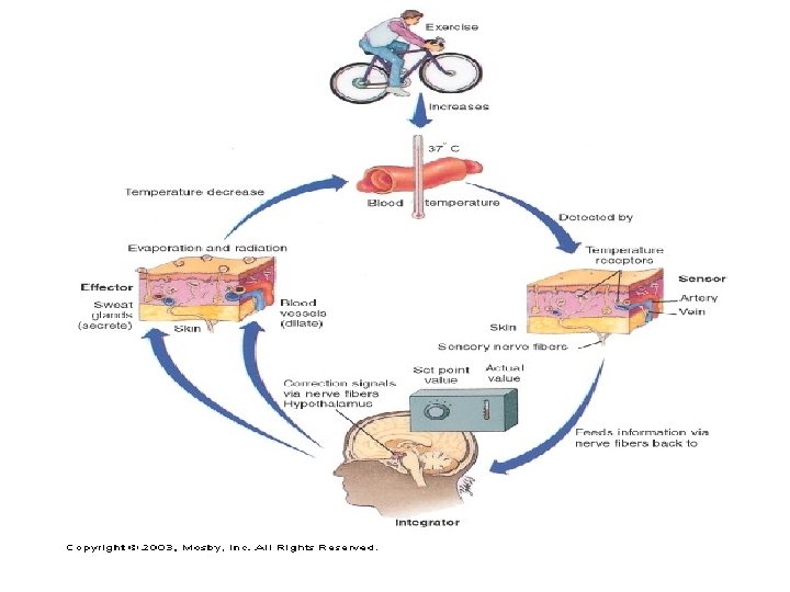
Thermoregulation • Body temperature fluctuates throughout the day. • Lowest in the morning; highest at night • Biochemical reactions and enzymes function within a narrow range • Hypothalamus is the body’s thermostat

Abnormal Body Temperature Heat Exhaustion • Body temperature remains normal • Loss of large amount of fluids and electrolytes • Vertigo(dizziness), nausea and loss of consciousness Heat Stroke • Body temp rises above 105 F • Tacycardia (rapid heart rate) • Hot dry skin (stop sweating!) • Confusion, convulsions • Body must be cooled immediately or death can result (No sufdden changes in temp) Hypothermia (hypo mean under) • Body temp below 95 F • Slowed heart rate • Treated by slowly warming persons body Frosbite • Damage results from ice crystals forming in skin • Necrosis results and possible gangrene due to lack of blood flow

Disorders of the Skin Albinism • Every race has about the same number of melanocytes • Skin color is determined by the amount of melanin produced(genetic) • Carotene, yellow pigment that also contributes to skin color(fat cells) • Sun exposure cause melanocyte to produce more melanin (AKA: a tan) • In albinism they lack pigment in hair, skin and eyes

Disorders of the Skin Epidermolysis bullosa • Mutation in the keratin gene. – Epidermis and dermis is not held together. – any friction causes them to separate and leave open sores and blister. – Bone marrow transplants have proven beneficial.

Disorders of the Skin Vitiligo • Charaterized by white patches of skin • Melanocytes no longer produce pigment

Disorders of the Skin Onycholysis • Separation of nails from the nail bed • Trauma to nail or fungal infection

Disorders of the Skin Impetigo • Caused by staph or strep infections • Blisters with yellowish or dark scabs • Common with children and around pools Tinea • Fungal infections • Ringworm, athletes foot, thrush, jock itch (ID by round circle patches) Warts • Bumpy projections caused by papilloma viruses • Easily removed Boils • Staph infection of follicles • Large pus filled lesions

Disorders of the Skin Acne • Result of overactive secretion of sebaceous glands(5 -10 X more than normal) • Pores are clogged and can form infected pustules Blisters • Desmosomes are damaged by injury or irritation • Immune response to protect skin Calluses • Hardened skin formed by continual pressure/rubbing Corns • Thick hardened skin usually form in bony areas as a result of friction

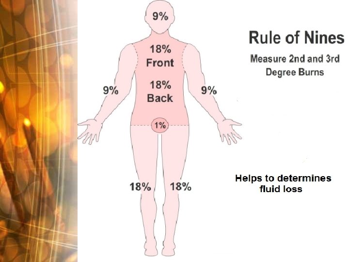
BURNS Can be caused by 1. Heat 2. Electricity 3. UV Rays 4. Chemicals First Degree Effects only epidermis Mild sunburns Second Degree Epidermis & top layer of dermis are damaged blisters form, shiny appearance Entire depth of skin destroyed, Charred skin ( black or grayish) Skin cannot heal itself Graphs normally needed Third Degree
 Basal Cell Carcinoma Melanoma Squamous Cell Carcinoma – can show")
Skin Cancer Benign =Non-Malignant(cancerous) Basal Cell Carcinoma Melanoma Squamous Cell Carcinoma – can show up anywhere – 95% of all skin cancers. – Metastasizes easily – Rarely metastasize – Severe burns increase your risk – sun exposed areas – Lifetime daily exposure

Basal Cell Carcinoma Squamous Cell Carcinoma

Malignant Melanoma

Prevention • Limit exposure to sun • Wear sunscreen • Screen body and map moles by your doctor • Know your ABCD’s
- Slides: 41