Integumentary System Skin and Its Appendages Anatomy Physiology


 Flat thin squamous cells n Surface cells dead & continually")
 n Nuclei absent n Cells contain eleidin which will be")
 n Keratinization begins n 2 -4 layers deep n Maybe")
 n 8 -10 layers with prominent desmosomes which appear spiny")
 n Single layer of columnar cells n Only cells which")


- Slides: 36
Integumentary System Skin and Its Appendages Anatomy & Physiology
Skin or Integument n Largest organ in the body n Integumentary System: denotes the skin and its appendages
Structure of the skin
Layers of the skin n Epidermis: Outer, thinner layer n Dermis: Thicker layer, connective tissue n Hypodermis: Subcutaneous layer, superficial fascia
Thick Skin Refers to epidermal layer only n Found: palms of hands, soles of feet, fingertips n Each of the 5 layers present n Dermal papillae: fingerprints n No hair n
Cell Types of Epidermis n Keratinocytes: contain keratin, make up 90% of epidermal cells n Melanocytes: contribute color to skin n Langerhans cells: immunological reactions in skin
Cell Layers of Epidermis n Stratum corneum n Stratum lucidum n Stratum granulosum n Stratum spinosum n Stratum basale
Stratum corneum (horny layer) Flat thin squamous cells n Surface cells dead & continually being shed n Cytoplasm in cells replaced by keratin n Desmosomes hold cells together n Barrier layer of the n
Stratum lucidum (clear layer) n Nuclei absent n Cells contain eleidin which will be transformed to keratin n Blocks water penetration or loss n Absent from thin skin
Stratum granulosum (granular layer) n Keratinization begins n 2 -4 layers deep n Maybe absent in thin skin n Cells filled with granules called keratohyalin
Stratum spinosum (spiny layer) n 8 -10 layers with prominent desmosomes which appear spiny under a microscope n Cells rich in RNA
Stratum basale (base layer) n Single layer of columnar cells n Only cells which undergo mitosis n Cells migrate from basal layer thru the outer layers
Dermal-Epidermal Junction n Contains basement membrane n Also contains a polysaccharide gel that “glues” 2 layers together
Dermis n Thin papillary layer & thick reticular layer n Thickest on soles & palms n Thinnest on eyelids & penis n Mechanical strength of skin
Papillary layer n Forms the bumps, dermal papillae which project into epidermis n Allows us to grip surfaces & creates fingerprints
Reticular layer n More dense collagen & elastic fibers n Serves as point of attachment for muscle fibers n Skeletal muscle: muscles of facial expression n Smooth muscle: arrector pili muscles on hair follicles
Skin Color n Determined by quantity of melanin in cells of epidermis n All races have about the same number of melanocytes but differ in amount of melanin produced n Sun can increase melanin production
Functions of skin n Protection n Sensation n Movement without injury n Vitamin D production n Excretion n Immunity n Temperature regulation
Heat Loss n Evaporation n Radiation n Conduction n Convection
Burns n Predict body surface area to determine how much fluid to replace: n Rule of palms (1%) n Rule of nines
First degree burn n Involves only the epidermis n No blistering or scarring n Sunburn n Reddening of the skin, mild discomfort
Second degree burn n Involves epidermis & dermis n Blistering, pain, swelling n May scar
Third degree burn n Destruction of epidermis & dermis, may involve underlying tissue n Severe scarring
Appendages of the skin n Hair n Nails n Skin glands
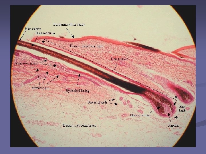
Hair n Lanugo hair: fine hair covering fetus n Vellus hair: replacement for lanugo hair, first appears on scalp, eyelids, eyebrows n Terminal hair: Coarse hair that replaces vellus hair-axillary, pubic, beard, chest & hair on arms &
Hair follicle n Stratum germinativum develops into follicle's inner layer and forms the germinal matrix n Small mound of dermis protruding into germinal matrix
Nails
Parts of the Nail n Matrix- the thickened, proximal area of the nail that is responsible for growth n Bed- the hard translucent visible part of the nail n Root- the point of attachment under the skin n Cuticle- the layer of skin that prevents dirt and bacteria from getting into the nail bed n Free Body- the end of the nail that is not
Glands n Sweat or sudoriferous glands n Eccrine sweat gland n Apocrine sweat glands n Sebaceous glands n Ceruminous glands
Eccrine sweat glands n Most numerous n Over most of the body n Secretory portion located in the subcutaneous tissue n Simple coiled tubular gland
Apocrine Sweat glands n Found in armpit, areola of breast, around the anus n Large than eccrine n Connected with hair follicles
Sebaceous glands n Secrete sebum into each follicle
Ceruminous glands n Modification of apocrine sweat glands n Open into ears n Produce cerumen
Image Citations n n n n Slide 1: cross section of skin, 7/12/06, http: //vilenski. org/science/humanbody/hb_html/skin. html Slide 3: Anthony’s Textbook of Anatomy & Physiology, Seventeenth Edition by Thibodeau & Patton, Chapter 6. Slide 5: Thick skin, 7/30/06, http: //erl. pathology. iupui. edu/HISTO/LABE 151. HTM Slide 6: Melanocytes, 7/30/06, http: //www. lab. anhb. uwa. edu. au/mb 140/Core. Pages/Integumentary/Integum. htm Slide 7: Thick skin trichrome, 7/30/06, http: //www. lab. anhb. uwa. edu. au/mb 140/Core. Pages/Integumentary/Integum. htm Slide 8: Slide 43, Thick skin, 7/30/06, http: //w 3. ouhsc. edu/histology/Glass%20 slides/43_09. jpg Slide 9: Stratum lucidum human foot, 7/30/06, http: //oregonstate. edu/~hanba/Projector%20 Slides/Skin%20 Strat um%20 Lucidum%20 Human%20 Foot-2. jpg Slide 10: Stratum granulosum, 7/30/06, http: //anatomy. iupui. edu/courses/histo_D 502/D 502 f 04/Labs. f 04/Lab 14/s 31. 100 x. i 3. jp g Slide 11: Stratum spinosum, 7/30/06, http: //anatomy. iupui. edu/courses/histo_D 502/D 502 f 04/Labs. f 04/Lab 14/s 31. 100 x. i 2. jp g Slide 12: Stratum basale, 7/30/06, http: //online-media. unimarburg. de/histologie/introhis/HIS/skin 06. gif Slide 14: Dermis, 7/30/06, http: //sprojects. mmi. mcgill. ca/dermatology/dermis. htm Slide 15: Dermis, 7/30/06, http: //neuromedia. neurobio. ucla. edu/campbell/skin/wp_images/7%20 dermis. jpg Slide 16: 7/30/06, http: //www. potterleague. org/Potter_Kids_Final/pet_body_lang. htm
Image Citations n n n n n Slide 20: Wallace’s rule of nines, 7/30/06, http: //www. sunmed. org/burns. html Slide 21: First degree burn, 7/30/06, http: //www. grossmanburncenter. com/orig-site/web/care/causes. htm Slide 22: Burn symptoms, 7/30/06, http: //www. maggiessecret. com. au/burnsscalds. aspx Slide 23: Third degree burn, 7/30/06, http: //www. grossmanburncenter. com/orig-site/web/care/causes. htm Slide 26: Thibodeau & Patton, Anthony’s Textbook of Anatomy & Physiology, Seventeenth Edition. Slide 27: Thin Skin, 7/30/06, http: //biology. clc. uc. edu/fankhauser/Labs/Anatomy_&_Physiology/A&P 201/I ntegumentary/hair_follicle_100 x_PA 112040 labeled. JPG Slide 28: Sebaceous gland shaft of hair follicle, 7/30/06, http: //www. keele. ac. uk/depts/ms/resources/anatomy/histologyimages/t 146. h tml Slide 29: “Structure of nails”, Thibodeau & Patton, Anthony’s Textbook of Anatomy & Physiology, Seventeenth Edition. Slide 31: “Skin Glands”, Thibodeau & Patton, Anthony’s Textbook of Anatomy & Physiology, Seventeenth Edition. Slide 32: Dermis (Apocrine sweat glands), 7/30/06, http: //www 3. umdnj. edu/histsweb/lab 11 apocrine. html