Integumentary System Integumentary System 7 of total body

Integumentary System

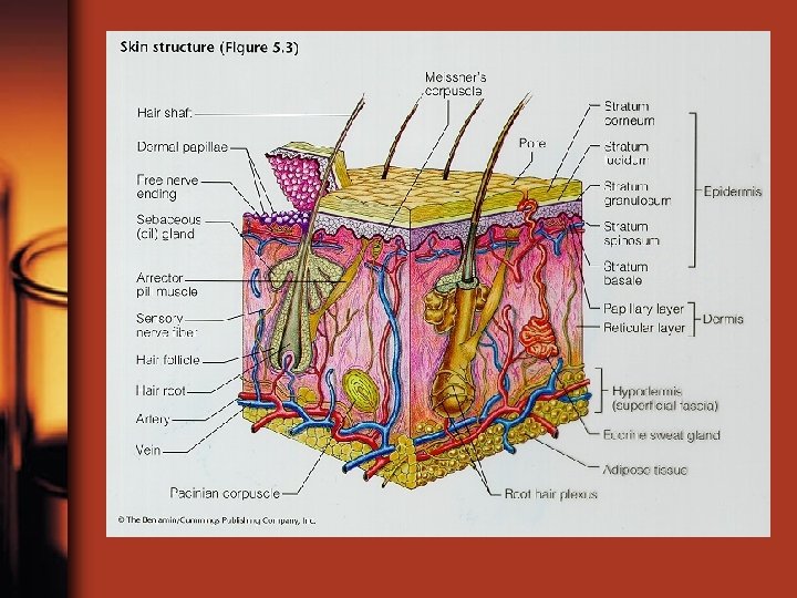
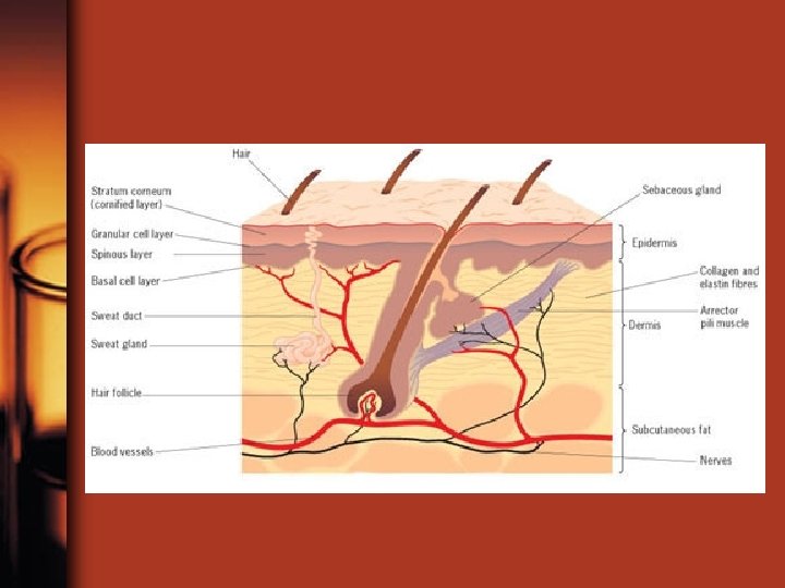
Integumentary System • • 7% of total body weight Components: 1. Cutaneous Membrane a. Epidermis b. Dermis c. Subcutaneous layer • 2. Accessory structures – – Hair Nails Glands Sensory receptors

Functions • • • 1. protection, cushion, insulate 2. excretion of salts, water, wastes 3. maintenance of temp. 4. synthesis of Vitamin D 3 5. storage of nutrients 6. detection of touch, pressure, pain, and temperature

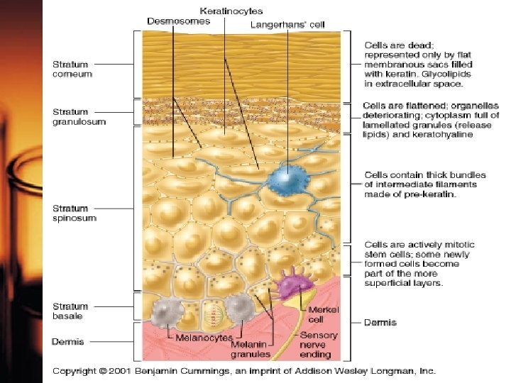
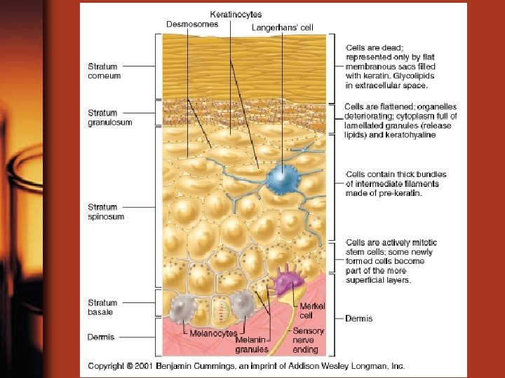
Epidermis • Stratified squamous Epithelium – 4 -5 layers thick –. 08 -. 5 mm thick
 • Innermost layer (deepest) • 3 -5 cells thick • Contains")
Stratum Germinativum (Basale) • Innermost layer (deepest) • 3 -5 cells thick • Contains Merkel cells- sensory for touch • Contains melanocytes (pigment cells) 10 -25% • Cells reproduce in this layer (mitosis)

 • 8 -10 cells thick • Contains Langerhans cells which")
Stratum Spinosum (spiny layer) • 8 -10 cells thick • Contains Langerhans cells which defend against microorganisms and some skin cancers • Mitosis occurs, but less than in Basale layer
 • 3 -5 cells in thickness • Most cells stop dividing")
Stratum Granulosum (grainy) • 3 -5 cells in thickness • Most cells stop dividing in this layer • Produce keratohyalin to start the hardening/waterproofing of cells (keratinization)

 • 3 -5 cells")
Stratum Lucidum • ONLY in THICK skin (palms & soles) • 3 -5 cells in thickness • Flattened cells with eleidin

Stratum Corneum • Outermost layer • Cell membrane thicken; less permeable • Cells die, dehydrate, and keratinize • Keratinization make skin water resistant, less permeable • Average person sheds 40 lbs (18 kg) in a lifetime
 –")
Skin Color • • Due to pigments and blood supply 1. Pigments a) – – b) – – – Carotene- orange/yellow Can be converted into Vitamin A Obtained from plant foods (carrot/tomato) Melanin- brown, yellow/brown, or black Produced by melanocytes Protects cells from UV rays UV exposure = melanocyte activity = color (tan) – ≈ 1000 melanocytes/ mm²
 Circulation - - blood in dermis gives skin a reddish tint")
Skin Color c) Circulation - - blood in dermis gives skin a reddish tint due to the hemoglobin Hematoma = bruise Poor 02 = bluish tint (cyanosis) Decubitis Ulcers (bed sores) are caused by lack of blood supply to an area

Vitamin D • UV exposure causes epidermal cells to produce Vitamin D, a vital hormone, which ensures proper bone growth and development • Vitamin D enables calcium absorption

Dermis • Binds epidermis to underlying tissue • Contains blood vessels, neurons, hair follicles, sweat glands, fibroblast, macrophages, and white blood cells • Strong flexible connective tissue • 2 layers
 – Loose connective tissue")
Dermis • 1. Papillary layer ( superficial 20% of Dermis) – Loose connective tissue (areolar) – Contains dermal papillae • Fingerlike pegs form fingerprints, palmprints, footprints
 – – Dense c. t. /collagen")
Dermis • 2. Reticular layer (80% of Dermis) – – Dense c. t. /collagen and elastic fibers collagen fibers give tensile strength/resilience Elastic fibers provide stretch/recoil properties Separations or less-dense regions between the collagen bundles form the lines of cleavage or tension lines of the skin— important to surgeons – Flexure lines-deep creases where dermis attaches tightly to underlying structures – Site of tatoos
 (superficial fascia) • Deep to the skin • Loose c. t.")
Subcutaneous layer (hypodermis) (superficial fascia) • Deep to the skin • Loose c. t. (areolar) and adipose • Stabilize the position of the skin/anchors to underlying structures (mostly muscle), but allows slide • Contains blood vessels • No organs • Insulator • Thickens with weight gain – ♀ Breast/Thighs – ♂ anterior abdomen “beer belly”

Hair • Present on all skin surfaces except palms, soles, lips , some external reproductive organs • Develops from hair follicle (epidermal cells) • Sense things that lightly touch our skin, protect scalp, shield eyes, filter air we breathe

Hair • Grows 2 -6 yrs, rests 2 months, then falls out • Cells divide at the base and push others up which keratinize (hard) and die • Root: embedded • Shaft: above skin surface • Three layers: • medulla • Cortex • cuticle

hair • Color is determined by two types of melanin and amount • Arrector pili muscle (smooth) is attached to each follicle; when contracts = hair is on end • Vellus- fine “peach fuzz” (women/children) • Terminal – long, dark, typical (scalp/puberty) • Average growth of 2 mm per/week

nails • Composed of keratinized stratified squamous epithelium • Free edge, body, root • Cell division occurs in nail root (matrix) under cuticle; extends out in half moon shape (lunula) • As it develops, slides forward over a layer of epithelium (nail bed)
")
Glands • A. Sebaceous – Usually around hair follicles – Produce oily secretion (sebum) – Keeps hair and nails smooth and soft – Stimulated by hormones, especially androgens

Sweat • 99% water, some salts, and traces of metabolic wastes • Normally produce 500 ml per day and up to 12 L on hot days and during vigorous exercise
 – Originate deep in dermis or subcutaneous layer –")
glands • B. Sudoriferous (sweat) – Originate deep in dermis or subcutaneous layer – Ball shaped coil with tube leading to surface
 – Some sweat glands (eccrine) respond to high body temperature and are")
Sudoriferous (sweat) – Some sweat glands (eccrine) respond to high body temperature and are scattered all over the body – Most abundant on palms, soles, forehead, back, neck – Watery
 – Some sweat glands (apocrine) respond to stress and are located in")
Sudoriferous (sweat) – Some sweat glands (apocrine) respond to stress and are located in the axillary and groin regions; functional after puberty – Thick, sticky, odorous

There are two types of sweat glands that are modified: • 1. Ceruminous glands are in the external ear canal and secrete cerumen (ear wax) • 2. Mammary glands which are in the breasts and secrete milk

Regulation of Body temp. • Excessive heat production – Blood vessels in dermis dialate – Eccrine glands activated-sweat heat lost – Respiration may increase; heat lost thru exhalation – Heart may beat faster; more blood moves into the skin

• Excessive heat loss – Blood vessels in dermis constrict; decreases blood flow to skin; conserves heat – Sweat glands inactive – Nerves may stimulate muscles to contract rhythmically – shiver; generates heat.

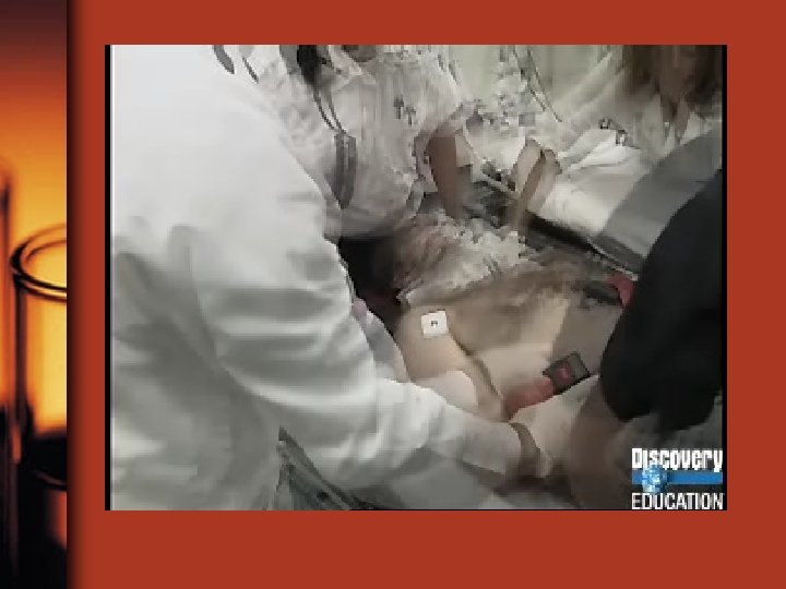
burns • A burn is tissue damage inflicted by heat, electricity, radiation, extreme friction, or certain harmful chemicals • The immediate threat to life from serious burns is a catastrophic loss of body fluids

• Fluid seeps from the burned surfaces • The body quickly loses water and salts • Dehydration leads to fatal circulatory shock • After the initial crisis has passed, infection becomes the main threat.
 as first, second, or third")
• Burns are classified by their severity (depth) as first, second, or third degree burns

First degree burns • • Only the epidermis is damaged Redness, swelling, and pain Heal in a few days Ex. sunburn

Second degree burns • Injury to the epidermis and the upper part of the dermis • Redness, swelling, blisters, and pain • Skin regenerates in 3 -4 weeks w/ little or no scarring

Third degree burns • Consume the entire thickness of the skin • White, red, or blackened • The skin might eventually regenerate, but it is impossible to wait for this because of fluid loss and infection • Skin from other parts of the patients body must be grafted

• Burns are considered critical if any of the following conditions exist: • 1. Over 10% of the body has thirddegree burns • 2. 25% of the body has seconddegree burns • 3. There are third degree burns on the face, hands, or feet.

Rule of nines • Body surface divided into 11 regions, each accounting for 9% (or a multiple of 9%) of total body area

Skin cancer • Skin cancer is the most common type of cancer, w/ about 1 million new cases appearing each year in the U. S. • Most important risk factor: overexposure to the UV rays in sunlight

Basal cell carcinoma • Least malignant • Most common • Cells of the stratum basale proliferate— invade the dermis and hypodermis causing tissue erosions • Dome shaped, shiny nodules • Develop a central ulcer and a “pearly” beaded edge • Grows slowly, metastasis seldom occurs

Squamous Cell Carcinoma • • • Keratinocytes of the stratum spinosum Scaly, irregular, reddened papule Grows rapidly Will metastasize if not removed If treated early, chance of complete cure is good. • Radiation therapy, surgery, or skin creams w/ anticancer drugs

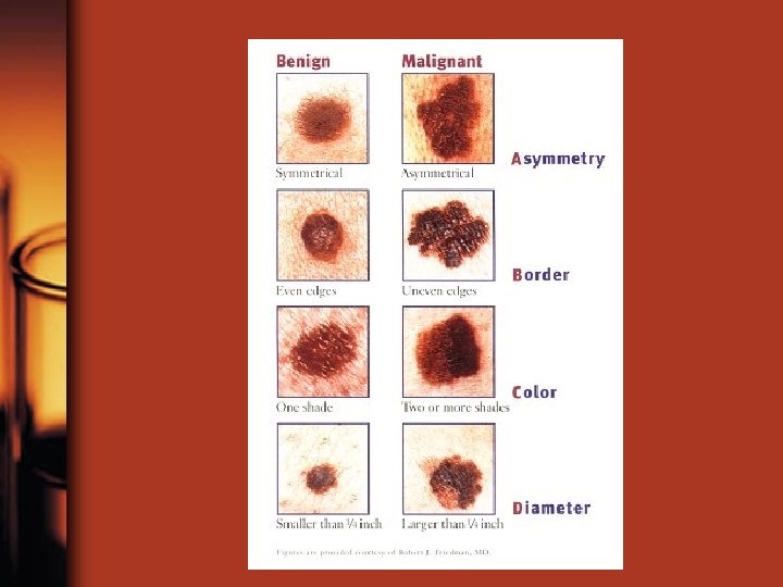
Melanoma • Cancer of melanocytes • Most dangerous, 1 of every 20 skin cancers • Melanoma can originate wherever there is pigment, often from existing moles • Appears as an expanding dark patch • Metastasize rapidly • Key—early detection • Resistant to chemo and current immunotherapy treatment, vaccines being tested.
 rule • A: asymmetry: the two halves of the spot or mole")
ABCD (E) rule • A: asymmetry: the two halves of the spot or mole don’t match • B: Border irregularity: the borders have indentions and notches • C: Color: the pigment contains several colors, blacks, browns, tans, blues, reds • D: Diameter: larger than 6 mm (pencil eraser) • E: Elevation: above the skin surface
- Slides: 51