INTEGUMENTARY SYSTEM HONORS ANATOMY PHYSIOLOGY CHAPTER 5 HUMAN


2, CO")


: exteroceptors: sense stimuli from outside")


 eyelid: 0. 02")
 �produced by melanocytes most are in stratum")


 �protects")


 filtrate of blood �also has: salts vitamin")


 cerumen")


 �#1 risk")


 5% of skin cancers")


- Slides: 91
INTEGUMENTARY SYSTEM HONORS ANATOMY & PHYSIOLOGY CHAPTER 5 HUMAN ANATOMY & PHYSIOLOGY
Integumentary System �Includes: Skin Nails Hair Sweat & Oil Glands
FUNCTIONS 1. Protection: � chemical barriers: skin secretions: acidic: hostile to most bacteria some bactericidal melanin: chemical pigment shield protects DNA from damage by UV rays � physical barrier: continuity of skin & hardness of keratinized cells stratum corneum: multiple layers of dead cells (brick) and water-resistantglycolipids (mortar)
Substances that Penetrate Skin 1. 2. 3. 4. 5. 6. lipid-soluble substances: )2, CO 2, Vit A, D, E, K oleoresins: poison ivy, poison oak organic solvents: acetone, dry-cleaning fluid, paint thinner all dissolve cell lipids heavy metal salts: Pb, Hg selected drugs: nitroglycerine, nicotine, fentanyl, seasickness meds, estrogen penetration enhancers: help carry other drugs into body thru skin
Functions – Protection cont’d 3. Biological Barriers: dendritic cells of epidermis �active elements of immune system �agn presenting cells dermal macrophages �phagocytes viruses or bacteria �agn presenting cells DNA �e- can absorb some UV rays heat
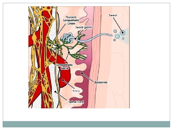
Functions of Skin -2 �body temperature homeostasis when body overheated blood flow to skin increases & some heat radiates off body, sweating (sensible losses) when body cold less blood flows to skin, more to trunk, goose bumps @normal temps sweat glands lose ~500 m. L/dthru insensible losses
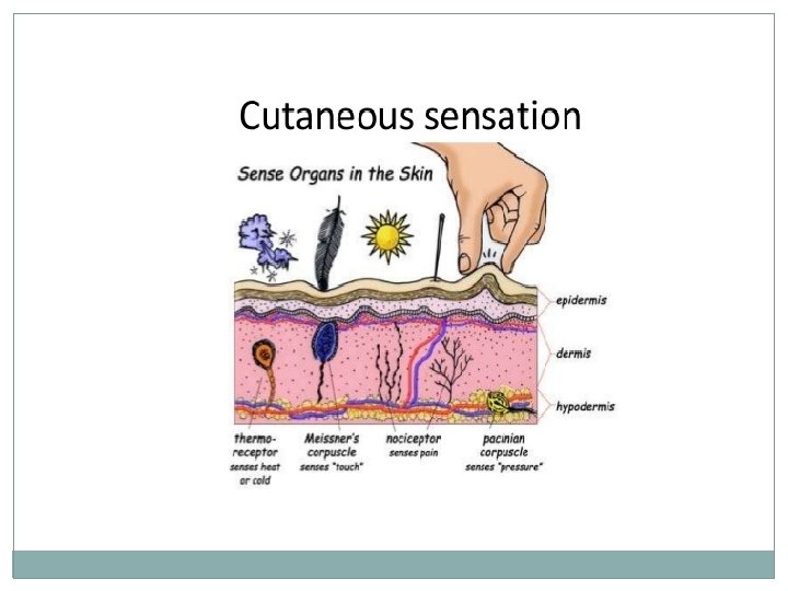
Functions of Skin - 3 �Cutaneous Sensation (sensory receptors): exteroceptors: sense stimuli from outside body Meissner’s corpuscles: in dermal papilla, feel light touch Pacinian corpuscles: deep dermis or hypodermis, feel deep touch hair follicle receptors: feel breezes, insect free nerve endings: pain from irritants or extreme cold or heat
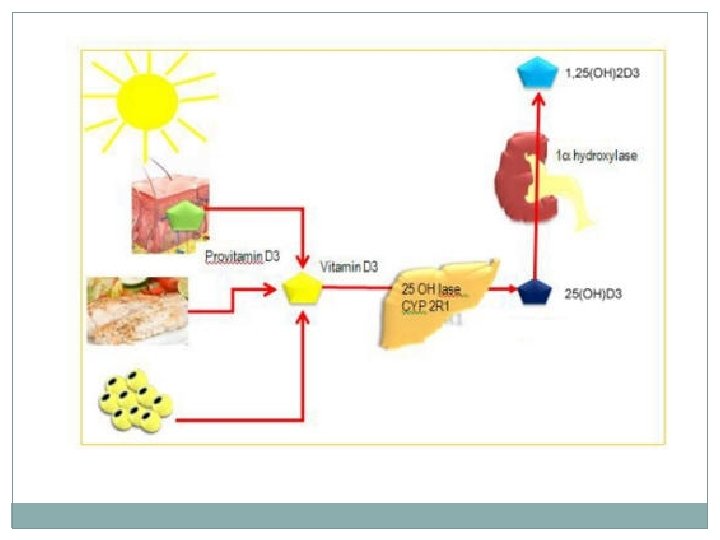
Functions of Skin - 4 �Metabolic Functions: 1. 2. 3. Sun light nrg powers some chemical reactions: modified cholesterol Vit D precursor keratinocyte enzymes � “disarm” cancer-causing chemicals � activate some steroid hormones (cortisone cream cortisol a more potent anti-inflammatory) synthesize important proteins � collagenase
Functions of Skin - 5 �Blood Reservoir dermal vessels can hold up to 5%of blood vol. vessels constricted when �other organs need extra blood �constriction shunts blood to areas in need
Functions of Skin- 6 �excretory function: sweat contains urea, uric acid (breakdown products of proteins) Na. Cl + water lost with profuse sweating
Structure of the Skin �made of 2 kinds of tissues 1. Epidermis 2. Dermis
Epidermis �made of stratified squamous epithelium �some keratinized, some not �avascular �Cells: Keratinocytes �majority of cells �make keratin Melanocytes Langerhans �Immune Cells System �aka dendritic cells
Cells of the Epidermis
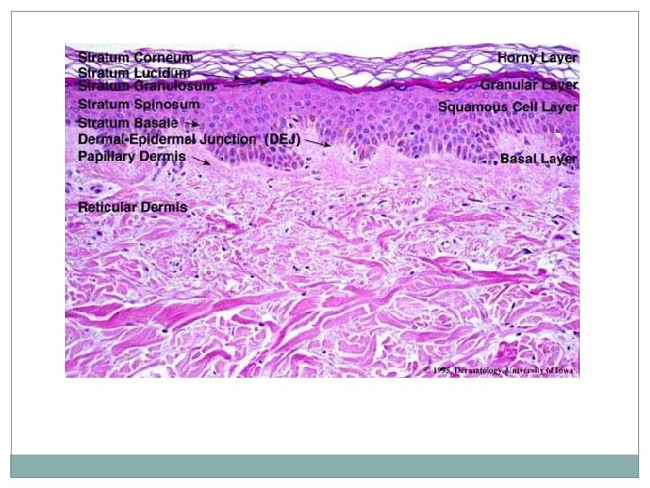
Epidermal Layers 1. stratum basale deepest layer constantly undergoing cell division/ cells pushed upward 2. stratum spinosum 3. stratum granulosum 4. stratum lucidum (only in thick skin) clear, flatter, more keratin 5. stratum corneum (cornified = keratinized) outermost layer/ 20 -30 dead cells thick
Layers of Epidermis
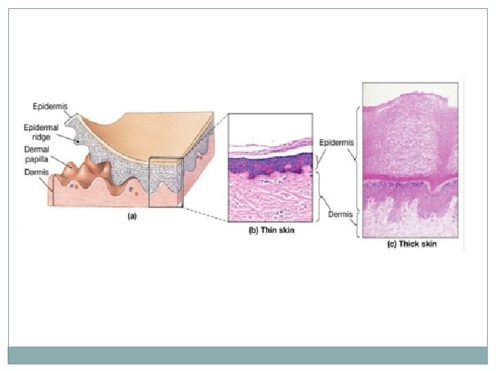
Thick Skin �areas subject to abrasion palms soles finger tips
Thin Skin �covers rest of body �thinnest skin (thinner subcutaneous layer) eyelid: 0. 02 mm bridge of nose ankles knuckles
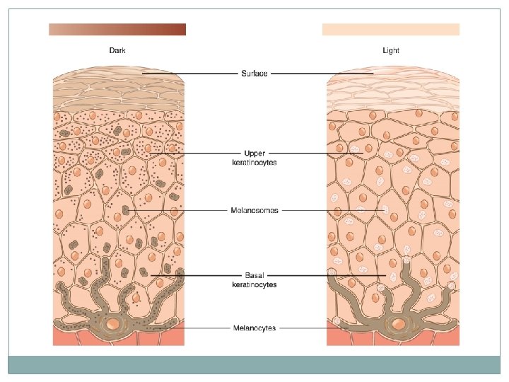
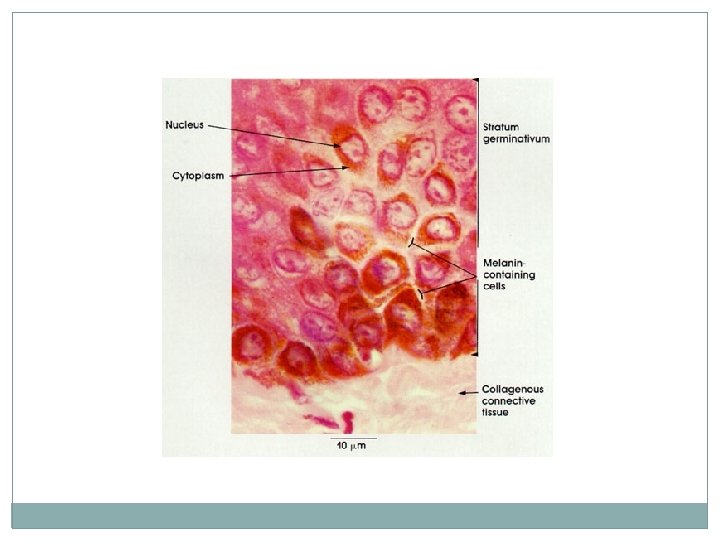
Melanin �pigment (yellow to brown to black) �produced by melanocytes most are in stratum basale �cells stimulated to make more melanin when skin exposed to sunlight shields DNA from damaging effects of UV radiation freckles & moles: seen where melanin concentrated in 1 spot
Freckle
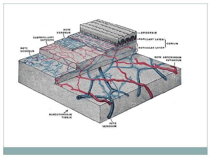
Dermis �a strong, stretchy envelope that helps to hold the body together leather is the dermis of whatever animal it was made from �made of dense CT: strong& flexible �rich blood supply � 2 regions: 1. Papillary 2. Reticular
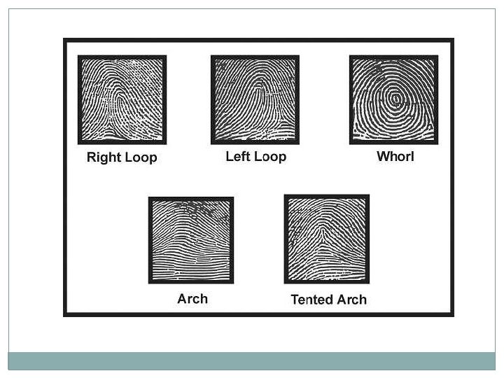
Papillary Layer �upper dermis �dermal papillae: uneven projections into lower epidermis that contain: 1. capillaries 2. pain receptors 3. touch receptors: Meissner’s. Corpuscles 4. in thick skin: form ridges (fingerprints) that improve gripping ability genetically determined
Dermal Papillae
Dermis: Reticular Layer �deepest skin layer �Contains: 1. sweat & oil glands, hair follicles, blood vessels 2. Pacinian corpuscles (deep touch receptors) 3. many phagocytes 4. fibers: elastic: give young skin elasticity collagen: make dermis tough & keeps skin hydrated by binding to water
Dermis
Striae �extreme stretching of skin white scars �aka stretch marks
Blisters �fluid –filled pocket separating epidermis/dermis
Skin Color � 3 pigments contribute to skin color: 1. Melanin amount & kind (yellow black) 2. Carotene orange – yellow pigment stratum corneum & subcutaneous layers 3. Hemoglobin amount O 2 bound to it in RBCs in dermal blood vessels has greater affect in light skinned people
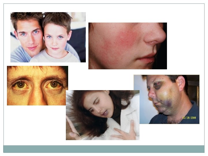
Skin Color in Sickness & in Health �cyanosis: blue hue to skin; due to poorly oxygenated blood �erythema: redness, due to increased blood flow (infection, inflammation); burn, HT, blushing �pallor: paleness, due to emotions, anemia, low BP, decreased blood flow �jaundice: yellow; usually from liver disease (not clearing bilirubin) �hematomas: bruising (bleeding under skin)
Appendages of the Skin �derivatives of epidermis: hair& hair follicles nails sweat glands sebaceous glands
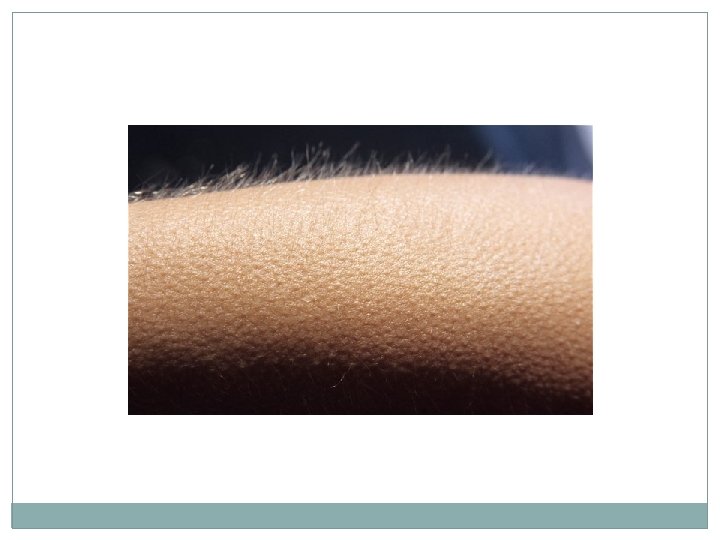
Hair & Hair Follicles �areas of skin with no hair: palms soles lips nipples parts of external genitalia
Functions of Hair �sense insects b/4 they bite �protects against heat loss (scalp) �protects against sun �shield eyes �filter air inhaled through nose
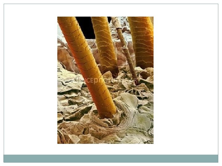
Hair �“pili” �produced by hair follicles �consist of dead keratinized cells �keratin here compared to keratin in epidermis: tougher & more durable individual cells do not flake off
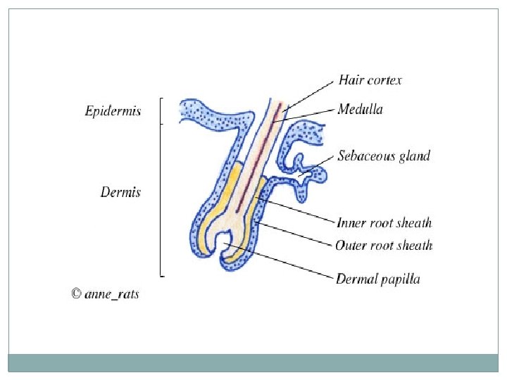
Hair � 3 concentric layers of keratinized cells 1. Medulla innermost large cells & air space absent in fine hair 2. Cortex surrounds medulla layers of flattened cells 3. Cuticle 1 layer overlapping cells provides strength
Hair Pigment �melanocytes @ base of follicle to cortical cells �differing proportions of melanins of different colors give different hair colors �melanin production decreases (delayed-action genes) replaced by air bubbles gray white
Hair Follicle �fold down from epidermis forming hair bulb �sensory nerve ending : root hair plexus surrounds hair bulb bending hair stimulates plexus sensitive touch receptors
Hair Follicle �dermal papilla: bit of dermal tissue protruding into base of follicle capillaries � matrix: actively dividing cells produces hair � arrector pili: smooth muscle from dermal part of follicle epidermis @ angle contraction goose bumps
Types of Hair VELLUS TERMINAL �pale, fine hair �coarse long hairs �children and females �eyebrows, scalp �@ puberty:
Factors the Influence Hair Growth Nutrition poor nutrition poor growth 2. Hormones 3. local inflammation increased growth 1. � normal growth~2. 5 mm/wk � fastest growth teens 40’s then slows � each follicle goes thru cycle: 1. active growth phase wks yrs 2. regressive phase 3. matrix cells die resting phase 1 – 3 mos
Hirsutism �excessive hairiness �could be from ovarian or adrenal gland tumor secreting high levels of androgens
Alopecia �hair thinning �starts @ anterior hairline posteriorly �males: by age 35: 40% by age 60: 85%
Male Pattern Baldness �most common type of baldness �X-linked recessive inheritance: delayed-action gene switches on in adult & changes hair follicle response to testosterone follicles very short growth cycle may get vellus hair
Alopecia Areata �immune system attacks hair follicles �hair falls out in patches
Nails �scalelike modification of epidermis forming clear protective covering on dorsal surface of distal digits �hard keratin �Functions: 1. pick up small objects 2. scratch 3. protect tips of digits
Parts of a Nail 1. free edge 2. nail plate or body 3. nail bed proximal portion = matrix: nail growth 4. proximal root 5. nail folds: proximally : eponychium (cuticle)
Nail Abnormalities �yellow: thyroid or respiratory problem �thickened yellow: fungus infection �outward concavity: iron deficiency (spoon nail) �horizontal lines: malnutrition
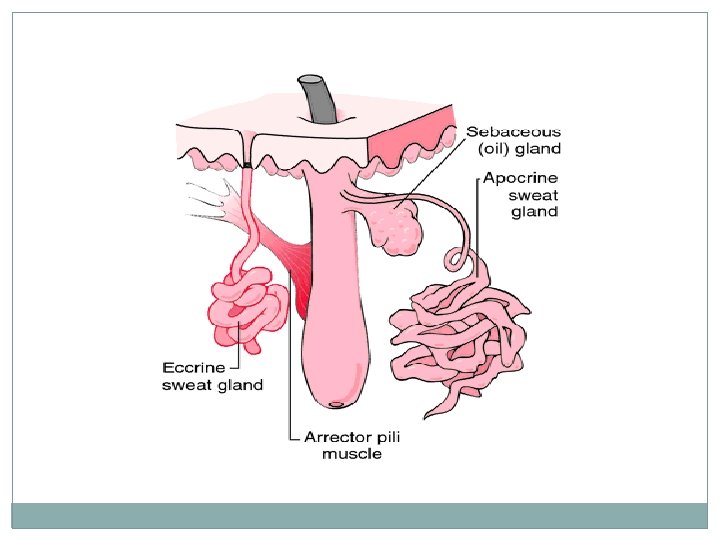
Sweat Glands �aka sudoriferous glands �over entire skin surface except: nipples parts of external genitalia � 2 types: 1. Eccrine Sweat Glands 2. Apocrine Sweat Glands �secretory cells surrounded by myoepithelial cells: contract when stimulated by nervous system sweat goes thru duct to skin
Eccrine Sweat Glands �more numerous than apocrine �abundant on palms & soles of feet �secretory cells in dermis �sweat released thru pore (not skin pore)
Eccrine Sweat Gland �secretion: hypotonic (99% water) filtrate of blood �also has: salts vitamin C abys wastes: urea, uric acid, NH 3
Eccrine Sweat Gland �regulated by sympathetic division of ANS �Functions: 1. prevent overheating forehead spreads inferiorly 2. “emotional” sweating fright or extreme nervousness palms, soles, or axilla over body
Apocrine Sweat. Glands �axillary & anogenital areas only �larger than eccrine glands �found deeper in dermis/hypoderrmis �empty onto hair follicle �odorless until released onto skinwhere bacteria decompose molecules musky, unpleasant odor �in addition to what is found in eccrine sweat: fatty acids proteins
Apocrine Sweat Glands �begin functioning @ puberty �activated by sympathetic division of ANS during: pain stress �exact function uncertain but likely analogous to animals glands that secrete pheromones
Ceruminous Glands �modified apocrine glands �secretion mixes with sebum (made by sebaceous gland) cerumen (ear wax) �Function: �deters insects �blocks entry of foreign objects
Mammary Glands �specialized sweat glands that secrete milk
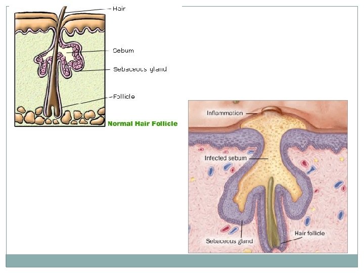
Sebaceous Glands �oil glands �found with hair follicles smaller on trunk & limbs large on face, neck, upper chest �secrete sebum: oily substance �develop as outgrowths of hair follicle �arrector pili contractions force sebum to skin surface �androgens stimulate secretion (activity starts in puberty)
Sebum �Functions: 1. softens & lubricates skin & hair 2. bactericidal action
Whitehead �created by: sebum blocks sebaceous gland duct Blackhead �sebum in whitehead oxidizes
Acne �active inflammation of sebaceous glands �see: pustules or cysts on skin surface �associated with bacterial infection (staph)
Skin Cancer �Benign tumors such as warts and moles are not serious. �Malignant tumors can start on the skin and invade other body areas. �Crucial risk factor- overexposure to UV radiation
Skin Cancer � 20% in USA will develop skin cancer (↑ing #s) �#1 risk factor: Sun UV radiation DNA damaged suppresses tumor suppressor gene new skin lotions contain liposomes initiate repairof damaged DNA
Types of Skin Cancer �Basal cell carcinoma- most common, 30% of all white skin people get it. Arises from the stratum basale layer of the skin 99% curable if caught early Dome shaped nodules that form an ulcer in the center. �Squamous Cell carcinoma Arise from stratum spinosum Grows rapidly and metastasizes if not removed Small red rounded elevation on the skin
Basal Cell Carcinoma
Lesion removed from patient Basal Cell Carcinoma
Squamous cell carcinoma
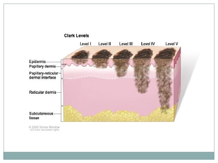
Skin Cancer Types cont. �Melanoma Cancer of melanocytes (very dangerous) 5% of skin cancers but rising fast Can arise from preexisting moles Appears as a spreading brown or black patch Chance of survival is poor if the lesion is greater than 4 mm thick
Melanoma
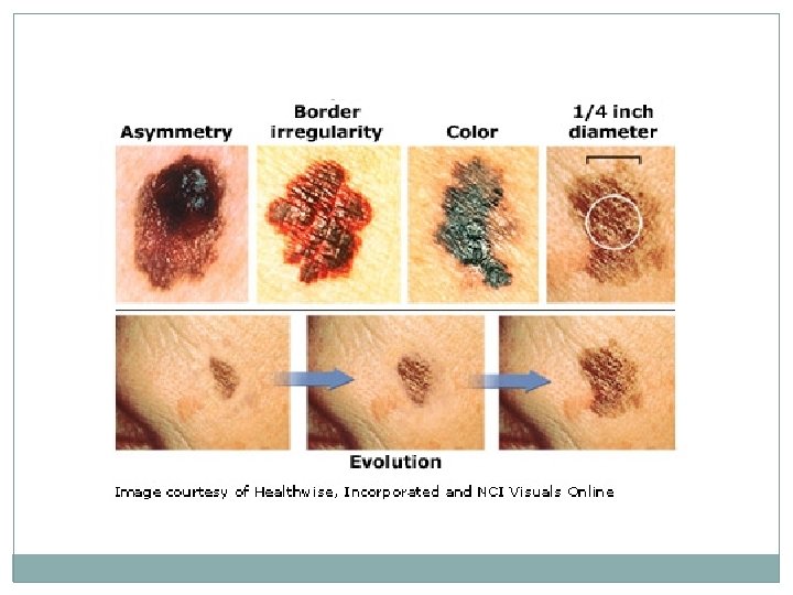
What is the ABCD rule? �Used for recognizing melanoma �A-Asymmetry: two sides of the pigmented mole do not match �B-Border irregularity: borders are not smooth �C- Color: lesion has a multiple of colors �D- Diameter the spot is larger than 6 mm in diameter (size of a pencil eraser)
What are the 3 types of burns? �First-degree burns: only the epidermis is damaged. Redness, swelling and pain are common. (sunburn) 2 -3 days to heal �Second-degree burns: epidermis and upper layers of dermis. Blistering can occur. 3 -4 weeks to heal. �Third-degree burns: involves the entire thickness of the skin. (pg. 164 -165)
Second-degree burns Third-degree burn
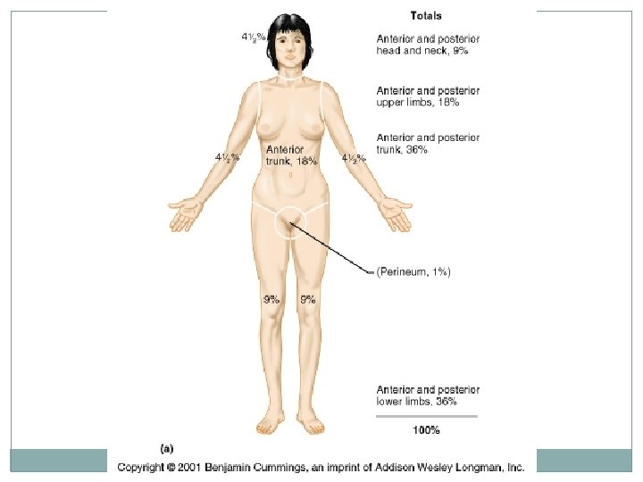
Burn Evaluation �Rule of 9’s: �divides body into 11 areas each 9% of body surface area + 1% for genitalia
Burn Treatment �patients require extra calories to replace lost proteins & allow tissue repair must add parenteral nutrition (IV) � 2◦ infections main threat 24 hrs after burn wound is sterile fungi, bacteria, & other pathogens multiply rapidly Immune system deficient infection leading cause of death in burn victims
Burn Treatment: Skin Grafts � 1 st debride burned tissue �wash area with antibiotic in solution �apply synthetic membrane temporarily �skin graft: healthy skin applied to area autograft: taken from the patient synthetic skin: silicone “epidermis” bound to spongy “dermis” made of collagen fibers & ground cartilage patients own cells absorb &replace synthetic * no rejection from immune system
Think About It