Integumentary System Dr Michael P Gillespie Integumentary System

 covers the external surface of the body.")


 n n Keratino = hornlike; cytes =")


 – indent the")
 – deeper part of dermis w")


 – oil glands - typically connected to hair")


 w Third-degree burn")


- Slides: 54
Integumentary System Dr. Michael P. Gillespie
Integumentary System w The skin (cutaneous membrane) covers the external surface of the body. w It is the largest organ of the body in terms of both surface area and weight.
Functions of the Integumentary System w Thermoregulation w Reservoir for blood w Protection from external environment w Cutaneous sensations w Excretion and absorption w Vitamin D synthesis
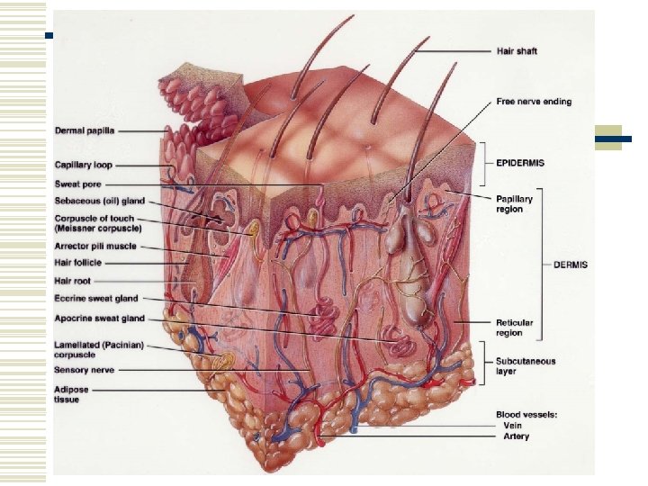
Structure of the Skin w Two main parts n n Epidermis – epithelial tissue Dermis – connective tissue w Subcutaneous layer (hypodermis) – not part of the skin – areolar and adipose tissue n Contains nerve endings called lamellated (Pacinian) corpuscles
Epidermis w Keratinized squamous epithelium w 4 types of cells w 5 basic layers of the epidermis
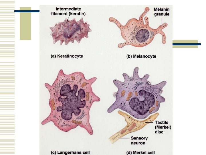
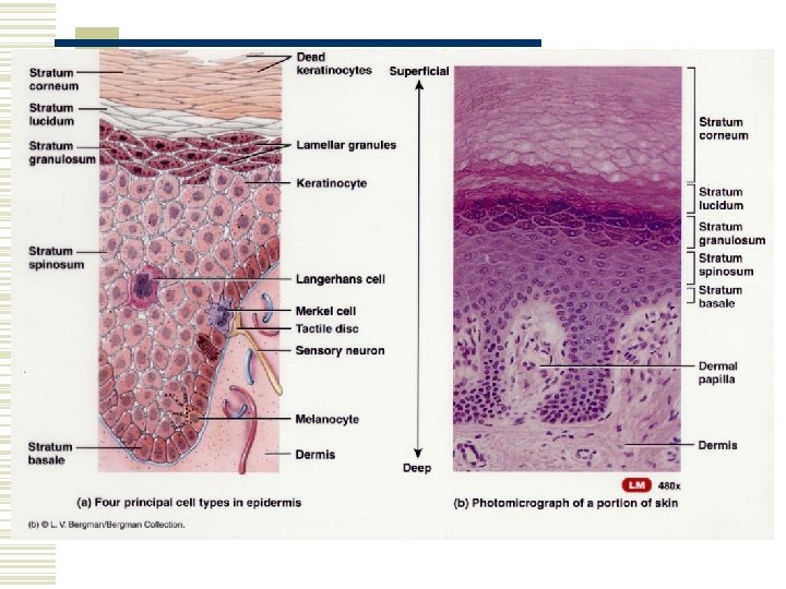
Cell Types in Epidermis w Keratinocytes (90%) n n Keratino = hornlike; cytes = cells Keratin –protects from heat, microbes, and chemicals w Melanocytes (8%) n n Melano = black Produce pigment melanin – absorbs UV light w Langerhans cells n n Arise from red bone marrow – migrate to epidermis Immune response w Merkel cells n Tactile (Merkel) disc
Layers of Epidermis w Stratum Basale w Stratum Spinosum w Stratum Granulosum w Stratum Lucidum w Stratum Corneum
Stratum Basale w Deepest layer w Single row of columnar or cuboidal keratinocytes w Keratin protects the deep layers from injury w Stem cells w Also known as the stratum germinativum (germ = sprout)
Stratum Spinosum w Spinos = thornlike w 8 – 10 layers of many sided keratinocytes close together w Provides strength and flexibility to the skin
Stratum Granulosum w Middle of the epidermis w Granulos – little grains w 3 -5 layers of flattened keratinocytes w Undergoing apoptosis (cell death) w Lamellar Granules – release a lipid-rich secretion
Stratum Lucidum w Lucid = clear w Present only in the thick skin of the fingertips, palms, and soles w 3 -5 layers of flattened clear, dead keratinocytes
Stratum Corneum w w w Corne = horn or horny 25 – 30 layers of flattened dead keratinocytes Shed and replaced by cells from deeper strata Mostly keratin Between the cells are lipids from lamellar granules – creates water repellent barrier w Protects deep layers from injury w Friction creates a callus – abnormal thickening
Growth of Epidermis w Newly formed cells are pushed to the surface w Accumulate more keratin (keratinization) w Undergo apoptosis w Keratinized cells slough off w 4 weeks
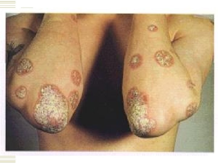
Psoriasis w Common skin disorder w Keratinocytes divide more quickly than normal and shed prematurely (7 -10 days) w Immature keratinocytes make abnormal keratin w Forms flaky, silvery scales w Knees, elbows, and scalp (dandruff) w Tx. Topical ointments, UV phototherapy (decreases cell division)
Dermis w Deeper layer w Mainly connective tissue w Collagen and elastic tissue w Fibroblasts, macrophages, and adipocytes
Dermis Continued… w Papillary region – dermal papillae (papillae = nipples) – indent the epidermis – capillary loops w Corpuscles of touch (Meissner corpuscles) w Free nerve endings – warmth, coolness, pain, tickling, and itching
Dermis continued… w Reticular region (reticul = netlike) – deeper part of dermis w Dense irregular CT w Adipose cells, hair follicles, nerves, sebaceous (oil) glands, sudoriferous (sweat) glands w Striae – streaks – stretch marks w Epidermal ridges – grip / friction – palms, fingers, soles, toes
Types of Skin w Thin skin – covers all surfaces of the body except for the palms, palmar surfaces of the digits, and soles. n Lacks a stratum lucidum w Thick skin – covers the palms, palmar surfaces of the fingers, and soles n Distinct stratum lucidum
Accessory Structure of the Skin w Hair w Skin glands w Nails
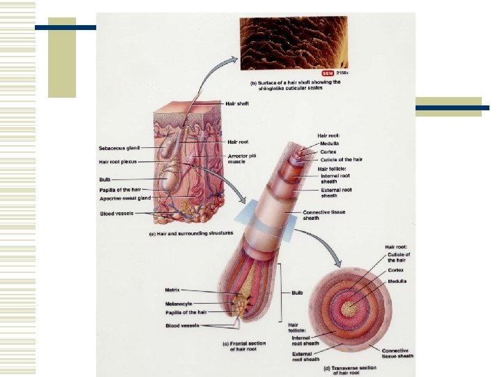
Hair w Hair or pili – present on most surfaces except the palms, palmar surfaces of fingers, soles, and plantar surfaces of feet w Shaft – superficial portion – projects from skin w Root – deeper portion – penetrates the dermis and sometimes into the subcutaneous layer w Arrector pili – muscle which pulls on hair shaft causing it to raise – emotional stress (cold or fright)
Conditions w Hirsutism = excessive body hair due to excessive androgens – tumor of the adrenal glands, testes, or ovaries w Androgenic alopecia – male-pattern baldness
Skin Glands w Sebaceous Glands (greasy) – oil glands - typically connected to hair follicles n Secrete sebum – coats hair and keeps it from becoming dry and brittle – keeps skin soft and pliable w Sudoriferous Glands – sweat glands n n Eccrine – throughout skin Apocrine – skin of axilla, groin, areolae and bearded regions w Ceruminous Glands – cer = wax – external ear n Cerumen = earwax – creates sticky body to impede entrance of foreign substances
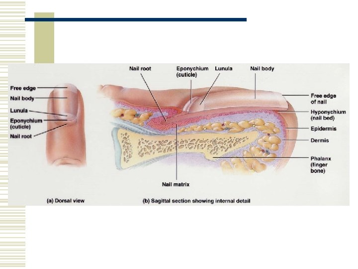
Nails w Tightly packed, hard, keratinized epidermal cells w Nail body, free edge and nail root w Lunula w Hyponychium – beneath free edge w Eponychium (cuticle) adheres to the lateral margin of the nail wall.
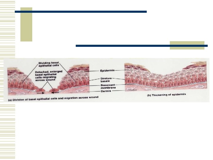
Epidermal Wound Healing w Cells enlarge and migrate across the wound w Contact inhibition – when migrating cells touch one another they stop due a this cellular response
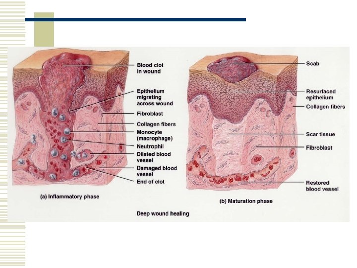
Deep Wound Healing w The injury extends to the dermis and subcutaneous layer w Inflammatory phase n n Blood clot forms Inflammation eliminates wastes and microbes w Migratory phase n Damaged blood vessels begin to regrow w Proliferative phase n n Extensive growth of epithelial cells Deposition of fibroblasts w Maturation phase
Contributions of the Integumentary System w The Integumentary System contributes to the functioning of all other body systems. w Refer to the table on page 155.
Skin Disorders w Skin Cancer w Burns w Pressure Ulcers
Skin Cancer w Almost exclusively caused by excessive exposure to the sun. w Basal cell carcinomas w Squamous cell carcinomas w Malignant Melanomas
Basal Cell Carcinoma
Squamous Cell Carcinoma
Detection of Malignant Melanoma w A Asymmetry n MM lack symmetry w B Border n MM have notched, indented, scalloped, or indistinct borders w C Color n MM have uneven coloration, may contain several colors w D Diameter n MM are typically greater than 6 mm (0. 25 in. ) w E Elevation
Normal Nevus & Malignant Melanoma
Risk Factors for Malignant Melanoma w 1. Skin type n Light skinned individuals who burn, but don’t tan w 2. Skin exposure n Sunny areas, high altitude (UV light), outdoor occupation w 3. Family Hx. w 4. Age w 5. Immunological status n Immunosuppressed individuals have a higher risk of skin cancer
Burns w Tissue damage caused by n n Excessive heat Electricity Radioactivity Corrosive chemicals w Destroy some of the skin’s contributions to homeostasis
Grading of Burns w First-degree burn w Second-degree burn (partial thickness) w Third-degree burn (full thickness)
Systemic Effects of Burns w 1. Large loss of water, plasma, and plasma proteins (causes shock) w 2. Bacterial infection w 3. Reduced circulation of blood w 4. Decreased production of urine w 5. Diminished immune response
Severity of Burns w Determined by the depth of the burn and the extent of the area involved. w According to the American Burn Association a major burn includes: n n n Third-degree burns over 10% Second-degree burns over 25% Third-degree burns over face, hands, feet, or perineum w When the burn area exceeds 70%, more than half of the victims die
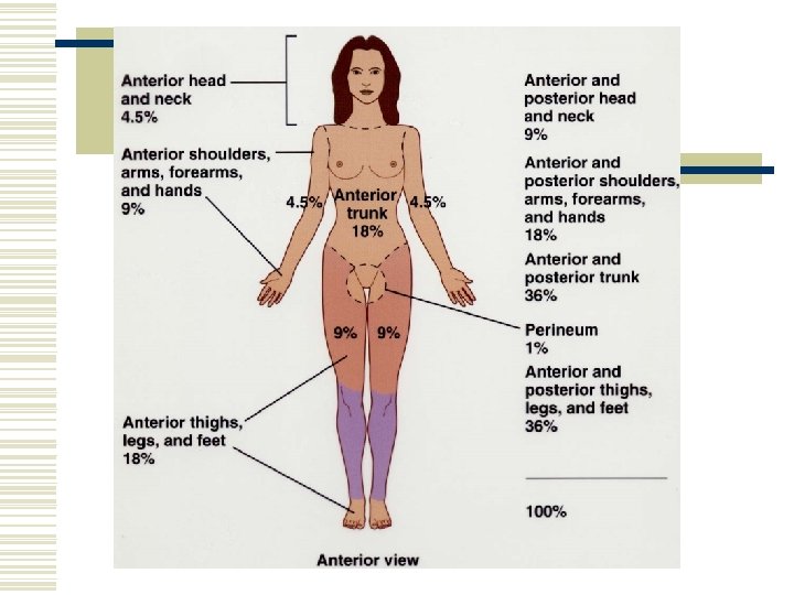
Determining the Extent of a Burn w Rule of nines – a quick method for estimating the surface area affected by burns w Lund-Browder method – a more accurate method for assessing the extent of burns
Skin Color w Melanin – causes skin color from pale yellow to black n n Melanocytes produce melanin Freckles and liver spots are accumulations of melanin w Carotene – yellow-orange pigment w Hemoglobin – imparts a red color w Albinism – inability to produce melanin - missing from the hair, eyes, and skin w Vitiligo – loss of melanocytes from patches of skin
Skin Color Clues w Cyanotic – blue - hemoglobin is depleted of oxygen w Jaundice – yellow – buildup of the yellow pigment bilirubin in the blood – usually indicates liver disease w Erythema – red – engorgement of capillaries in the skin – skin injury, heat, infection, inflammation, allergies
Carotonemia
Cyanosis
Jaundice
Vitiligo
Scabies