Insulin Glucagon and Diabetes Mellitus Dr Noori M

Insulin, Glucagon, and Diabetes Mellitus Dr. Noori M Luaibi

Insulin, Glucagon, and Diabetes Mellitus The pancreas, in addition to its digestive functions, secretes two important hormones, insulin and glucagon, that are crucial for normal regulation of glucose, lipid, and protein metabolism. Although the pancreas secretes other hormones, such as amylin, somatostatin, and pancreatic polypeptide, their functions are not as well established. The main purpose of this chapter is to discuss the physiologic roles of insulin and glucagon and the pathophysiology of diseases, especially diabetes mellitus, caused by abnormal secretion or activity of these hormones.

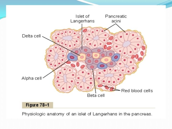
Physiologic Anatomy of the Pancreas. The pancreas is composed of two major types of tissues, as shown in Figure 78– 1: (1) the acini, which secrete digestive juices into the duodenum, and (2) the islets of Langerhans, which secrete insulin and glucagon directly into the blood. The human pancreas has 1 to 2 million islets of Langerhans, each only about 0. 3 millimeter in diameter and organized around small capillaries into which its cells secrete their hormones. The islets contain three major types of cells, alpha, beta, and delta cells, which are distinguished from one another by their morphological and staining characteristics. The beta cells, constituting about 60 per cent of all the cells of the islets, lie mainly in the middle of each islet and secrete insulin and amylin, a hormone that is often secreted in parallel with insulin, although its function is unclear. The alpha cells, about 25 per cent of the total, secrete glucagon. And the delta cells, about 10 per cent of the total, secrete somatostatin. In addition, at least one other type of cell, the PP cell, is present in small numbers in the islets and secretes a hormone of uncertain function called pancreatic polypeptide. The close interrelations among these cell types in the islets of Langerhans allow cell-to-cell communication and direct control of secretion of some of the hormones by the other hormones. For instance, insulin inhibits glucagon secretion, amylin inhibits insulin secretion, and somatostatin inhibits the secretion of both insulin and glucagon.

Insulin and Its Metabolic Effects Insulin was first isolated from the pancreas in 1922 by Banting and Best, and almost overnight the outlook for the severely diabetic patient changed from one of rapid decline and death to that of a nearly normal person. Historically, insulin has been associated with “blood sugar, ” and true enough, insulin has profound effects on carbohydrate metabolism. Yet it is abnormalities of fat metabolism, causing such conditions as acidosis and arteriosclerosis, that are the usual causes of death in diabetic patients. Also, in patients with prolonged diabetes, diminished ability to synthesize proteins leads to wasting of the tissues as well as many cellular functional disorders. Therefore, it is clear that insulin affects fat and protein metabolism almost as much as it does carbohydrate metabolism.

Insulin Is a Hormone Associated with Energy Abundance As we discuss insulin in the next few pages, it will become apparent that insulin secretion is associated with energy abundance. That is, when there is great abundance of energy -giving foods in the diet, especially excess amounts of carbohydrates, insulin is secreted in great quantity. In turn, the insulin plays an important role in storing the excess energy. In the case of excess carbohydrates, it causes them to be stored as glycogen mainly in the liver and muscles. Also, all the excess carbohydrates that cannot be stored as glycogen are converted under the stimulus of insulin into fats and stored in the adipose tissue. In the case of proteins, insulin has a direct effect in promoting amino acid uptake by cells and conversion of these amino acids into protein. In addition, it inhibits the breakdown of the proteins that are already in the cells.

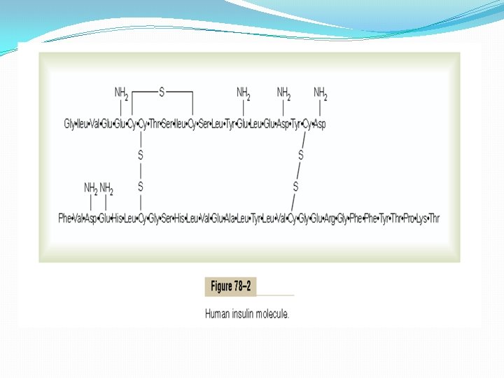
Insulin Chemistry and Synthesis Insulin is a small protein; human insulin has a molecular weight of 5808. It is composed of two amino acid chains, shown in Figure 78– 2, connected to each other by disulfide linkages. When the two amino acid chains are split apart, the functional activity of the insulin molecule is lost. Insulin is synthesized in the beta cells by the usual cell machinery for protein synthesis, beginning with translation of the insulin RNA by ribosomes attached to the endoplasmic reticulum to form an insulin preprohormone. This initial preprohormone has a molecular weight of about 11, 500, but it is then cleaved in the endoplasmic reticulum to form a proinsulin with a molecular weight of about 9000; most of this is further cleaved in the Golgi apparatus to form insulin and peptide fragments before being packaged in the secretory granules. However, about one sixth of the final secreted product is still in the form of proinsulin. The proinsulin has virtually no insulin activity. When insulin is secreted into the blood, it circulates almost entirely in an unbound form; it has a plasma half-life that averages only about 6 minutes, so that it is mainly cleared from the circulation within 10 to 15 minutes. Except for that portion of the insulin that combines with receptors in the target cells, the remainder is degraded by the enzyme insulinase mainly in the liver, to a lesser extent in the kidneys and muscles, and slightly in most other tissues. This rapid removal from the plasma is important, because, at times, it is as important to turn off rapidly as to turn on the control functions of insulin.

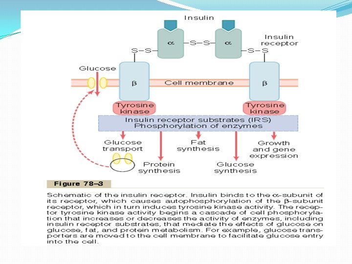
Activation of Target Cell Receptors by Insulin and the Resulting Cellular Effects To initiate its effects on target cells, insulin first binds with and activates a membrane receptor protein that has a molecular weight of about 300, 000 (Figure 78– 3). It is the activated receptor, not the insulin, that causes the subsequent effects. The insulin receptor is a combination of four subunits held together by disulfide linkages: two alpha subunits that lie entirely outside the cell membrane and two beta subunits that penetrate through the membrane, protruding into the cell cytoplasm. The insulin binds with the alpha subunits on the outside of the cell, but because of the linkages with the beta subunits, the portions of the beta subunits protruding into the cell become autophosphorylated. Thus, the insulin receptor is an example of an enzyme-linked receptor, Auto phosphorylation of the beta subunits of the receptor activates a local tyrosine kinase, which in turn causes phosphorylation of multiple other intracellular enzymes including a group called insulin-receptor substrates (IRS). Different types of IRS (e. g. IRS-1, IRS-2, IRS-3) are expressed in different tissues. The net effect is to activate some of these enzymes while inactivating others. In this way, insulin directs the intracellular metabolic machinery to produce the desired effects on carbohydrate, fat, and protein metabolism.

The end effects of insulin stimulation are the following: 1. Within seconds after insulin binds with its membrane receptors, the membranes of about 80 per cent of the body’s cells markedly increase their uptake of glucose. This is especially true of muscle cells and adipose cells but is not true of most neurons in the brain. The increased glucose transported into the cells is immediately phosphorylated and becomes a substrate for all the usual carbohydrate metabolic functions. The increased glucose transport is believed to result from translocation of multiple intracellular vesicles to the cell membranes; these vesicles carry in their own membranes multiple molecules of glucose transport proteins, which bind with the cell membrane and facilitate glucose uptake into the cells. When insulin is no longer available, these vesicles separate from the cell membrane within about 3 to 5 minutes and move back to the cell interior to be used again and again as needed. 2. The cell membrane becomes more permeable to many of the amino acids, potassium ions, and phosphate ions, causing increased transport of these substances into the cell. 3. Slower effects occur during the next 10 to 15 minutes to change the activity levels of many more intracellular metabolic enzymes. These effects result mainly from the changed states of phosphorylation of the enzymes. 4. Much slower effects continue to occur for hours and even several days. They result from changed rates of translation of messenger RNAs at the ribosomes to form new proteins and still slower effects from changed rates of transcription of DNA in the cell nucleus. In this way, insulin remolds much of the cellular enzymatic machinery to achieve its metabolic goals.

Effect of Insulin on Carbohydrate Metabolism Immediately after a high-carbohydrate meal, the glucose that is absorbed into the blood causes rapid secretion of insulin, The insulin in turn causes rapid uptake, storage, and use of glucose by almost all tissues of the body, but especially by the muscles, adipose tissue, and liver. Insulin Promotes Muscle Glucose Uptake and Metabolism During much of the day, muscle tissue depends not on glucose for its energy but on fatty acids. The principal reason for this is that the normal resting muscle membrane is only slightly permeable to glucose, except when the muscle fiber is stimulated by insulin; between meals, the amount of insulin that is secreted is too small to promote significant amounts of glucose entry into the muscle cells. However, under two conditions the muscles do use large amounts of glucose. One of these is during moderate or heavy exercise. This usage of glucose does not require large amounts of insulin, because exercising muscle fibers become more permeable to glucose even in the absence of insulin because of the contraction process itself. The secondition for muscle usage of large amounts of glucose is during the few hours after a meal. At this time the blood glucose concentration is high and the pancreas is secreting large quantities of insulin. The extra insulin causes rapid transport of glucose into the muscle cells. This causes the muscle cell during this period to use glucose preferentially over fatty.

Storage of Glycogen in Muscle If the muscles are not exercising after a meal and yet glucose is transported into the muscle cells in abundance, then most of the glucose is stored in the form of muscle glycogen instead of being used for energy, up to a limit of 2 to 3 per cent concentration. The glycogen can later be used for energy by the muscle. It is especially useful for short periods of extreme energy use by the muscles and even to provide spurts of anaerobic energy for a few minutes at a time by glycolytic breakdown of the glycogen to lactic acid, which can occur even in the absence of oxygen.

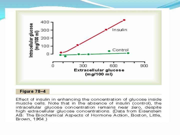
Quantitative Effect of Insulin to Facilitate Glucose Transport Through the Muscle Cell Membrane The quantitative effect of insulin to facilitate glucose transport through the muscle cell membrane is demonstrated by the experimental results shown in Figure 78– 4. The lower curve labeled “control” shows the concentration of free glucose measured inside the cell, demonstrating that the glucose concentration remained almost zero despite increased extracellular glucose concentration up to as high as 750 mg/100 ml. In contrast, the curve labeled “insulin” demonstrates that the intracellular glucose concentration rose to as high as 400 mg/100 ml when insulin was added. Thus, it is clear that insulin can increase the rate of transport of glucose into the resting muscle cell by at least 15 -fold.

Insulin Promotes Liver Uptake, Storage, and Use of Glucose One of the most important of all the effects of insulin is to cause most of the glucose absorbed after a meal to be stored almost immediately in the liver in the form of glycogen. Then, between meals, when food is not available and the blood glucose concentration begins to fall, insulin secretion decreases rapidly and the liver glycogen is split back into glucose, which is released back into the blood to keep the glucose concentration from falling too low. The mechanism by which insulin causes glucose uptake and storage in the liver includes several almost simultaneous steps:

1. Insulin inactivates liver phosphorylase, the principal enzyme that causes liver glycogen to split into glucose. This prevents breakdown of the glycogen that has been stored in the liver cells. 2. Insulin causes enhanced uptake of glucose from the blood by the liver cells. It does this by increasing the activity of the enzyme glucokinase, which is one of the enzymes that causes the initial phosphorylation of glucose after it diffuses into the liver cells. Once phosphorylated, the glucose is temporarily trapped inside the liver cells because phosphorylated glucose cannot diffuse back through the cell membrane. 3. Insulin also increases the activities of the enzymes that promote glycogen synthesis, including especially glycogen synthase, which is responsible for polymerization of the monosaccharide units to form the glycogen molecules. The net effect of all these actions is to increase the amount of glycogen in the liver. The glycogen can increase to a total of about 5 to 6 per cent of the liver mass, which is equivalent to almost 100 grams of stored glycogen in the whole liver.

Glucose Is Released from the Liver Between Meals When the blood glucose level begins to fall to a low level between meals, several events transpire that cause the liver to release glucose back into the circulating blood: 1. The decreasing blood glucose causes the pancreas to decrease its insulin secretion. 2. The lack of insulin then reverses all the effects listed earlier for glycogen storage, essentially stopping further synthesis of glycogen in the liver and preventing further uptake of glucose by the liver from the blood. 3. The lack of insulin (along with increase of glucagon, which is discussed later) activates the enzyme phosphorylase, which causes the splitting of glycogen into glucose phosphate. 4. The enzyme glucose phosphatase, which had been inhibited by insulin, now becomes activated by the insulin lack and causes the phosphate radical to split away from the glucose; this allows the free glucose to diffuse back into the blood. Thus, the liver removes glucose from the blood when it is present in excess after a meal and returns it to the blood when the blood glucose concentration falls between meals. Ordinarily, about 60 per cent of the glucose in the meal is stored in this way in the liver and then returned later.

Insulin Promotes Conversion of Excess Glucose into Fatty Acids and Inhibits Gluconeogenesis in the Liver When the quantity of glucose entering the liver cells is more than can be stored as glycogen or can be used for local hepatocyte metabolism, insulin promotes the conversion of all this excess glucose into fatty acids. These fatty acids are subsequently packaged as triglycerides in very-low-density lipoproteins and transported in this form by way of the blood to the adipose tissue and deposited as fat. Insulin also inhibits gluconeogenesis. It does this mainly by decreasing the quantities and activities of the liver enzymes required for gluconeogenesis. However, part of the effect is caused by an action of insulin that decreases the release of amino acids from muscle and other extrahepatic tissues and in turn the availability of these necessary precursors required for gluconeogenesis.

Lack of Effect of Insulin on Glucose Uptake and Usage by the Brain The brain is quite different from most other tissues of the body in that insulin has little effect on uptake or use of glucose. Instead, the brain cells are permeable to glucose and can use glucose without the intermediation of insulin. The brain cells are also quite different from most other cells of the body in that they normally use only glucose for energy and can use other energy substrates, such as fats, only with difficulty. Therefore, it is essential that the blood glucose level always be maintained above a critical level, which is one of the most important functions of the blood glucose control system. When the blood glucose falls too low, into the range of 20 to 50 mg/100 ml, symptoms of hypoglycemic shock develop, characterized by progressive nervous irritability that leads to fainting, seizures, and even coma.

Effect of Insulin on Carbohydrate Metabolism in Other Cells Insulin increases glucose transport into and glucose usage by most other cells of the body (with the exception of the brain cells, as noted) in the same way that it affects glucose transport and usage in muscle cells. The transport of glucose into adipose cells mainly provides substrate for the glycerol portion of the fat molecule. Therefore, in this indirect way, insulin promotes deposition of fat in these cells. Effect of Insulin on Fat Metabolism Although not quite as visible as the acute effects of insulin on carbohydrate metabolism, insulin’s effects on fat metabolism are, in the long run, equally important. Especially dramatic is the long-term effect of insulin lack in causing extreme atherosclerosis, often leading to heart attacks, cerebral strokes, and other vascular accidents. But first, let us discuss the acute effects of insulin on fat metabolism.

Insulin Promotes Fat Synthesis and Storage Insulin has several effects that lead to fat storage in adipose tissue. First, insulin increases the utilization of glucose by most of the body’s tissues, which automatically decreases the utilization of fat, thus functioning as a fat sparer. However, insulin also promotes fatty acid synthesis. This is especially true when more carbohydrates are ingested than can be used for immediate energy, thus providing the substrate for fat synthesis. Almost all this synthesis occurs in the liver cells, and the fatty acids are then transported from the liver by way of the blood lipoproteins to the adipose cells to be stored. The different factors that lead to increased fatty acid synthesis in the liver include the following:

1. Insulin increases the transport of glucose into the liver cells. After the liver glucogen concentration reaches 5 to 6 per cent, this in itself inhibits further glycogen synthesis. Then all the additional glucose entering the liver cells becomes available to form fat. The glucose is first split to pyruvate in the glycolytic pathway, and the pyruvate subsequently is converted to acetyl coenzyme A (acetyl-Co. A), the substrate from which fatty acids are synthesized. 2. An excess of citrate and isocitrate ions is formed by the citric acid cycle when excess amounts of glucose are being used for energy. These ions then have a direct effect in activating acetyl. Co. A carboxylase, the enzyme required to carboxylate acetyl-Co. A to form malonyl-Co. A, the first stage of fatty acid synthesis. 3. Most of the fatty acids are then synthesized within the liver itself and used to form triglycerides, the usual form of storage fat. They are released from the liver cells to the blood in the lipoproteins. Insulin activates lipoprotein lipase in the capillary walls of the adipose tissue, which splits the triglycerides again into fatty acids, a requirement for them to be absorbed into the adipose cells,

Role of Insulin in Storage of Fat in the Adipose Cells Insulin has two other essential effects that are required for fat storage in adipose cells: 1. Insulin inhibits the action of hormone-sensitive lipase. This is the enzyme that causes hydrolysis of the triglycerides already stored in the fat cells. Therefore, the release of fatty acids from the adipose tissue into the circulating blood is inhibited. 2. Insulin promotes glucose transport through the cell membrane into the fat cells in exactly the same ways that it promotes glucose transport into muscle cells. Some of this glucose is then used to synthesize minute amounts of fatty acids, but more important, it also forms large quantities of a-glycerol phosphate. This substance supplies the glycerol that combines with fatty acids to form the triglycerides that are the storage form of fat in adipose cells. Therefore, when insulin is not available, even storage of the large amounts of fatty acids transported from the liver in the lipoproteins is almost blocked.

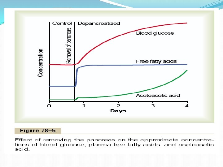
Insulin Deficiency Increases Use of Fat for Energy All aspects of fat breakdown and use for providing energy are greatly enhanced in the absence of insulin. This occurs even normally between meals when secretion of insulin is minimal, but it becomes extreme in diabetes mellitus when secretion of insulin is almost zero. The resulting effects are as follows. Insulin Deficiency Causes Lipolysis of Storage Fat and Release of Free Fatty Acids. In the absence of insulin, all the effects of insulin noted earlier that cause storage of fat are reversed. The most important effect is that the enzyme hormone-sensitive lipase in the fat cells becomes strongly activated. This causes hydrolysis of the stored triglycerides, releasing large quantities of fatty acids and glycerol into the circulating blood. Consequently, the plasma concentration of free fatty acids begins to rise within minutes. This free fatty acid then becomes the main energy substrate used by essentially all tissues of the body besides the brain. Figure 78– 5 shows the effect of insulin lack on the plasma concentrations of free fatty acids, glucose, and acetoacetic acid. Note that almost immediately after removal of the pancreas, the free fatty acid concentration in the plasma begins to rise, more rapidly even than the concentration of glucose.

Insulin Deficiency Increases Plasma Cholesterol and Phospholipid Concentrations. The excess of fatty acids in the plasma associated with insulin deficiency also promotes liver conversion of some of the fatty acids into phospholipids and cholesterol, two of the major products of fat metabolism. These two substances, along with excess triglycerides formed at the same time in the liver, are then discharged into the blood in the lipoproteins. Occasionally the plasma lipoproteins increase as much as threefold in the absence of insulin, giving a total concentration of plasma lipids of several per cent rather than the normal 0. 6 per cent. This high lipid concentration— especially the high concentration of cholesterol—promotes the development of atherosclerosis in people with serious diabetes.

Excess Usage of Fats During Insulin Lack Causes Ketosis and Acidosis. Insulin lack also causes excessive amounts of acetoacetic acid to be formed in the liver cells. This results from the following effect: In the absence of insulin but in the presence of excess fatty acids in the liver cells, the carnitine transport mechanism for transporting fatty acids into the mitochondria becomes increasingly activated. In the mitochondria, beta oxidation of the fatty acids then proceeds very rapidly, releasing extreme amounts of acetyl-Co. A. A large part of this excess acetyl-Co. A is then condensed to form acetoacetic acid, which in turn is released into the circulating blood. Most of this passes to the peripheral cells, where it is again converted into acetyl-Co. A and used for energy in the usual manner. At the same time, the absence of insulin also depresses the utilization of acetoacetic acid in the peripheral tissues. Thus, so much acetoacetic acid is released from the liver that it cannot all be metabolized by the tissues. Therefore, as shown in Figure 78– 5, its concentration rises during the days after cessation of insulin secretion, sometimes reaching concentrations of 10 m. Eq/L or more, which is a severe state of body fluid acidosis. As explained in Chapter 68, some of the acetoacetic acid is also converted into b-hydroxybutyric acid and acetone. These two substances, along with the acetoacetic acid, are called ketone bodies, and their presence in large quantities in the body fluids is called ketosis. We see later that in severe diabetes the acetoacetic acid and the b-hydroxybutyric acid can cause severe acidosis and coma, which often leads to death.

Effect of Insulin on Protein Metabolism and on Growth Insulin Promotes Protein Synthesis and Storage. During the few hours after a meal when excess quantities of nutrients are available in the circulating blood, not only carbohydrates and fats but proteins as well are stored in the tissues; insulin is required for this to occur. The manner in which insulin causes protein storage is not as well understood as the mechanisms for both glucose and fat storage. Some of the facts follow. 1) Insulin stimulates transport of many of the amino acids into the cells. Among the amino acids most strongly transported are valine, leucine, isoleucine, tyrosine, and phenylalanine. Thus, insulin shares with growth hormone the capability of increasing the uptake of amino acids into cells. However, the amino acids affected are not necessarily the same ones. 2) Insulin increases the translation of messenger RNA, thus forming new proteins. In some unexplained way, insulin “turns on” the ribosomal machinery. In the absence of insulin, the ribosomes simply stop working, almost as if insulin operates an “on-off” mechanism. 3) Over a longer period of time, insulin also increases the rate of transcription of selected DNA genetic sequences in the cell nuclei, thus forming increased quantities of RNA and still more protein synthesis— especially promoting a vast array of enzymes for storage of carbohydrates, fats, and proteins.
 Insulin inhibits the catabolism of proteins, thus decreasing the rate of amino acid")
4) Insulin inhibits the catabolism of proteins, thus decreasing the rate of amino acid release from the cells, especially from the muscle cells. Presumably this results from the ability of insulin to diminish the normal degradation of proteins by the cellular lysosomes. 5) In the liver, insulin depresses the rate of gluconeogenesis. It does this by decreasing the activity of the enzymes that promote gluconeogenesis. Because the substrates most used for synthesis of glucose by gluconeogenesis are the plasma amino acids, this suppression of gluconeogenesis conserves the amino acids in the protein stores of the body. In summary, insulin promotes protein formation and prevents the degradation of proteins.

Insulin Lack Causes Protein Depletion and Increased Plasma Amino Acids. Virtually all protein storage comes to a halt when insulin is not available. The catabolism of proteins increases, protein synthesis stops, and large quantities of amino acids are dumped into the plasma. The plasma amino acid concentration rises considerably, and most of the excess amino acids are used either directly for energy or as substrates for gluconeogenesis. This degradation of the amino acids also leads to enhanced urea excretion in the urine. The resulting protein wasting is one of the most serious of all the effects of severe diabetes mellitus. It can lead to extreme weakness as well as many deranged functions of the organs.

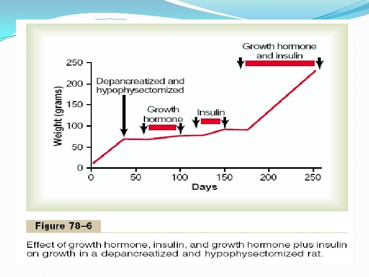
Insulin and Growth Hormone Interact Synergistically to Promote Growth. Because insulin is required for the synthesis of proteins, it is as essential for growth of an animal as growth hormone is. This is demonstrated in Figure 78– 6, which shows that a depancreatized, hypophysectomized rat without therapy hardly grows at all. Furthermore, the administration of either growth hormone or insulin one at a time causes almost no growth. Yet a combination of these hormones causes dramatic growth. Thus, it appears that the two hormones function synergistically to promote growth, each performing a specific function that is separate from that of the other. Perhaps a small part of this necessity for both hormones results from the fact that each promotes cellular uptake of a different selection of amino acids, all of which are required if growth is to be achieved.

Mechanisms of Insulin Secretion Figure 78– 7 shows the basic cellular mechanisms for insulin secretion by the pancreatic beta cells in response to increased blood glucose concentration, the primary controller of insulin secretion. The beta cells have a large number of glucose transporters (GLUT 2) that permit a rate of glucose influx that is proportional to the blood concentration in the physiologic range. Once inside the cells, glucose is phosphorylated to glucose-6 phosphate by glucokinase. This step appears to be the rate limiting for glucose metabolism in the beta cell and is considered the major mechanism for glucose sensing and adjustment of the amount of secreted insulin to the blood glucose levels. The glucose-6 -phosphate is subsequently oxidized to form adenosine triphosphate (ATP), which inhibits the ATP-sensitive potassium channels of the cell. Closure of the potassium channels depolarizes the cell membrane, thereby opening voltage-gated calcium channels, which are sensitive to changes in membrane voltage. This produces an influx of calcium that stimulates fusion of the docked insulin-containing vesicles with the cell membrane and secretion of insulin into the extracellular fluid by exocytosis. Other nutrients, such as certain amino acids, can also be metabolized by the beta cells to increase intracellular ATP levels and stimulate insulin secretion. Some hormones, such as glucagon and gastric inhibitory peptide, as well as acetylcholine increase intracellular calcium levels through other signaling pathways and enhance the effect of glucose, although they do not have major effects on insulin secretion in the absence of glucose. Other hormones, including somatostatin and norepinephrine (by activating aadrenergic receptors), inhibit exocytosis of insulin. Sulfonylurea drugs stimulate insulin secretion by binding to the ATP-sensitive potassium channels and blocking their activity. This results in a depolarizing effect that triggers insulin secretion, making these drugs very useful in stimulating insulin secretion in patients with type II diabetes, as we will discuss later.
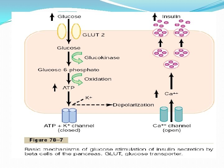

Table 78– 1 summarizes some of the factors that can increase or decrease insulin secretion. Table 78– 1 Factors and Conditions That Increase or Decrease Insulin Secretion Increase Insulin Secretion • Increased blood glucose • Increased blood free fatty acids • Increased blood amino acids • Gastrointestinal hormones (gastrin, cholecystokinin, secretin, gastric inhibitory peptide) • Glucagon, growth hormone, cortisol • Parasympathetic stimulation; acetylcholine • b-Adrenergic stimulation • Insulin resistance; obesity • Sulfonylurea drugs (glyburide, tolbutamide) Decrease Insulin Secretion • Decreased blood glucose • Fasting • Somatostatin • a-Adrenergic activity • Leptin

Control of Insulin Secretion Formerly, it was believed that insulin secretion was controlled almost entirely by the blood glucose concentration. However, as more has been learned about the metabolic functions of insulin for protein and fat metabolism, it has become apparent that blood amino acids and other factors also play important roles in controlling insulin secretion (see Table 78– 1). Increased Blood Glucose Stimulates Insulin Secretion. At the normal fasting level of blood glucose of 80 to 90 mg/100 ml, the rate of insulin secretion is minimal— on the order of 25 ng/min/kg of body weight, a level that has only slight physiologic activity. If the blood glucose concentration is suddenly increased to a level two to three times normal and kept at this high level thereafter, insulin secretion increases markedly in two stages, as shown by the changes in plasma insulin concentration seen in Figure 78– 8.
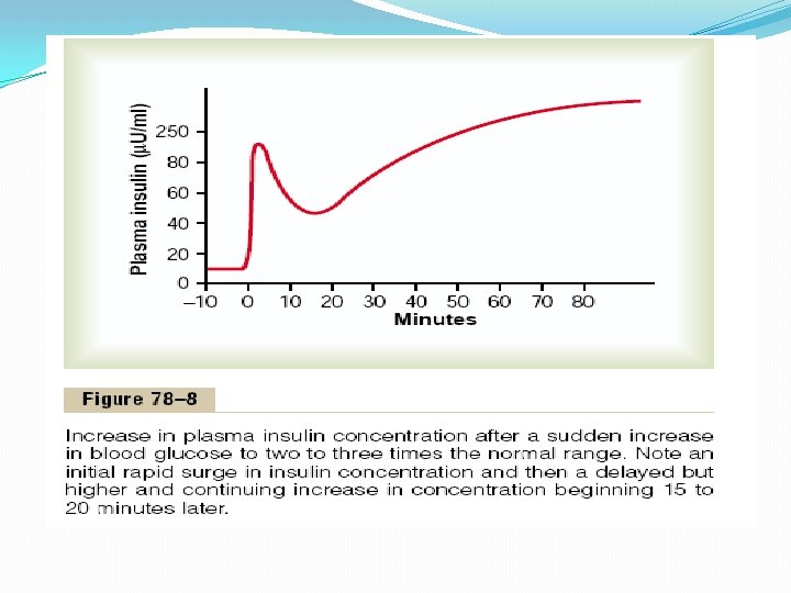

1. Plasma insulin concentration increases almost 10 -fold within 3 to 5 minutes after the acute elevation of the blood glucose; this results from immediate dumping of preformed insulin from the beta cells of the islets of Langerhans. However, the initial high rate of secretion is not maintained; instead, the insulin concentration decreases about halfway back toward normal in another 5 to 10 minutes. 2. Beginning at about 15 minutes, insulin secretion rises a second time and reaches a new plateau in 2 to 3 hours, this time usually at a rate of secretion even greater than that in the initial phase. This secretion results both from additional release of preformed insulin and from activation of the enzyme system that synthesizes and releases new insulin from the cells.

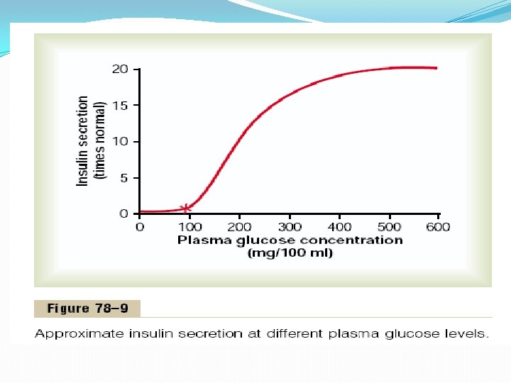
Feedback Relation Between Blood Glucose Concentration and Insulin Secretion Rate. As the concentration of blood glucose rises above 100 mg/100 ml of blood, the rate of insulin secretion rises rapidly, reaching a peak some 10 to 25 times the basal level at blood glucose concentrations between 400 and 600 mg/100 ml, as shown in Figure 78– 9. Thus, the increase in insulin secretion under a glucose stimulus is dramatic both in its rapidity and in the tremendous level of secretion achieved. Furthermore, the turn-off of insulin secretion is almost equally as rapid, occurring within 3 to 5 minutes after reduction in blood glucose concentration back to the fasting level. This response of insulin secretion to an elevated blood glucose concentration provides an extremely important feedback mechanism for regulating blood glucose concentration. That is, any rise in blood glucose increases insulin secretion, and the insulin in turn increases transport of glucose into liver, muscle, and other cells, thereby reducing the blood glucose concentration back toward the normal value.

Other Factors That Stimulate Insulin Secretion Amino Acids. In addition to the stimulation of insulin secretion by excess blood glucose, some of the amino acids have a similar effect. The most potent of these arginine and lysine. This effect differs from glucose stimulation of insulin secretion in the following way: Amino acids administered in the absence of a rise in blood glucose cause only a small increase in insulin secretion. However, when administered at the same time that the blood glucose concentration is elevated, the glucoseinduced secretion of insulin may be as much as doubled in the presence of the excess amino acids. Thus, the amino acids strongly potentiate the glucose stimulus for insulin secretion. The stimulation of insulin secretion by amino acids is important, because the insulin in turn promotes transport of amino acids into the tissue cells as well as intracellular formation of protein. That is, insulin is important for proper utilization of excess amino acids in the same way that it is important for the utilization of carbohydrates.

Gastrointestinal Hormones. A mixture of several important gastrointestinal hormones— gastrin, secretin, cholecystokinin, and gastric inhibitory peptide (which seems to be the most potent)—causes a moderate increase in insulin secretion. These hormones are released in the gastrointestinal tract after a person eats a meal. They then cause an “anticipatory” increase in blood insulin in preparation for the glucose and amino acids to be absorbed from the meal. These gastrointestinal hormones generally act the same way as amino acids to increase the sensitivity of insulin response to increased blood glucose, almost doubling the rate of insulin secretion as the blood glucose level rises.

Other Hormones and the Autonomic Nervous System. Other hormones that either directly increase insulin secretion or potentiate the glucose stimulus for insulin secretion include glucagon, growth hormone, cortisol, and, to a lesser extent, progesterone and estrogen. The importance of the stimulatory effects of these hormones is that prolonged secretion of any one of them in large quantities can occasionally lead to exhaustion of the beta cells of the islets of Langerhans and thereby increase the risk for developing diabetes mellitus. Indeed, diabetes often occurs in people who are maintained on high pharmacological doses of some of these hormones. Diabetes is particularly common in giants or acromegalic people with growth hormone-secreting tumors, or in people whose adrenal glands secrete excess glucocorticoids. Under some conditions, stimulation of the parasympathetic nerves to the pancreas can increase insulin secretion. However, it is doubtful that this effect is of physiologic significance for regulating insulin secretion.
 in “Switching” Between Carbohydrate and Lipid Metabolism From")
Role of Insulin (and Other Hormones) in “Switching” Between Carbohydrate and Lipid Metabolism From the preceding discussions, it should be clear that insulin promotes the utilization of carbohydrates for energy, whereas it depresses the utilization of fats. Conversely, lack of insulin causes fat utilization mainly to the exclusion of glucose utilization, except by brain tissue. Furthermore, the signal that controls this switching mechanism is principally the blood glucose concentration. When the glucose concentration is low, insulin secretion is suppressed and fat is used almost exclusively for energy everywhere except in the brain. When the glucose concentration is high, insulin secretion is stimulated and carbohydrate is used instead of fat, and the excess blood glucose is stored in the form of liver glycogen, liver fat, and muscle glycogen. Therefore, one of the most important functional roles of insulin in the body is to control which of these two foods from moment to moment will be used by the cells for energy.

At least four other known hormones also play important roles in this switching mechanism: growth hormone from the anterior pituitary gland, cortisol from the adrenal cortex, epinephrine from the adrenal medulla, and glucagon from the alpha cells of the islets of Langerhans in the pancreas. Glucagon is discussed in the next section of this chapter. Both growth hormone and cortisol are secreted in response to hypoglycemia, and both inhibit cellular utilization of glucose while promoting fat utilization. However, the effects of both of these hormones develop slowly, usually requiring many hours for maximal expression. Epinephrine is especially important in increasing plasma glucose concentration during periods of stress when the sympathetic nervous system is excited. However, epinephrine acts differently from the other hormones in that it increases the plasma fatty acid concentration at the same time. The reasons for these effects are as follows:
 epinephrine has the potent effect of causing glycogenolysis in the liver, thus releasing")
(1) epinephrine has the potent effect of causing glycogenolysis in the liver, thus releasing within minutes large quantities of glucose into the blood; (2) it also has a direct lipolytic effect on the adipose cells because it activates adipose tissue hormone-sensitive lipase, thus greatly enhancing the blood concentration of fatty acids as well. Quantitatively, the enhancement of fatty acids is far greater than the enhancement of blood glucose. Therefore, epinephrine especially enhances the utilization of fat in such stressful states as exercise, circulatory shock, and anxiety.

Glucagon and Its Functions Glucagon, a hormone secreted by the alpha cells of the islets of Langerhans when the blood glucose concentration falls, has several functions that are diametrically opposed to those of insulin. Most important of these functions is to increase the blood glucose concentration, an effect that is exactly the opposite that of insulin. Like insulin, glucagon is a large polypeptide. It has a molecular weight of 3485 and is composed of a chain of 29 amino acids. On injection of purified glucagon into an animal, a profound hyperglycemic effect occurs. Only 1 mg/kg of glucagon can elevate the blood glucose concentration about 20 mg/100 ml of blood (a 25 per cent increase) in about 20 minutes. For this reason, glucagon is also called the hyperglycemic hormone.
")
Effects on Glucose Metabolism The major effects of glucagon on glucose metabolism are (1) breakdown of liver glycogen (glycogenolysis) (2) increased gluconeogenesis in the liver. Both of these effects greatly enhance the availability of glucose to the other organs of the body. Glucagon Causes Glycogenolysis and Increased Blood Glucose Concentration. The most dramatic effect of glucagon is its ability to cause glycogenolysis in the liver, which in turn increases the blood glucose concentration within minutes. It does this by the following complex cascade of events: 1. Glucagon activates adenylyl cyclase in the hepatic cell membrane, 2. Which causes the formation of cyclic adenosine monophosphate, 3. Which activates protein kinase regulator protein, 4. Which activates protein kinase, 5. Which activates phosphorylase b kinase, 6. Which converts phosphorylase b into phosphorylase a, 7. Which promotes the degradation of glycogen into glucose-1 phosphate, 8. Which then is dephosphorylated; and the glucose is released from the liver cells.

This sequence of events is exceedingly important for several reasons. First, it is one of the most thoroughly studied of all the second messenger functions of cyclic adenosine monophosphate. Second, it demonstrates a cascade system in which each succeeding product is produced in greater quantity than the preceding product. Therefore, it represents a potent amplifying mechanism; this type of amplifying mechanism is widely used throughout the body for controlling many, if not most, cellular metabolic systems, often causing as much as a millionfold amplification in response. This explains how only a few micrograms of glucagon cause the blood glucose level to double or increase even more within a few minutes. Infusion of glucagon for about 4 hours can cause such intensive liver glycogenolysis that all the liver stores of glycogen become depleted.

Glucagon Increases Gluconeogenesis. Even after all the glycogen in the liver has been exhausted under the influence of glucagon, continued infusion of this hormone still causes continued hyperglycemia. This results from the effect of glucagon to increase the rate of amino acid uptake by the liver cells and then the conversion of many of the amino acids to glucose by gluconeogenesis. This is achieved by activating multiple enzymes that are required for amino acid transport and gluconeogenesis, especially activation of the enzyme system for converting pyruvate to phosphoenolpyruvate, a rate-limiting step in gluconeogenesis.

Other Effects of Glucagon Most other effects of glucagon occur only when its concentration rises well above the maximum normally found in the blood. Perhaps the most important effect is that glucagon activates adipose cell lipase, making increased quantities of fatty acids available to the energy systems of the body. Glucagon also inhibits the storage of triglycerides in the liver, which prevents the liver from removing fatty acids from the blood; this also helps make additional amounts of fatty acids available for the other tissues of the body. Glucagon in very high concentrations also (1)enhances the strength of the heart; (2) increases blood flow in some tissues, especially the kidneys; (3)enhances bile secretion; (4) inhibits gastric acid secretion. All these effects are probably of minimal importance in the normal function of the body.

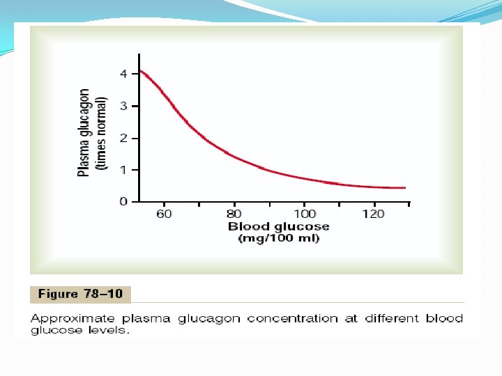
Regulation of Glucagon Secretion Increased Blood Glucose Inhibits Glucagon Secretion. The blood glucose concentration is by far the most potent factor that controls glucagon secretion. Note specifically, however, that the effect of blood glucose concentration on glucagon secretion is in exactly the opposite direction from the effect of glucose on insulin secretion. This is demonstrated in Figure 78– 10, showing that a decrease in the blood glucose concentration from its normal fasting level of about 90 mg/100 ml of blood down to hypoglycemic levels can increase the plasma concentration of glucagon severalfold. Conversely, increasing the blood glucose to hyperglycemic levels decreases plasma glucagon. Thus, in hypoglycemia, glucagon is secreted in large amounts; it then greatly increases the output of glucose from the liver and thereby serves the important function of correcting the hypoglycemia.

Increased Blood Amino Acids Stimulate Glucagon Secretion. High concentrations of amino acids, as occur in the blood after a protein meal (especially the amino acids alanine and arginine), stimulate the secretion of glucagon. This is the same effect that amino acids have in stimulating insulin secretion. Thus, in this instance, the glucagon and insulin responses are not opposites. The importance of amino acid stimulation of glucagon secretion is that the glucagon then promotes rapid conversion of the amino acids to glucose, thus making even more glucose available to the tissues. Exercise Stimulates Glucagon Secretion. In exhaustive exercise, the blood concentration of glucagon often increases fourfold to fivefold. What causes this is not understood, because the blood glucose concentration does not necessarily fall. A beneficial effect of the glucagon is that it prevents a decrease in blood glucose. One of the factors that might increase glucagon secretion in exercise is increased circulating amino acids. Other factors, such as b-adrenergic stimulation of the islets of Langerhans, may also play a role.

Somatostatin Inhibits Glucagon and Insulin Secretion The delta cells of the islets of Langerhans secrete the hormone somatostatin, a polypeptide containing only 14 amino acids that has an extremely short half-life of only 3 minutes in the circulating blood. Almost all factors related to the ingestion of food stimulate somatostatin secretion. They include (1) increased blood glucose, (2) increased amino acids, (3) increased fatty acids, (4) increased concentrations of several of the gastrointestinal hormones released from the upper gastrointestinal tract in response to food intake.

In turn, somatostatin has multiple inhibitory effects as follows: 1. Somatostatin acts locally within the islets of Langerhans themselves to depress the secretion of both insulin and glucagon. 2. Somatostatin decreases the motility of the stomach, duodenum, and gallbladder. 3. Somatostatin decreases both secretion and absorption in the gastrointestinal tract. Putting all this information together, it has been suggested that the principal role of somatostatin is to extend the period of time over which the food nutrients are assimilated into the blood. At the same time, the effect of somatostatin to depress insulin and glucagon secretion decreases the utilization of the absorbed nutrients by the tissues, thus preventing rapid exhaustion of the food and therefore making it available over a longer period of time. It should also be recalled that somatostatin is the same chemical substance as growth hormone inhibitory hormone, which is secreted in the hypothalamus and suppresses anterior pituitary gland growth hormone secretion.

Summary of Blood Glucose Regulation In a normal person, the blood glucose concentration isnarrowly controlled, usually between 80 and 90 mg/ 100 ml of blood in the fasting person each morning before breakfast. This concentration increases to 120 to 140 mg/100 ml during the first hour or so after a meal, but the feedback systems for control of blood glucose return the glucose concentration rapidly back to the control level, usually within 2 hours after the last absorption of carbohydrates. Conversely, in starvation, the gluconeogenesis function of the liver provides the glucose that is required to maintain the fasting blood glucose level. The mechanisms for achieving this high degree of control have been presented in this chapter. Let us summarize them.

1. The liver functions as an important blood glucose buffer system. That is, when the blood glucose rises to a high concentration after a meal and the rate of insulin secretion also increases, as much as two thirds of the glucose absorbed from the gut is almost immediately stored in the liver in the form of glycogen. Then, during the succeeding hours, when both the blood glucose concentration and the rate of insulin secretion fall, the liver releases the glucose back into the blood. In this way, the liver decreases the fluctuations in blood glucose concentration to about one third of what they would otherwise be. In fact, in patients with severe liver disease, it becomes almost impossible to maintain a narrow range of blood glucose concentration. 2. Both insulin and glucagon function as important feedback control systems for maintaining a normal blood glucose concentration. When the glucose concentration rises too high, insulin is secreted; the insulin in turn causes the blood glucose concentration to decrease toward normal. Conversely, a decrease in blood glucose stimulates glucagon secretion; the glucagon then functions in the opposite direction to increase the glucose toward normal. Under most normal conditions, the insulin feedback mechanism is much more important than the glucagon mechanism, but in instances of starvation or excessive utilization of glucose during exercise and other stressful situations, the glucagon mechanism also becomes valuable.

3. Also, in severe hypoglycemia, a direct effect of low blood glucose on the hypothalamus stimulates the sympathetic nervous system. In turn, the epinephrine secreted by the adrenal glands causes still further release of glucose from the liver. This, too, helps protect against severe hypoglycemia. 4. And finally, over a period of hours and days, both growth hormone and cortisol are secreted in response to prolonged hypoglycemia, and they both decrease the rate of glucose utilization by most cells of the body, converting instead to greater amounts of fat utilization. This, too, helps return the blood glucose concentration toward normal.

Importance of Blood Glucose Regulation. One might ask the question: Why is it so important to maintain a constant blood glucose concentration, particularly because most tissues can shift to utilization of fats and proteins for energy in the absence of glucose? The answer is that glucose is the only nutrient that normally can be used by the brain, retina, and germinal epithelium of the gonads in sufficient quantities to supply them optimally with their required energy. Therefore, it is important to maintain the blood glucose concentration at a sufficiently high level to provide this necessary nutrition. Most of the glucose formed by gluconeogenesis during the inter digestive period is used for metabolism in the brain. Indeed, it is important that the pancreas not secrete any insulin during this time; otherwise, the scant supplies of glucose that are available would all go into the muscles and other peripheral tissues, leaving the brain without a nutritive source. It is also important that the blood glucose concentration not rise too high for four reasons:
 Glucose can exert a large amount of osmotic pressure in the extracellular fluid,")
(1) Glucose can exert a large amount of osmotic pressure in the extracellular fluid, and if the glucose concentration rises to excessive values, this can cause considerable cellular dehydration. (2) An excessively high level of blood glucose concentration causes loss of glucose in the urine. (3) Loss of glucose in the urine also causes osmotic diuresis by the kidneys, which can deplete the body of its fluids and electrolytes. (4) Long-term increases in blood glucose may cause damage to many tissues, especially to blood vessels. Vascular injury, associated with uncontrolled diabetes mellitus, leads to increased risk for heart attack, stroke, end-stage renal disease, and blindness.

Diabetes Mellitus Diabetes mellitus is a syndrome of impaired carbohydrate, fat, and protein metabolism caused by either lack of insulin secretion or decreased sensitivity of the tissues to insulin. There are two general types of diabetes mellitus: 1. Type I diabetes, also called insulin-dependent diabetes mellitus (IDDM), is caused by lack of insulin secretion. 2. Type II diabetes, also called non–insulin-dependent diabetes mellitus (NIDDM), is caused by decreased sensitivity of target tissues to the metabolic effect of insulin. This reduced sensitivity to insulin is often called insulin resistance. In both types of diabetes mellitus, metabolism of all the main foodstuffs is altered. The basic effect of insulin lack or insulin resistance on glucose metabolism is to prevent the efficient uptake and utilization of glucose by most cells of the body, except those of the brain. As a result, blood glucose concentration increases, cell utilization of glucose falls increasingly lower, and utilization of fats and proteins increases.

Type I Diabetes—Lack of Insulin Production by Beta Cells of the Pancreas Injury to the beta cells of the pancreas or diseases that impair insulin production can lead to type I diabetes. Viral infections or autoimmune disorders may be involved in the destruction of beta cells in many patients with type I diabetes, although heredity also plays a major role in determining the susceptibility of the beta cells to destruction by these insults. In some instances, there may be a hereditary tendency for beta cell degeneration even without viral infections or autoimmune disorders. The usual onset of type I diabetes occurs at about 14 years of age in the United States, and for this reason it is often called juvenile diabetes mellitus. Type I diabetes may develop very abruptly, over a period of a few days or weeks, with three principal sequelae: (1) increased blood glucose, (2) increased utilization of fats for energy and formation of cholesterol by the liver, (3) depletion of the body’s proteins.

Blood Glucose Concentration Rises to Very High Levels in Diabetes Mellitus. The lack of insulin decreases the efficiency of peripheral glucose utilization and augments glucose production, raising plasma glucose to 300 to 1200 mg/100 ml. The increased plasma glucose then has multiple effects throughout the body. Increased Blood Glucose Causes Loss of Glucose in the Urine The high blood glucose causes more glucose to filter into the renal tubules than can be reabsorbed, and the excess glucose spills into the urine. This normally occurs when the blood glucose concentration rises above 180 mg/100 ml, a level that is called the blood “threshold” for the appearance of glucose in the urine. When the blood glucose level rises to 300 to 500 mg/100 ml— common values in people with severe untreated diabetes— 100 or more grams of glucose can be lost into the urine each day.

Increased Blood Glucose Causes Dehydration The very high levels of blood glucose (sometimes as high as 8 to 10 times normal in severe untreated diabetes) can cause severe cell dehydration throughout the body. This occurs partly because glucose does not diffuse easily through the pores of the cell membrane, and the increased osmotic pressure in the extracellular fluids causes osmotic transfer of water out of the cells. In addition to the direct cellular dehydrating effect of excessive glucose, the loss of glucose in the urine causes osmotic diuresis. That is, the osmotic effect of glucose in the renal tubules greatly decreases tubular reabsorption of fluid. The overall effect is massive loss of fluid in the urine, causing dehydration of the extracellular fluid, which in turn causes compensatory dehydration of the intracellular fluid, Thus, polyuria (excessive urine excretion), intracellular and extracellular dehydration, and increased thirst are classic symptoms of diabetes.

Chronic High Glucose Concentration Causes Tissue Injury When blood glucose is poorly controlled over long periods in diabetes mellitus, blood vessels in multiple tissues throughout the body begin to function abnormally and undergo structural changes that result in inadequate blood supply to the tissues. This in turn leads to increased risk for heart attack, stroke, end-stage kidney disease, retinopathy and blindness, and ischemia and gangrene of the limbs. Chronic high glucose concentration also causes damage to many other tissues. For example, peripheral neuropathy, which is abnormal function of peripheral nerves, and autonomic nervous system dysfunction are frequent complications of chronic, uncontrolled diabetes mellitus. These abnormalities can result in impaired cardiovascular reflexes, impaired bladder control, decreased sensation in the extremities, and other symptoms of peripheral nerve damage. The precise mechanisms that cause tissue injury in diabetes are not well understood but probably involve multiple effects of high glucose concentrations and other metabolic abnormalities on proteins of endothelial and vascular smooth muscle cells, as well as other tissues. In addition, hypertension, secondary to renal injury, and atherosclerosis, secondary to abnormal lipid metabolism, often develop in patients with diabetes and amplify the tissue damage caused by the elevated glucose.

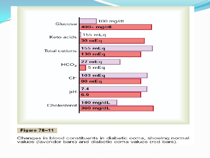
Diabetes Mellitus Causes Increased Utilization of Fats and Metabolic Acidosis. The shift from carbohydrate to fat metabolism in diabetes increases the release of keto acids, such as acetoacetic acid and b-hydroxybutyric acid, into the plasma more rapidly than they can be taken up and oxidized by the tissue cells. As a result, the patient develops severe metabolic acidosis from the excess keto acids, which, in association with dehydration due to the excessive urine formation, can cause severe acidosis. This leads rapidly to diabetic coma and death unless the condition is treated immediately with large amounts of insulin. All the usual physiologic compensations that occur in metabolic acidosis take place in diabetic acidosis. They include rapid and deep breathing, which causes increased expiration of carbon dioxide; this buffers the acidosis but also depletes extracellular fluid bicarbonate stores. The kidneys compensate by decreasing bicarbonate excretion and generating new bicarbonate that is added back to the extracellular fluid. Although extreme acidosis occurs only in the most severe instances of uncontrolled diabetes, when the p. H of the blood falls below about 7. 0, acidotic coma and death can occur within hours. The overall changes in the electrolytes of the blood as a result of severe diabetic acidosis are shown in Figure 78– 11. Excess fat utilization in the liver occurring over a long time causes large amounts of cholesterol in the circulating blood and increased deposition of cholesterol in the arterial walls. This leads to severe arteriosclerosis and other vascular lesions, as discussed earlier.

Diabetes Causes Depletion of the Body’s Proteins. Failure to use glucose for energy leads to increased utilization and decreased storage of proteins as well as fat. Therefore, a person with severe untreated diabetes mellitus suffers rapid weight loss and asthenia (lack of energy) despite eating large amounts of food (polyphagia). Without treatment, these metabolic abnormalities can cause severe wasting of the body tissues and death within a few weeks. Type II Diabetes—Resistance to the Metabolic Effects of Insulin Type II diabetes is far more common than type I, accounting for about 90 per cent of all cases of diabetes mellitus. In most cases, the onset of type II diabetes occurs after age 30, often between the ages of 50 and 60 years, and the disease develops gradually. Therefore, this syndrome is often referred to as adult-onset diabetes. In recent years, however, there has been a steady increase in the number of younger individuals, some less than 20 years old, with type II diabetes. This trend appears to be related mainly to the increasing prevalence of obesity, the most important risk factor for type II diabetes in children as well as in adults.

Obesity, Insulin Resistance, and “Metabolic Syndrome” Usually Precede Development of Type II Diabetes. Type II diabetes, in contrast to type I, is associated with increased plasma insulin concentration (hyperinsulinemia). This occurs as a compensatory response by the pancreatic beta cells for diminished sensitivity of target tissues to the metabolic effects of insulin, a condition referred to as insulin resistance. The decrease in insulin sensitivity impairs carbohydrate utilization and storage, raising blood glucose and stimulating a compensatory increase in insulin secretion. Development of insulin resistance and impaired glucose metabolism is usually a gradual process, beginning with excess weight gain and obesity. The mechanisms that link obesity with insulin resistance, however, are still uncertain. Some studies suggest that there are fewer insulin receptors, especially in the skeletal muscle, liver, and adipose tissue, in obese than in lean subjects. However, most of the insulin resistance appears to be caused by abnormalities of the signaling pathways that link receptor activation with multiple cellular effects. Impaired insulin signaling appears to be closely related to toxic effects of lipid accumulation in tissues such as skeletal muscle and liver secondary to excess weight gain.

Insulin resistance is part of a cascade of disorders that is often called the “metabolic syndrome. ” Some of the features of the metabolic syndrome include: (1) obesity, especially accumulation of abdominal fat; (2) insulin resistance; (3) fasting hyperglycemia; (4) lipid abnormalities such as increased blood triglycerides and decreased blood high-density lipoprotein-cholesterol; (5) hypertension. All of the features of the metabolic syndrome are closely related to excess weight gain, especially when it is associated with accumulation of adipose tissue in the abdominal cavity around the visceral organs. The role of insulin resistance in contributing to some of the components of the metabolic syndrome is unclear, although it is clear that insulin resistance is the primary cause of increased blood glucose concentration. The major adverse consequence of the metabolic syndrome is cardiovascular disease, including atherosclerosis and injury to various organs throughout the body. Several of the metabolic abnormalities associated with the syndrome are risk factors for cardiovascular disease, and insulin resistance predisposes to the development of type II diabetes mellitus, also a major cause of cardiovascular disease.

Other Factors That Can Cause Insulin Resistance and Type II Diabetes. Although most patients with type II diabetes are overweight or have substantial accumulation of visceral fat, severe insulin resistance and type II diabetes can also occur as a result of other acquired or genetic conditions that impair insulin signaling in peripheral tissues (Table 78– 2). Polycystic ovary syndrome (PCOS), for example, is associated with marked increases in ovarian androgen production and insulin resistance and is one of the most common endocrine disorders in women, affecting approximately 6 per cent of all women during their reproductive life. Although the pathogenesis of PCOS remains uncertain, insulin resistance and hyperinsulinemia are found in approximately 80 per cent of affected women. The long-term consequences include increased risk for diabetes mellitus, increased blood lipids, and cardiovascular disease. Excess formation of glucocorticoids (Cushing’s syndrome) or growth hormone (acromegaly) also decreases the sensitivity of various tissues to the metabolic effects of insulin and can lead to development of diabetes mellitus. Genetic causes of obesity and insulin resistance, if severe enough, also can lead to type II diabetes as well as many other features of the metabolic syndrome, including cardiovascular disease.

Development of Type II Diabetes During Prolonged Insulin Resistance. With prolonged and severe insulin resistance, even the increased levels of insulin are not sufficient to maintain normal glucose regulation. As a result, moderate hyperglycemia occurs after ingestion of carbohydrates in the early stages of the disease. Table 78– 2 Some Causes of Insulin Resistance • Obesity/overweight (especially excess visceral adiposity) • Excess glucocorticoids (Cushing’s syndrome or steroid therapy) • Excess growth hormone (acromegaly) • Pregnancy, gestational diabetes • Polycystic ovary disease • Lipodystrophy (acquired or genetic; associated with lipid accumulation in liver) • Autoantibodies to the insulin receptor • Mutations of the peroxisome proliferators’ activator receptor g (PPARg) • Mutations that cause genetic obesity (e. g. , melanocortin receptor mutations) • Hemochromatosis (a hereditary disease that causes tissue iron

In the later stages of type II diabetes, the pancreatic beta cells become “exhausted” and are unable to produce enough insulin to prevent more severe hyperglycemia, especially after the person ingests a carbohydrate -rich meal. Some obese people, although having marked insulin resistance and greater than normal increases in blood glucose after a meal, never develop clinically significant diabetes mellitus; apparently, the pancreas in these people produces enough insulin to prevent severe abnormalities of glucose metabolism. In others, however, the pancreas gradually becomes exhausted from secreting large amounts of insulin, and full-blown diabetes mellitus occurs. Some studies suggest that genetic factors play an important role in determining whether an individual’s pancreas can sustain the high output of insulin over many years that is necessary to avoid the severe abnormalities of glucose metabolism in type II diabetes. In many instances, type II diabetes can be effectively treated, at least in the early stages, with exercise, caloric restriction, and weight reduction, and no exogenous insulin administration is required. Drugs that increase insulin sensitivity, such as thiazolidinediones and metformin, or drugs that cause additional release of insulin by the pancreas, such as sulfonylureas, may also be used. However, in the later stages of type II diabetes, insulin administration is usually required to control plasma glucose.

Physiology of Diagnosis of Diabetes Mellitus Table 78– 3 compares some of clinical features of type I and type II diabetes mellitus. The usual methods for diagnosing diabetes are based on various chemical tests of the urine and the blood. Urinary Glucose. Simple office tests or more complicated quantitative laboratory tests may be used to determine the quantity of glucose lost in the urine. In general, a normal person loses undetectable amounts of glucose, whereas a person with diabetes loses glucose in small to large amounts, in proportion to the severity of disease and the intake of carbohydrates. Fasting Blood Glucose and Insulin Levels. The fasting blood glucose level in the early morning is normally 80 to 90 mg/100 ml, and 110 mg/100 ml is considered to be the upper limit of normal. A fasting blood glucose level above this value often indicates diabetes mellitus or a least marked insulin resistance. In type I diabetes, plasma insulin levels are very low or undetectable during fasting and even after a meal. In type II diabetes, plasma insulin concentration may be several fold higher than normal and usually increases to a greater extent after ingestion of a standard glucose load during a glucose tolerance test.

Glucose Tolerance Test. As demonstrated by the bottom curve in Figure 78– 12, called a “glucose tolerance curve, ” when a normal, fasting person ingests 1 gram of glucose per kilogram of body weight, the blood glucose level rises from about 90 mg/100 ml to 120 to 140 mg/100 ml and falls back to below normal in about 2 hours. In a person with diabetes, the fasting blood glucose concentration is almost always above 110 mg/100 ml and often above 140 mg/100 ml. Also, the glucose tolerance test is almost always abnormal. On ingestion of glucose, these people exhibit a much greater than normal rise in blood glucose level, as demonstrated by the upper curve in Figure 78– 12, and the glucose level falls back to the control value only after 4 to 6 hours; furthermore, it fails to fall below the control level. The slow fall of this curve and its failure to fall below the control level demonstrate that either (1) the normal increase in insulin secretion after glucose ingestion does not occur or (2) there is decreased sensitivity to insulin. A diagnosis of diabetes mellitus can usually be established on the basis of such a curve, and type II diabetes can be distinguished from each other by measurements of plasma insulin, with plasma insulin being low or undetectable in type I diabetes and increased in type II diabetes.
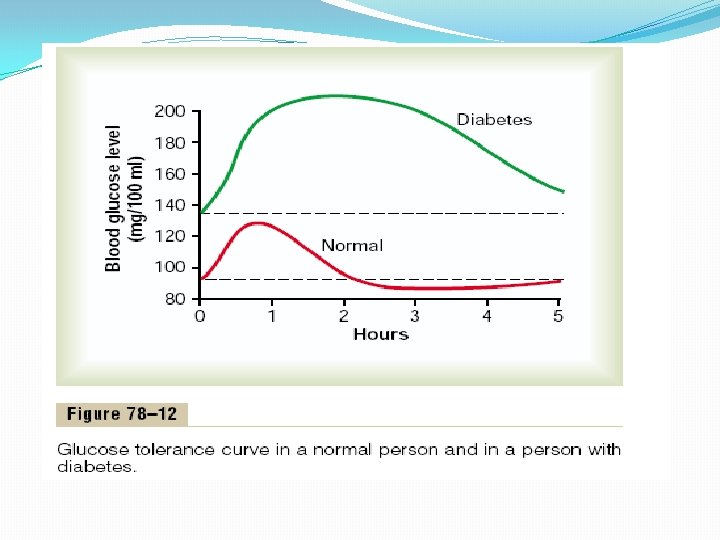

Table 78– 3 Clinical Characteristics of Patients with Type I and Type II Diabetes Mellitus Feature Type II Age at onset Usually <20 years Usually >30 years Body mass Low (wasted) to normal Obese Plasma insulin Low or absent Normal to high initially Plasma glucagon High, can be High, suppressed High, resistant to suppressed suppression Plasma glucose Increased Insulin sensitivity Normal Reduced Therapy insulin Weight loss, thiazolidinediones, metformin, sulfonylureas, insulin

Acetone Breath. As pointed out in Chapter 68, small quantities of acetoacetic acid in the blood, which increase greatly in severe diabetes, are converted to acetone. This is volatile and vaporized into the expired air. Consequently, one can frequently make a diagnosis of type I diabetes mellitus simply by smelling acetone on the breath of a patient. Also, keto acids can be detected by chemical means in the urine, and their quantitation aids in determining the severity of the diabetes. In the early stages of type II diabetes, however, keto acids are usually not produced in excess amounts. However, when insulin resistance becomes very severe and there is greatly increased utilization of fats for energy, keto acids are then produced in persons with type II diabetes.

Treatment of Diabetes The theory of treatment of type I diabetes mellitus is to administer enough insulin so that the patient will have carbohydrate, fat, and protein metabolism that is as normal as possible. Insulin is available in several forms. “Regular” insulin has a duration of action that lasts from 3 to 8 hours, whereas other forms of insulin (precipitated with zinc or with various protein derivatives) are absorbed slowly from the injection site and therefore have effects that last as long as 10 to 48 hours. Ordinarily, a patient with severe type I diabetes is given a single dose of one of the longer-acting insulins each day to increase overall carbohydrate metabolism throughout the day. Then additional quantities of regular insulin are given during the day at those times when the blood glucose level tends to rise too high, such as at mealtimes. Thus, each patient is provided with an individualized pattern of treatment.

In persons with type II diabetes, dieting and exercise are usually recommended in an attempt to induce weight loss and to reverse the insulin resistance. If this fails, drugs may be administered to increase insulin sensitivity or to stimulate increased production of insulin by the pancreas. In many persons, however, exogenous insulin must be used to regulate blood glucose. In the past, the insulin used for treatment was derived from animal pancreata. However, human insulin produced by the recombinant DNA process has become more widely used because some patients develop immunity and sensitization against animal insulin, thus limiting its effectiveness.

Relation of Treatment to Arteriosclerosis. Diabetic patients, mainly because of their high levels of circulating cholesterol and other lipids, develop atherosclerosis, arteriosclerosis, severe coronary heart disease, and multiple microcirculatory lesions far more easily than do normal people. Indeed, those who have poorly controlled diabetes throughout childhood are likely to die of heart disease in early adulthood. In the early days of treating diabetes, the tendency was to severely reduce the carbohydrates in the diet so that the insulin requirements would be minimized. This procedure kept the blood glucose from increasing too high and attenuated loss of glucose in the urine, but it did not prevent many of the abnormalities of fat metabolism. Consequently, the current tendency is to allow the patient an almost normal carbohydrate diet and to give large enough insulin to metabolize the carbohydrates. This decreases the rate of fat metabolism and depresses the high level of blood cholesterol. Because the complications of diabetes—such as atherosclerosis, greatly increased susceptibility to infection, diabetic retinopathy, cataracts, hypertension, and chronic renal disease—are closely associated with the level of blood lipids as well as the level of blood glucose, most physicians also use lipid-lowering drugs to help prevent these disturbances.

Insulinoma—Hyperinsulinism Although much rarer than diabetes, excessive insulin production occasionally occurs from an adenoma of an islet of Langerhans. About 10 to 15 per cent of these adenomas are malignant, and occasionally metastases from the islets of Langerhans spread throughout the body, causing tremendous production of insulin by both the primary and the metastatic cancers. Indeed, more than 1000 grams of glucose have had to be administered every 24 hours to prevent hypoglycemia in some of these patients.

Insulin Shock and Hypoglycemia. As already emphasized, the central nervous system normally derives essentially all its energy from glucose metabolism, and insulin is not necessary for this use of glucose. However, if high levels of insulin cause blood glucose to fall to low values, the metabolism of the central nervous system becomes depressed. Consequently, in patients with insulin-secreting tumors or in patients with diabetes who administer too much insulin to themselves, the syndrome called insulin shock may occur as follows. As the blood glucose level falls into the range of 50 to 70 mg/100 ml, the central nervous system usually becomes quite excitable, because this degree of hypoglycemia sensitizes neuronal activity. Sometimes various forms of hallucinations result, but more often the patient simply experiences extreme nervousness, trembles all over, and breaks out in a sweat. As the blood glucose level falls to 20 to 50 mg/100 ml, clonic seizures and loss of consciousness are likely to occur. As the glucose level falls still lower, the seizures cease and only a state of coma remains. Indeed, at times it is difficult by simple clinical observation to distinguish between diabetic coma as a result of insulin-lack acidosis and coma due to hypoglycemia caused by excess insulin. The acetone breath and the rapid, deep breathing of diabetic coma are not present in hypoglycemic coma. Proper treatment for a patient who has hypoglycemic shock or coma is immediate intravenous administration of large quantities of glucose. This usually brings the patient out of shock within a minute or more. Also, the administration of glucagon (or, less effectively, epinephrine) can cause glycogenolysis in the liver and thereby increase the blood glucose level extremely rapidly. If treatment is not effected immediately, permanent damage to the neuronal cells of the central nervous system often occurs.
- Slides: 85