
Institute for microbiology presents TRACING THE CRIMINAL L

Institute for microbiology presents: TRACING THE CRIMINAL L Part Five: Gram-Negative Criminals II
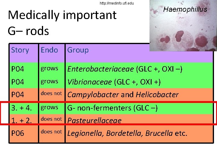
http: //medinfo. ufl. edu Medically important G– rods Haemophillus Story Endo Group P 04 grows Enterobacteriaceae (GLC +, OXI –) P 04 grows Vibrionaceae (GLC +, OXI +) P 04 does not Campylobacter and Helicobacter 3. + 4. 1. + 2. P 06 grows G- non-fermenters (GLC –) Pasteurellaceae Legionella, Bordetella, Brucella etc. does not
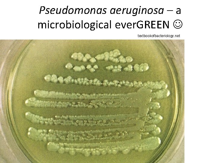
Pseudomonas aeruginosa – a microbiological ever. GREEN textbookofbacteriology. net
Summary Clinical characteristics – Pasteurellaceae Clinical characteristics – G– glucose non-fermenters Diagnostics of Pasteurellaceae Diagnostics of G– glucose non-fermenters
Clinical characteristics – Pasteurellaceae
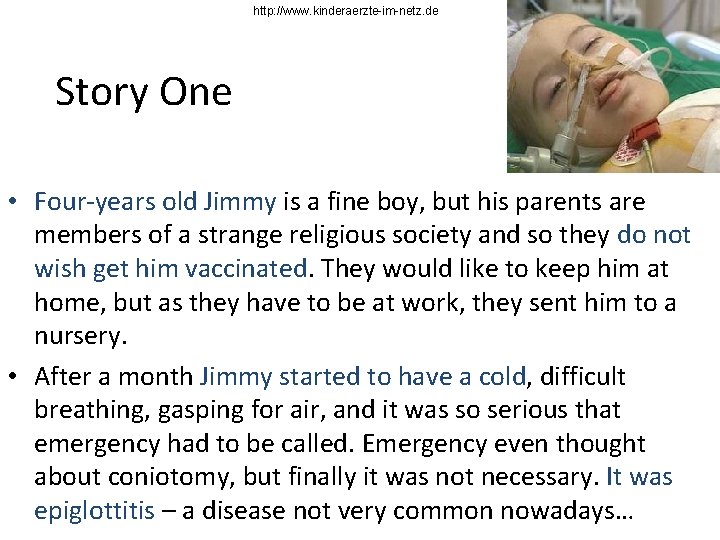
http: //www. kinderaerzte-im-netz. de Story One • Four-years old Jimmy is a fine boy, but his parents are members of a strange religious society and so they do not wish get him vaccinated. They would like to keep him at home, but as they have to be at work, they sent him to a nursery. • After a month Jimmy started to have a cold, difficult breathing, gasping for air, and it was so serious that emergency had to be called. Emergency even thought about coniotomy, but finally it was not necessary. It was epiglottitis – a disease not very common nowadays…
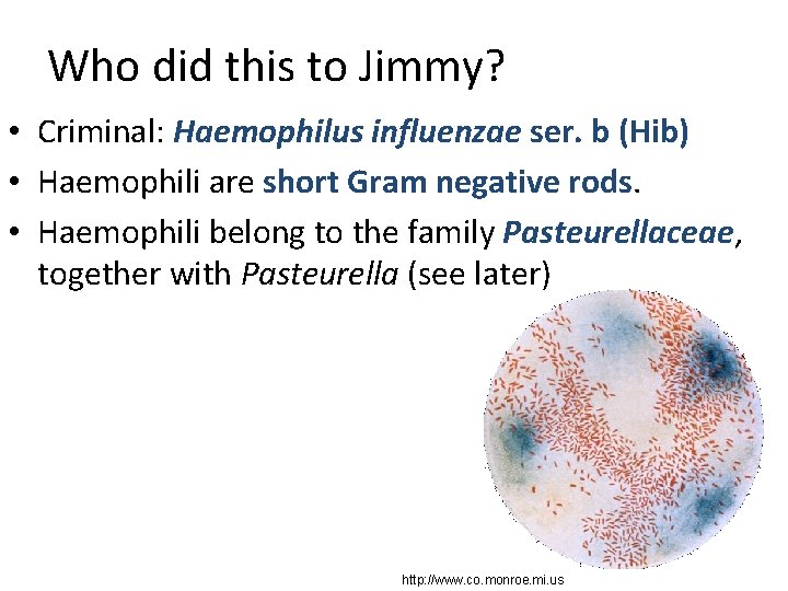
Who did this to Jimmy? • Criminal: Haemophilus influenzae ser. b (Hib) • Haemophili are short Gram negative rods. • Haemophili belong to the family Pasteurellaceae, together with Pasteurella (see later) http: //www. co. monroe. mi. us
Classification of haemophili • Haemophilus influenzae – capsullar type b (Hib) – preventable (vaccine) – capsullar types a, c, d, e, f – non-encapsulated strains • Haemophilus parainfluenzae (much more common, much less pathogenic) • Haemophilus aphrophilus and many other species • Haemophilus ducreyi, causative agent of a sexually transmitted disease ulcus molle
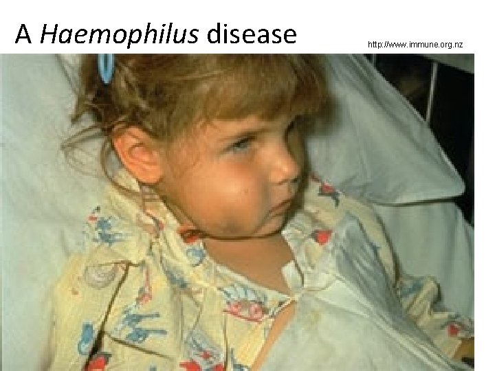
Pathogenicity of haemophili • The most severe diseases caused by haemophili are epiglottitis, meningitis and sepsis. This is mostly typical for Haemophilus influenzae, serotype b. • Other common diseases are otitis media and sinutisitis (after Streptococcus pneumoniae and together with Moraxella catarrhalis) • Their presence in throat is very common and their pathogenic role is very questionable. Especially in a case of Haemophilus parainfluenzae, we usually do not suppose them to be pathogen.
A Haemophilus disease http: //www. immune. org. nz
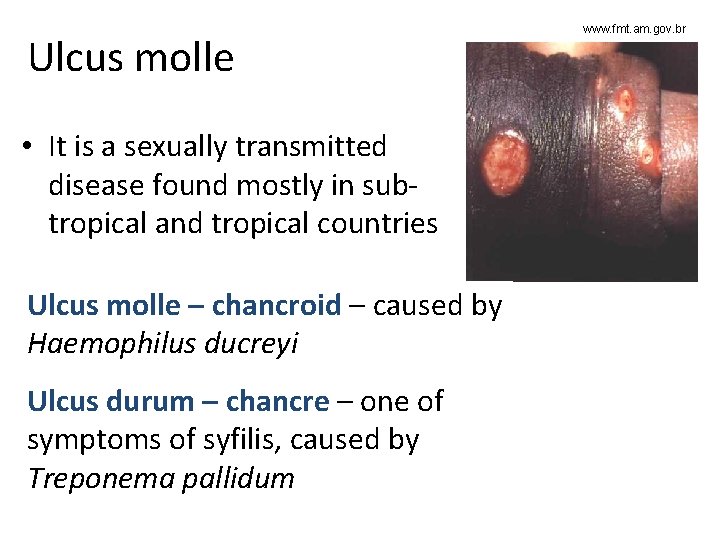
Ulcus molle • It is a sexually transmitted disease found mostly in subtropical and tropical countries Ulcus molle – chancroid – caused by Haemophilus ducreyi Ulcus durum – chancre – one of symptoms of syfilis, caused by Treponema pallidum www. fmt. am. gov. br
Story Two • Joana was walking in gardens as usual. Unfortunatelly, one garden fence was too old and rotten and the dog behind too strong. The dog run out and Joana was bitten into her leg. • The owners of the dog had proven that the dog has been vaccinated against rabies. Nevertheless, some pus soon occured in the wound. The pus was sent to the laboratory. And the criminal was…
Pasteurella multocida • Pasteurella multocida is normal respiratory microbiota in dogs. • In humans, it causes mainly pyogenic wound inflamations after being bitten by a dog or another animal. • It smells similarly as Haemophillus (some people say „like old rag“), but it grows on blood agar (not Endo agar). • The morphology of colonies: something between Streptococcus and Enterococcus, but it is vancomycin resistant and this is suspicious to the microbiologist (especially with parallel susceptibility to penicillin)
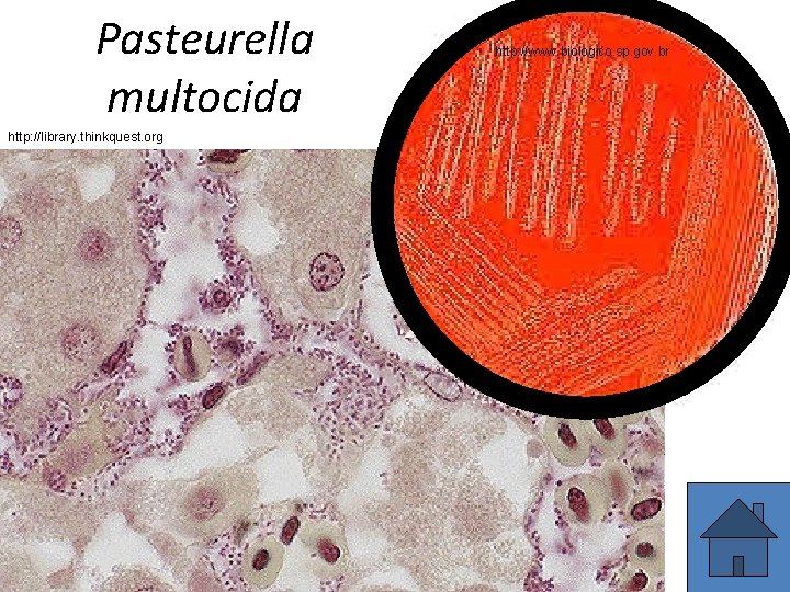
Pasteurella multocida http: //library. thinkquest. org http: //www. biologico. sp. gov. br
Clinical characteristics – Gram– glucose nonfermenting bacteria
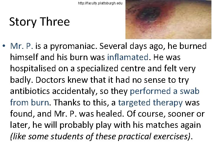
http: //faculty. plattsburgh. edu Story Three • Mr. P. is a pyromaniac. Several days ago, he burned himself and his burn was inflamated. He was hospitalised on a specialized centre and felt very badly. Doctors knew that it had no sense to try antibiotics accidentaly, so they performed a swab from burn. Thanks to this, a targeted therapy was found, and Mr. P. was healed. Of course, sooner or later, he will probably play with his matches again (like some students of these practical exercises).
Who is guilty this time? • Pseudomonas aeruginosa, the most common of „Gram– non-fermentation bacteria“ (G– NF) • On the other hand, the guilty one could be any of that group, e. g. Acinetobacter, Burkholderia cepacia or Stenotrophomonas maltophilia • Those bacteria are mostly strict aerobes, instead of fermentation of sugars, they utilize them by aerobic respiration, and their adaptation to outer environment is obvious also in other properties – they have low temperature optimum and they are often pigmented, so they fight with sunlight in outer environment
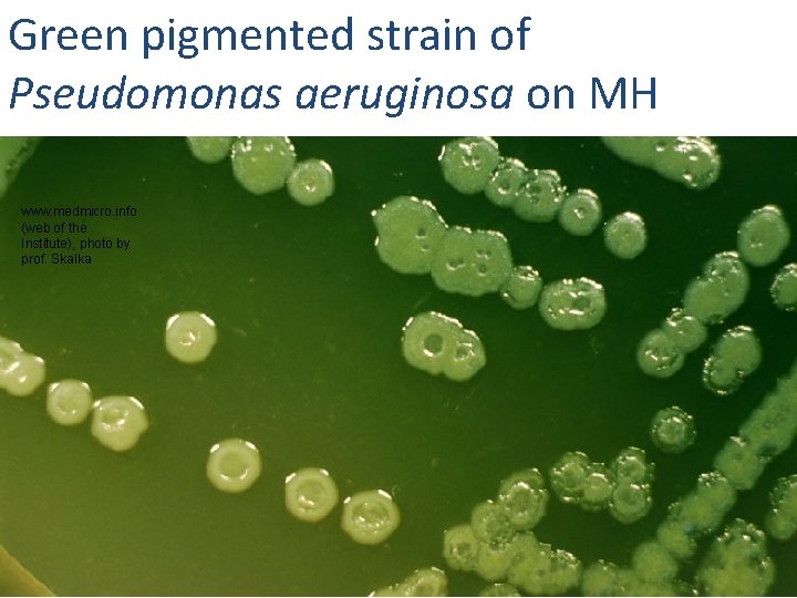
Green pigmented strain of Pseudomonas aeruginosa on MH www. medmicro. info (web of the Institute), photo by prof. Skalka
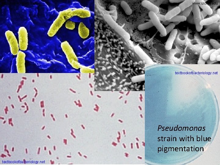
http: //de. wikipedia. org www. uiowa. edu textbookofbacteriology. net Pseudomonas strain with blue pigmentation textbookofbacteriology. net
Pathogenicity of G– NF • Commonly: bacteria from outer environment, often plant pathogens, „not-brave-bacteria“, which are not able to infect a healthy person. Their aims are patients with burns, clients of emergency units, transplant centers, e. t. c. • They often cause wound infections, can be found in respiratory ways, and even in the bloodstream of hospitalized persons. • So they are important causative agents of nosocomial infections • Sometimes it is difficult to differentiate between an infection and a colonisation – especially in superficial wounds it is often useless to use other than topical antibiotics
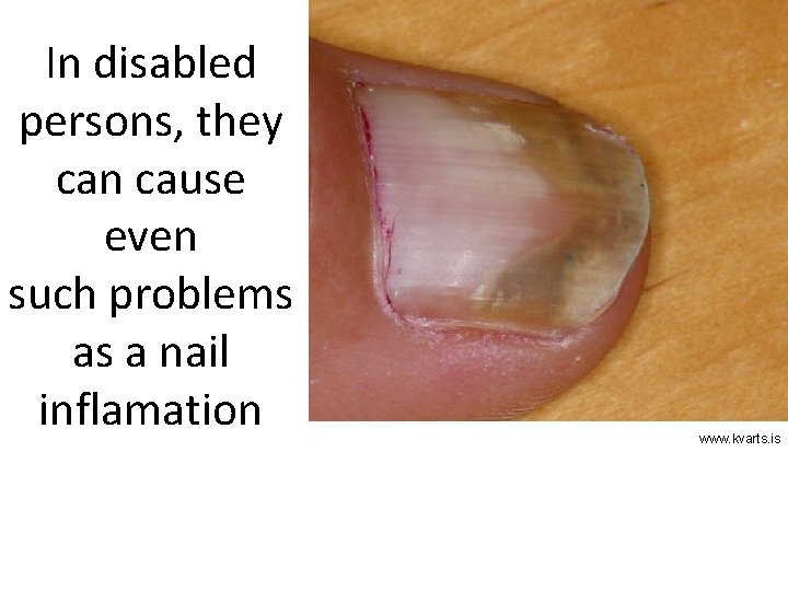
In disabled persons, they can cause even such problems as a nail inflamation www. kvarts. is
Dr. Zahradníček‘s autocauistics • Friday, 13 th January 2006: fall into a not-covered canalisation hole in the city of Padang, West Sumatera, Indonesia. Quite large wound, reaching tibial periost • Some three weeks later, the wound started to smell like Pseudomonas, and really, this bacterium was sucsesfully cultured from it. • Dr. Zahradníček decided for local treatment (gentamicin + polymyxine) – in such wound infections topic terapy uses to be more important than systemic treatmend (also because of likely presence of wound biofilm) • The therapy was successfull

Padang Photo O. Z.
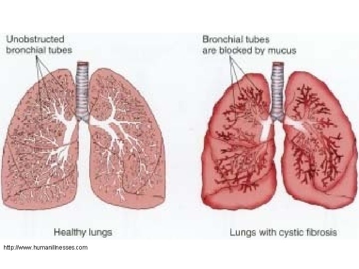
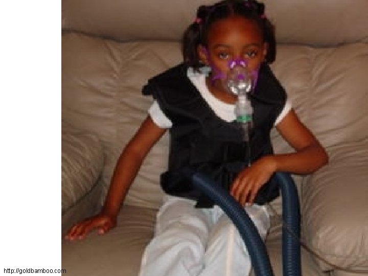
Story Four • Linda was a poor girl: she suffered from an inborne disease, cystic fibrosis. • Her lung surfactant was different from surfactant of healthy people. So, it was infected very often. • It was Staphylococcus aureus last time. This time it was different: the causative agent was Burkholderia cepacia, one of G– non-fermentating bacteria.
Non-fermenters and Cystic fibrosis • Cystic fibrosis is a severe, inborn lung disease, with failure of production of normal lung surfactant. This leads to changed characteristics of lungs, including many times increased risk of infection • Most common causative agents are Pseudomonas aeruginosa, Burkolderia cepacia and Staphylococcus aureus. Strains often become polyresistant and many children with cystic fibrosis die very young.
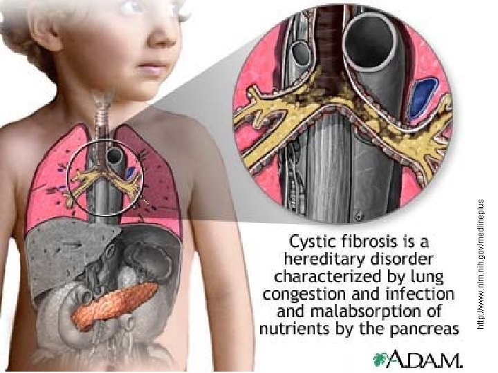
http: //www. nlm. nih. gov/medlineplus
http: //www. humanillnesses. com
http: //goldbamboo. com
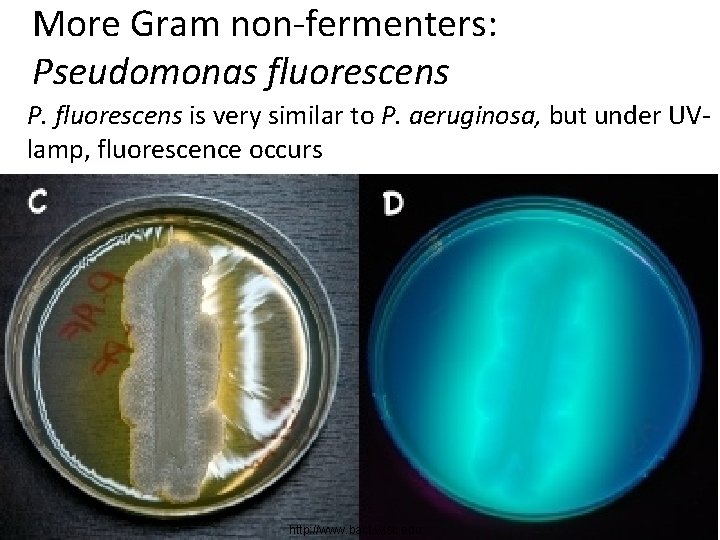
More Gram non-fermenters: Pseudomonas fluorescens P. fluorescens is very similar to P. aeruginosa, but under UVlamp, fluorescence occurs http: //www. bact. wisc. edu
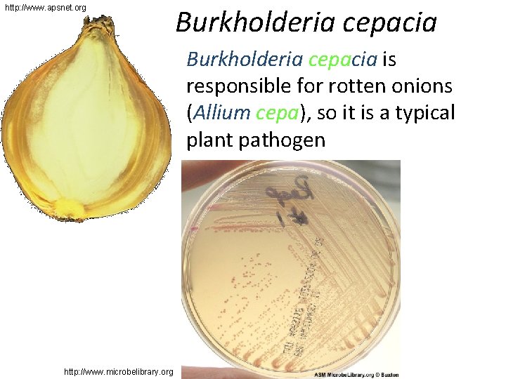
http: //www. apsnet. org Burkholderia cepacia is responsible for rotten onions (Allium cepa), so it is a typical plant pathogen http: //www. microbelibrary. org
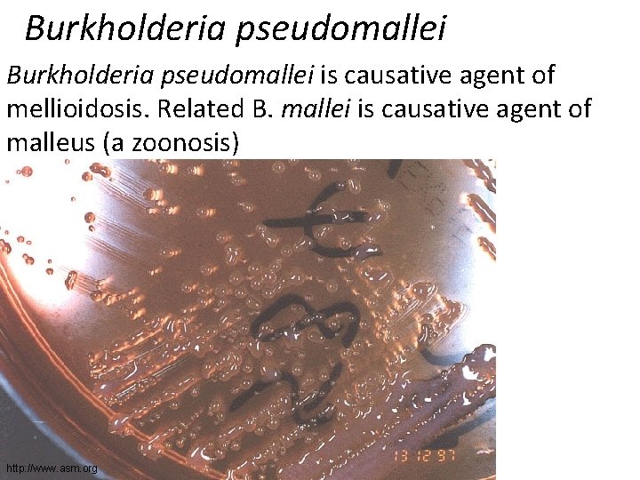
Burkholderia pseudomallei is causative agent of mellioidosis. Related B. mallei is causative agent of malleus (a zoonosis) http: //www. asm. org
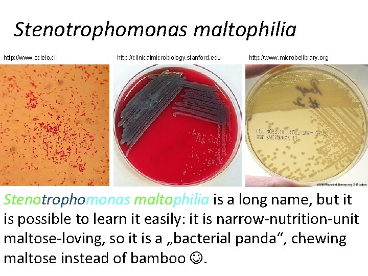
Stenotrophomonas maltophilia http: //www. scielo. cl http: //clinicalmicrobiology. stanford. edu http: //www. microbelibrary. org Stenotrophomonas maltophilia is a long name, but it is possible to learn it easily: it is narrow-nutrition-unit maltose-loving, so it is a „bacterial panda“, chewing maltose instead of bamboo .
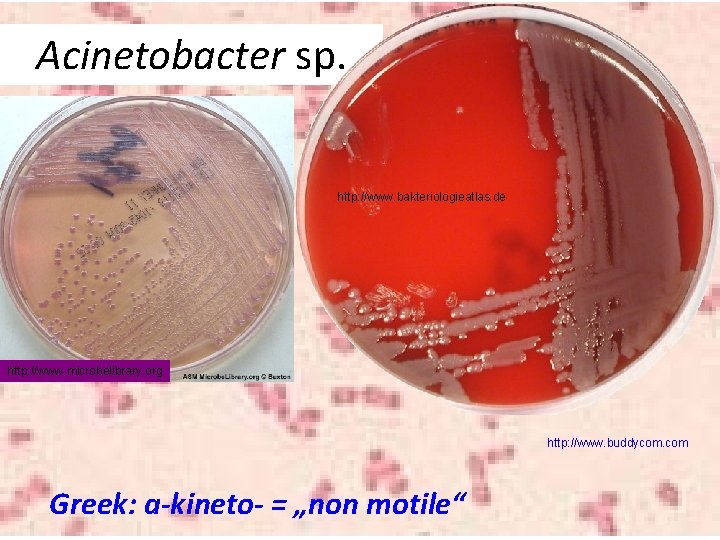
Acinetobacter sp. http: //www. bakteriologieatlas. de http: //www. microbelibrary. org http: //www. buddycom. com Greek: a-kineto- = „non motile“
Bacterial metabolism and relation of bacteria to oxygen We know already that G– non-fermenters are bacteria that do not ferment sugars, but they perform aerobic respiration. Let‘s compare two bacteria: • Escherichia coli lives in the intestine. It has enough nutrients, but not enough oxygen (unlike other gases ), so it prefers glucose (and other substrates) fermentation. Escherichia coli is a facultative anaerobe. Some other intestinal bacteria are strict anaerobes. • On the other hand, Pseudomonas has oxygen enough, but nutrients not enough. It uses aerobic respiration: enables better exploitation of nutrients. Pseudomonas is a strict aerobe.
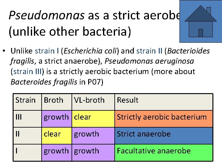
Pseudomonas as a strict aerobe (unlike other bacteria) • Unlike strain I (Escherichia coli) and strain II (Bacterioides fragilis, a strict anaerobe), Pseudomonas aeruginosa (strain III) is a strictly aerobic bacterium (more about Bacteroides fragilis in P 07) Strain Broth VL-broth Result III growth clear Strictly aerobic bacterium II clear Strict anaerobe I growth Facultative anaerobe
Diagnostics of Pasteurellaceae
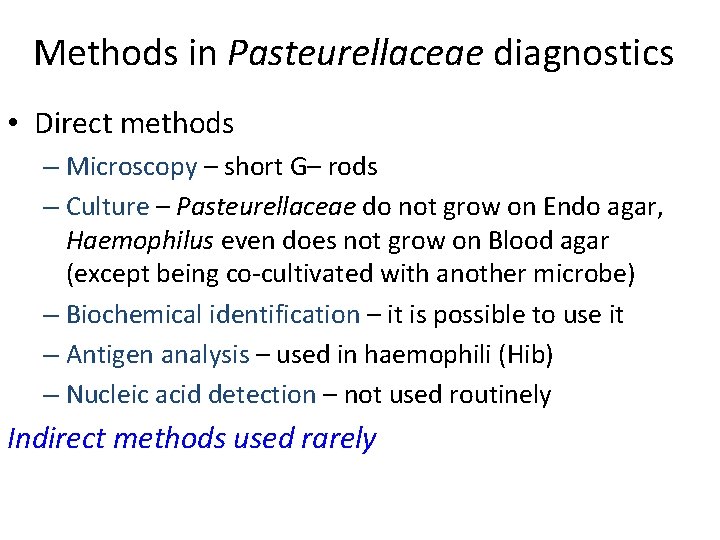
Methods in Pasteurellaceae diagnostics • Direct methods – Microscopy – short G– rods – Culture – Pasteurellaceae do not grow on Endo agar, Haemophilus even does not grow on Blood agar (except being co-cultivated with another microbe) – Biochemical identification – it is possible to use it – Antigen analysis – used in haemophili (Hib) – Nucleic acid detection – not used routinely Indirect methods used rarely
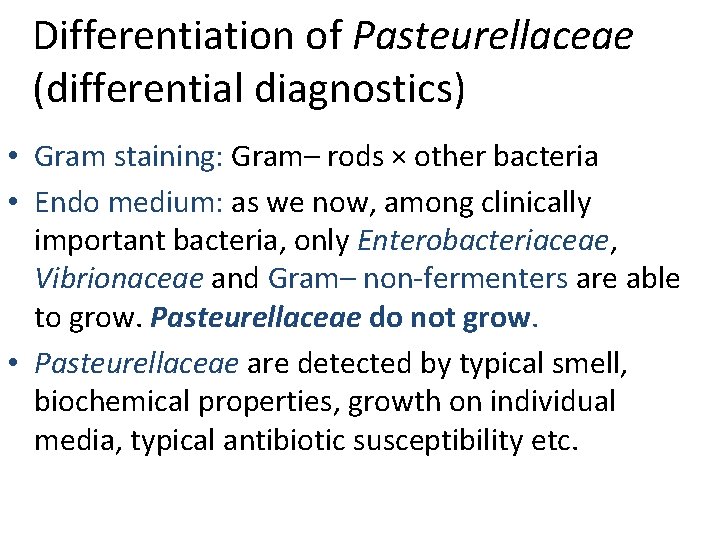
Differentiation of Pasteurellaceae (differential diagnostics) • Gram staining: Gram– rods × other bacteria • Endo medium: as we now, among clinically important bacteria, only Enterobacteriaceae, Vibrionaceae and Gram– non-fermenters are able to grow. Pasteurellaceae do not grow. • Pasteurellaceae are detected by typical smell, biochemical properties, growth on individual media, typical antibiotic susceptibility etc.
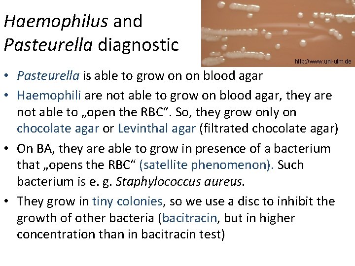
Haemophilus and Pasteurella diagnostic http: //www. uni-ulm. de • Pasteurella is able to grow on on blood agar • Haemophili are not able to grow on blood agar, they are not able to „open the RBC“. So, they grow only on chocolate agar or Levinthal agar (filtrated chocolate agar) • On BA, they are able to grow in presence of a bacterium that „opens the RBC“ (satellite phenomenon). Such bacterium is e. g. Staphylococcus aureus. • They grow in tiny colonies, so we use a disc to inhibit the growth of other bacteria (bacitracin, but in higher concentration than in bacitracin test)
Satellite phenomenon • As we already know, haemophilli need factors from RBC, but they are not able to break an RBC. They need the RBCs to be broken – by heating – chocolate agar – by presence of another microbe • Satellite phenomenon is an example of the second way how to make haemophilli be able to exploit blood factors. That means the growth of Haemophilus around Staphylococcus line only. • Presence of satellite phenomenon is a confirmation, that our bacterium is really a Haemophilus
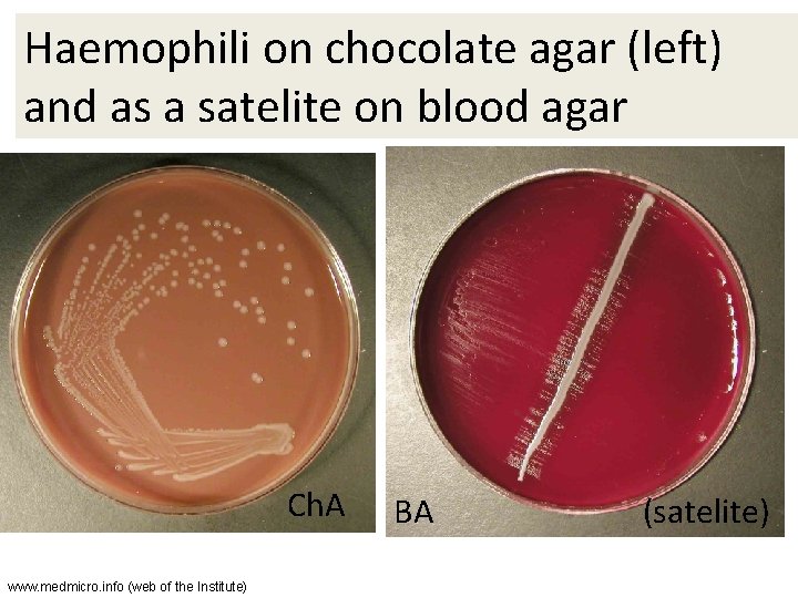
Haemophili on chocolate agar (left) and as a satelite on blood agar Ch. A www. medmicro. info (web of the Institute) BA (satelite)
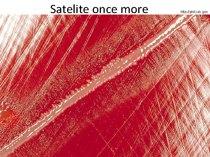
Satelite once more http: //phil. cdc. gov
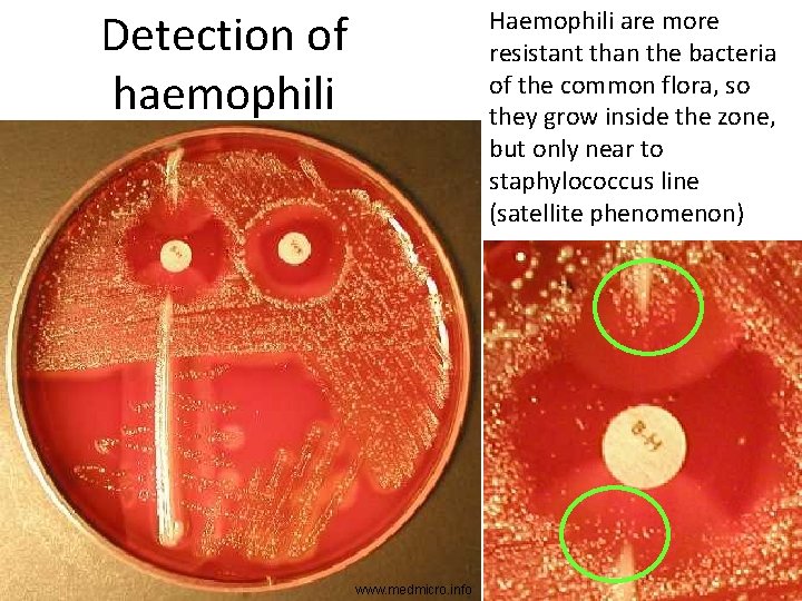
Detection of haemophili Haemophili are more resistant than the bacteria of the common flora, so they grow inside the zone, but only near to staphylococcus line (satellite phenomenon) www. medmicro. info
Growth factors of Haemophili (= determination of individual species) • Haemophilli need factors from blood, but the need of individual factors is species specific. – H. parainfluenzae needs factor V (= NAD) – H. aphrophilus needs factor X (= hemin) – H. influenzae needs both factors. • We use discs with these factors: one with X, another with V, and the third with a mixture of both of them.
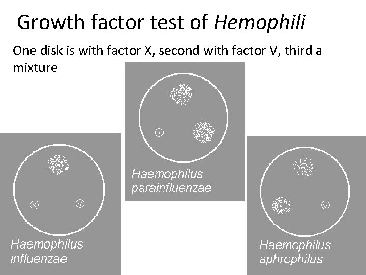
Growth factor test of Hemophili One disk is with factor X, second with factor V, third a mixture
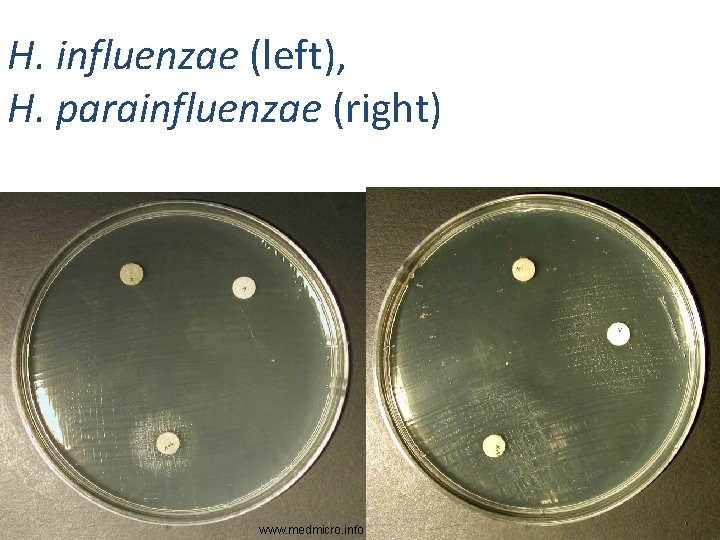
H. influenzae (left), H. parainfluenzae (right) www. medmicro. info
Haemophillus influenzae: antigen analysis (intra-species diagnostics • Antigen analysis in Haemophillus influenzae is performed like in other bacteria. The main goal is diferentiation of Hib. Today, we have comercially available sets, containing e. g. latex particles. We try to assess the capsular type of H. influenzae (a, b, c, d, e, or f). When the strain does not agglutinate with any sera, it is probably an non-encapsulated strain • Formerly, so named co-agglutination with Staphylococcus strain was used: agglutinate was more dense because of Staphylococcus binding the Fc-end of anti-haemophilus antibody
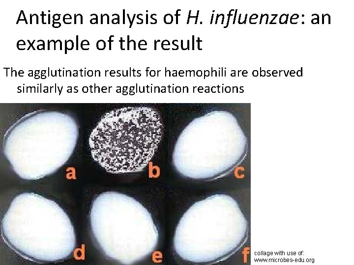
Antigen analysis of H. influenzae: an example of the result The agglutination results for haemophili are observed similarly as other agglutination reactions collage with use of: www. microbes-edu. org
Detection of Pasteurella using typical susceptibility pattern • No Gram-negative bacterium is susceptible to vancomycin. Vancomycin can be used for Grampositive bacteria only, it is very strong; all streptococi and majority of staphylococci and enterococci are susceptible • On the other hand, very little bacteria are susceptible to penicillin, escepically among G– bacilli. • So, susceptibility to penicillin and resistance to vancomycin is quite typical for Pasteurella.
Tests of atb susceptibility • Haemophilli do not grow on MH agar • Usually Levinthal agar (fitrated chocolate agar) is used for diffusion disc test – for this purpose, this agar is better than classical chocolate agar • Our laboratory uses „Haemophilus agar“, similar to Levinthal agar • Reading of the zones is the same as for any other bacteria

Antibiotic susceptibility testing: An example of Pasteurellaceae antibiotic set Antibiotikum Abbrev. Ampicilin (aminopeniciline) Co-amoxicilin (am. +inhib. ) Chloramphenicol Doxycycline (tetracycline) Co-trimoxazol (mixure) Azithromycin (macrolid) AMP AMC C DO SXT AZM Reference zone 22 mm 18 mm 29 mm 16 mm 12 mm
Diagnostics of Gram– non-fermenters
Methods for G– non-fermenters • Direct methods – Microscopy – mostly G– rods, but Acinetobacter is a G– coccus – Culture – non-fermenters grow on majority of media, including Endo agar. As glucose-non fermenters, they are mostly also lactose-non fermenters, but their colonies are sometimes quite dark, because of pigmentation – Biochemical identification – possible, but tests cheking aerobic respiration (not fermentation) should be used. We also use mostly decreased temperature and prolonged incubation – antigen analysis, nucleic acid detection – not used routinely Indirect methods used rarely
Differentiation of G– non-fermenters (differential diagnostics) • Gram staining: Gram– rods × other bacteria • Endo agar: they grow. As glucose-non fermenters, they are mostly also lactose-non fermenters, but their colonies are sometimes quite dark, because of pigmenation • Non-fermenters are differentiated from enterobacteria/vibria by no fermentation of glucose (e. g. Hajna medium remains completelly red after culture, no colour change; but eventual light brown colour does not matter, it is due to presence of pigments)
Further diagnostics of individual genera and species of G– NFs • Pseudomonas is usually detected by: – Presence of typical odour (young cultures) – Pigments, mostly green, sometiomes blue or maroon. Best visible on MH, worse on BA and Endo agar – Positive oxidase • Other non-fermenters, or not-sure Pseudomonas, shlould be differenciated biochemically, e. g. by NEFERMtest 24
Pseudomonas on MH agar and other media • Remember, that MH agar itself is nearly colourless (or slightly yellowish). • All green colour you see is product of Pseudomonas, or more precisely, of its pigment pyoverdin • On BA and Endo, pigment production is not so strong, but partially visible, too. Nevertheless, something more visible on these media is the typical pearl smooth surface of the colonies
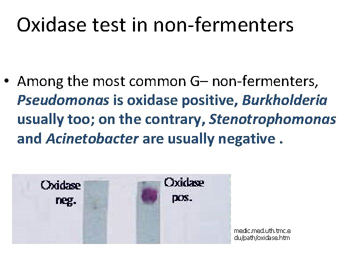
Oxidase test in non-fermenters • Among the most common G– non-fermenters, Pseudomonas is oxidase positive, Burkholderia usually too; on the contrary, Stenotrophomonas and Acinetobacter are usually negative. medic. med. uth. tmc. e du/path/oxidase. htm
NEFERMtest 24 • For precise biochemical identification of G– nonfermenters we use mostly NEFERMtest 24 (or a similar test). • It is a triple-strip (not double as last week) • There is a different way of code-formation than for (for example) ENTEROtest 16: – first number is 0 (oxidase –) or 1 (oxidase +) – next 6 numbers come from collumns H to C – collumns B and A are not counted (they are eventually used for more detailed determination)
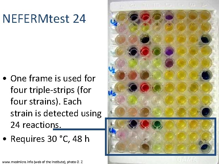
NEFERMtest 24 • One frame is used for four triple-strips (for four strains). Each strain is detected using 24 reactions. • Requires 30 °C, 48 h www. medmicro. info (web of the Institute), photo O. Z.
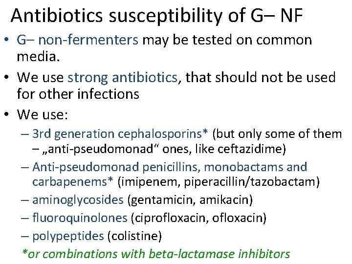
Antibiotics susceptibility of G– NF • G– non-fermenters may be tested on common media. • We use strong antibiotics, that should not be used for other infections • We use: – 3 rd generation cephalosporins* (but only some of them – „anti-pseudomonad“ ones, like ceftazidime) – Anti-pseudomonad penicillins, monobactams and carbapenems* (imipenem, piperacillin/tazobactam) – aminoglycosides (gentamicin, amikacin) – fluoroquinolones (ciprofloxacin, ofloxacin) – polypeptides (colistine) *or combinations with beta-lactamase inhibitors
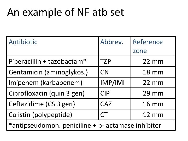
An example of NF atb set Antibiotic Abbrev. Reference zone Piperacillin + tazobactam* TZP 22 mm Gentamicin (aminoglykos. ) CN 18 mm Imipenem (karbapenem) IMP/IMI 22 mm Ciprofloxacin (quin 3 gen) CIP 29 mm Ceftazidime (CS 3 gen) CAZ 16 mm Colistin (polypeptide) CT 12 mm *antipseudomon. peniciline + b-lactamase inhibitor
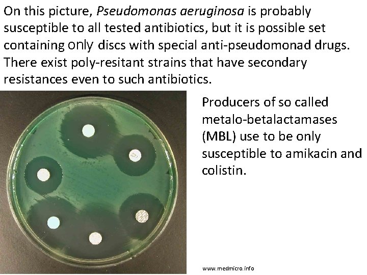
On this picture, Pseudomonas aeruginosa is probably susceptible to all tested antibiotics, but it is possible set containing only discs with special anti-pseudomonad drugs. There exist poly-resitant strains that have secondary resistances even to such antibiotics. Producers of so called metalo-betalactamases (MBL) use to be only susceptible to amikacin and colistin. www. medmicro. info
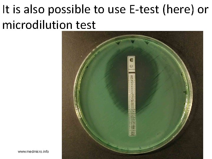
It is also possible to use E-test (here) or microdilution test www. medmicro. info

The End http: //www. scienceclarified. com
Inflamation of external ear – otitis media (bonus) • Common in children (short horizontal Eustach tube) • Caused by: Streptococcus pneumoniae, Haemophilus influenzae, Moraxella catarrhalis • In chronical cases also some G– rods It is necessary to differentiate otitis externa: here Staphylococcus aureus is the main pathogen (as in other skin inflamations), local therapy, e. g. Framycoin drops
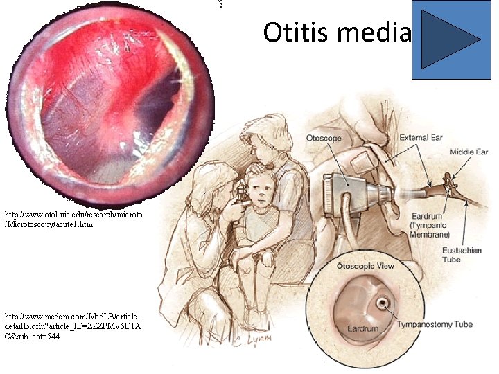
Otitis media http: //www. otol. uic. edu/research/microto /Microtoscopy/acute 1. htm http: //www. medem. com/Med. LB/article_ detaillb. cfm? article_ID=ZZZPMV 6 D 1 A C&sub_cat=544
Examination and treatment of otitis media • Therapy is indicated in case of a real inflamation (pain, redness, fever) and it does not react to antiinflamatory treatment • Drug of choice is amoxicilin (e. g. AMOCLEN), an alternative is co-trimoxazol • Ear swab examination is meaningfull only after paracentesis • Otherwise it is also possible to examinate pyogenic liquid taken during paracenthesis
- Slides: 67