Insert additional logo here if necessary otherwise delete


![Transport network Blood pumped by heart into • Arteries [arterial tree] into • Capillary](https://slidetodoc.com/presentation_image/5b45e7c79b219b051926aefca63e82c0/image-10.jpg "Transport network Blood pumped by heart into • Arteries [arterial tree] into • Capillary")
![Arterial tree • Branching system of tubes of reducing diameters called arteries [larger] and](https://slidetodoc.com/presentation_image/5b45e7c79b219b051926aefca63e82c0/image-11.jpg "Arterial tree • Branching system of tubes of reducing diameters called arteries [larger] and")


![So Second [pulmonary] set of capillaries must be inserted into the main circuit Because](https://slidetodoc.com/presentation_image/5b45e7c79b219b051926aefca63e82c0/image-18.jpg "So Second [pulmonary] set of capillaries must be inserted into the main circuit Because")


![Respiratory system Exchange takes place at thin membrane [alveolar-capillary membrane] Blood comes from pulmonary](https://slidetodoc.com/presentation_image/5b45e7c79b219b051926aefca63e82c0/image-27.jpg "Respiratory system Exchange takes place at thin membrane [alveolar-capillary membrane] Blood comes from pulmonary")


![Lungs • outer lining – the [visceral] pleura • air-conducting system – the bronchial](https://slidetodoc.com/presentation_image/5b45e7c79b219b051926aefca63e82c0/image-31.jpg "Lungs • outer lining – the [visceral] pleura • air-conducting system – the bronchial")


![Respiratory muscles • • Diaphragm External intercostals Internal intercostals Accessory muscles [sterno-cleidomastoid, trapezius, scalenes]](https://slidetodoc.com/presentation_image/5b45e7c79b219b051926aefca63e82c0/image-40.jpg "Respiratory muscles • • Diaphragm External intercostals Internal intercostals Accessory muscles [sterno-cleidomastoid, trapezius, scalenes]")


- Slides: 55
Insert additional logo here if necessary, otherwise delete this box by going to View Slide Master Introduction to cardio-vascular & respiratory anatomy
Plan of the afternoon • • Lecture 1. Explain basic physiology and anatomy of cardiovascular system Lecture 2. Explain basic physiology and anatomy of respiratory system Introduce surface anatomy • Group work. Examine anatomical models and answer some simple questions
Definitions: • Cardiovascular system: Transports oxygen and nutrition to the cells • Respiratory system: Provides oxygen to the blood stream
Sugar + oxygen = energy + carbon dioxide + water or C 6 H 12 O 6 + O 2 = e + CO 2 + H 20
Vascular System • Transport medium- blood- is circulated to the tissues by a • Pump – the heart - through a • Transport network - the vascular system [arteries, capillaries & veins] • Regulation mechanisms
Transport medium • • • Blood - fluid and cells Fluid - water and dissolved chemicals Cells – Red, which carry oxygen White of several types with a variety of functions, Platelets to aid clotting
Pump • Pump is the heart, which is a muscular organ. • Contraction of the muscular wall forces blood into the arterial system. • Valves ensure unidirectional flow.
Transport network Blood pumped by heart into • Arteries [arterial tree] into • Capillary network where diffusion to cells takes place and drains back into • Veins and back to heart
Arterial tree • Branching system of tubes of reducing diameters called arteries [larger] and arterioles [smaller] • Organs perfused in parallel • Thick, muscular walls, relatively indistensible. These tubes lead to the capillary network
Capillary network Thin walled tubes, which penetrate all tissues so that every active cell in the body is within diffusion range. Lead to the venous system.
The venous system Drains from the capillaries through tubes [smaller venules and larger veins ] of varying –increasing – diameter. Generally thin-walled and distensible. Returns blood to the heart
Cardiovascular system PUMP -> ARTERIAL TREE -> CAPILLARY NETWORK -> VENOUS SYSTEM -> PUMP
BUT!
Oxygen problems • Low solubility • Little storage • Arterial blood normally carries maximum quantity in haemoglobin • Very short time that some tissues can survive without it,
Oxygen transport • Arterial blood must be carrying the greatest possible amount of oxygen. • ALL the cardiac output must be oxygenated before delivery to the tissues. • All the cardiac output blood must pass through the lungs with each circulation
So Second [pulmonary] set of capillaries must be inserted into the main circuit Because of the resistance of the capillary network this in turn necessitates a second pump. PUMP 1 -> BODY-> PUMP 2 -> LUNGS -> PUMP 1 ->
Cardiovascular system LEFT HEART -> SYSTEMIC CIRCULATION -> RIGHT HEART -> PULMONARY CIRCULATION -> LEFT HEART ->
Just to complicate matters further Each pump has two chambers in series • The first chamber on each side is called the atrium [pl atria] • The second chamber is the ventricle and is the principal pumping mechanism
Heart Valves Between RIGHT atrium and RIGHT ventricle TRICUSPID Between RIGHT ventricle and PULMONARY ARTEY PULMONARY Between LEFT atrium and LEFT ventricle MITRAL/BICUSPID Between LEFT ventricle and aorta AORTIC
Double Circulation Pulmonary Systemic • Circulation to the alveoli • Low pressure • Oxygenated blood from the lungs to the left heart to the capillaries, and deoxygenated blood from right side of the heart to the lungs • Circulation to all the body except the alveoli • High pressure • Oxygenated blood from the left heart to the capillaries, and deoxygenated blood from the capillaries to the right side of the heart
Circulation of the blood
Heart
Respiratory system Function – To allow gas exchange between blood and external atmosphere at blood gas interface
Respiratory system
Respiratory system Exchange takes place at thin membrane [alveolar-capillary membrane] Blood comes from pulmonary system, [right ventricle and pulmonary artery] Air comes from exterior through airways to microscopic air sacs called alveoli
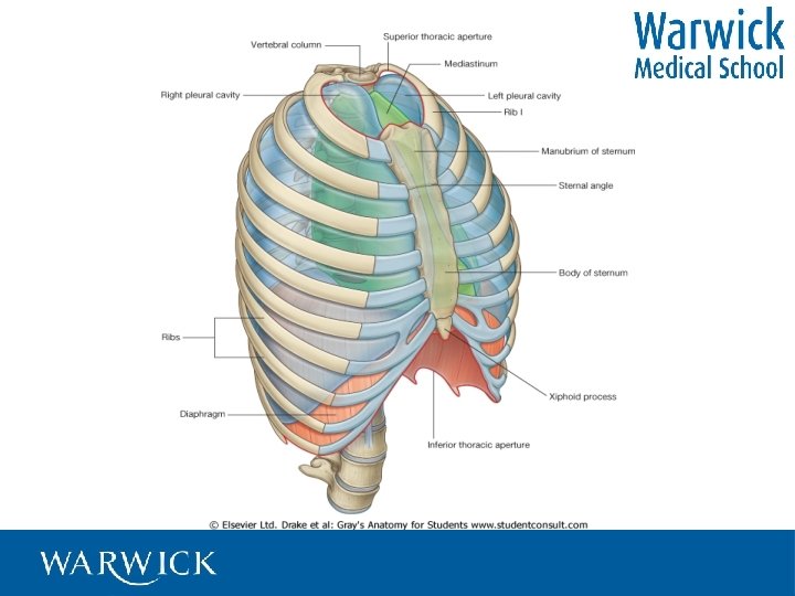
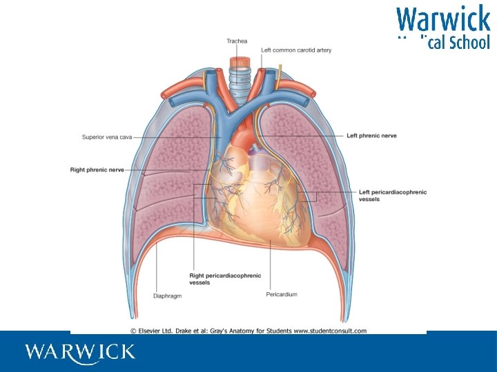
Respiratory system, • • • Supporting framework, - THORAX Air channels- AIRWAY Blood supply Bellows to give tidal flow –MUSCLES Interface –ALVEOLAR MEMBRANE, Regulation mechanisms.
Thorax
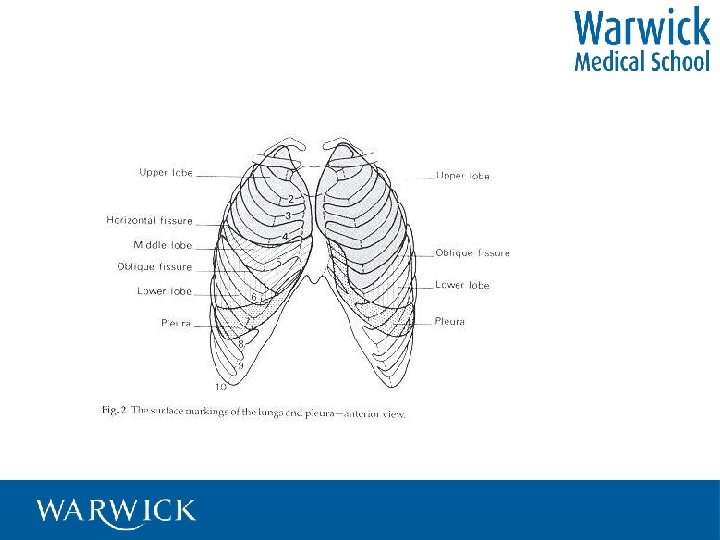
Lungs • outer lining – the [visceral] pleura • air-conducting system – the bronchial tree • air blood interface – the alveoli • blood supply from both sides of the heart • supporting structures
Airway Extends from exterior to alveoli • Mouth/nose pharynx • Larynx • Trachea & bronchi • Bronchioles • Alveoli
Upper airway
Extra-pulmonary airway Extends from exterior to lungs and consists of • Mouth/nose to pharynx • Larynx [voice-box] • Trachea divides into two major • Bronchi [s. bronchus] one of which enters each lung
Intra-pulmonary airway • Main bronchi enter lung and divide into • Lobar bronchi • Inter-lobar bronchi • Bronchioles • Broncho-alveolar airways • Alveoli
Lower airway
Lower airway and lungs
Alveolar-capillary membrane
Lung in situ
Respiratory muscles • • Diaphragm External intercostals Internal intercostals Accessory muscles [sterno-cleidomastoid, trapezius, scalenes]
Diaphragm
Respiratory cycle • • • Respiratory muscles contract Volume of thorax increases Air drawn in Respiratory muscles relax Air expelled
Respiratory cycle • Air is drawn into the lungs intermittently • Blood is pumped to the capillary network in the lungs continually • Gas exchange takes place at capillaries level – the in the lung
Respiratory system
Surface anatomy • Helps us identify deeper structures • Bony points better then soft tissue
Surface anatomy Some uses • In CPR - where to do compressions – xiphi-sternum • Shows us where to listen to the heart • And which parts of the lung we are examining • And where to find liver or feel for the appendix
Surface anatomy
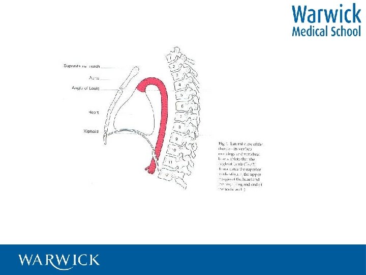
Manubrio-sternal angle “Angle of Louis” Second intercostal space Apex of right atrium Bifurcation of trachea Root of aorta T 4
Surface anatomy
Surface anatomy
Surface anatomy
Surface anatomy