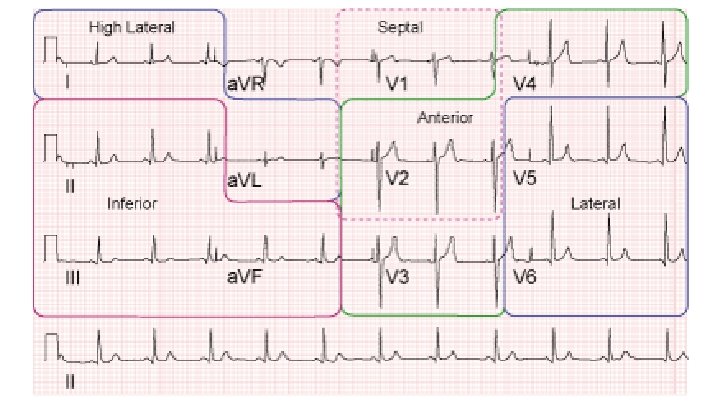
Injury Ischemia and Infarction patterns Basic EKG interpretation

Injury, Ischemia, and Infarction patterns Basic EKG interpretation

-ST Elevations -ST Depressions -T wave inversions -Non specific ST-T changes -Q waves

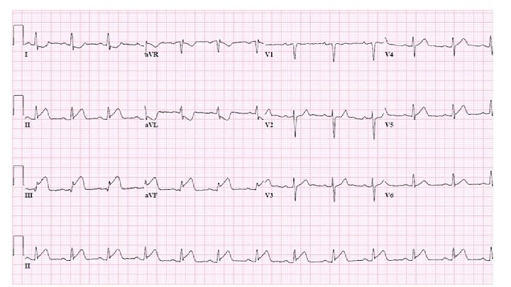
ST Elevations

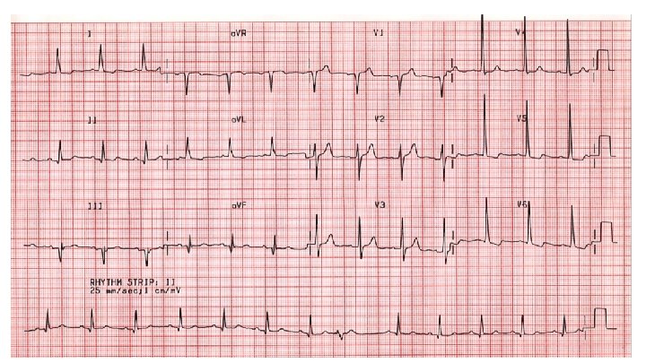
Definition ●New ST-segment elevation at the J-point in two contiguous leads with the cutpoints: ≥ 0. 1 m. V (1 mm) in all leads other than leads V 2 -V 3;

Definition ●New ST-segment elevation at the J-point in two contiguous leads with the cutpoints: ≥ 0. 1 m. V (1 mm) in all leads other than leads V 2 -V 3; ●For leads V 2 -V 3: ≥ 2 mm in men ≥ 40 years; ≥ 2. 5 mm in men <40 years, or ≥ 1. 5 mm in women regardless of age.

STEMI imitators -Pericarditis -Prinzmetel’s Angina -Early repolarization -Left Ventricular Hypertrophy -LBBB -Ventricular aneurysm -Ventricular paced -Increased intracranial pressure -Brugada syndrome

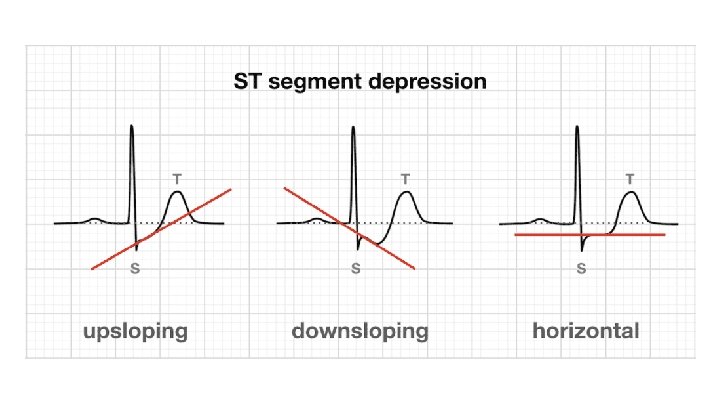
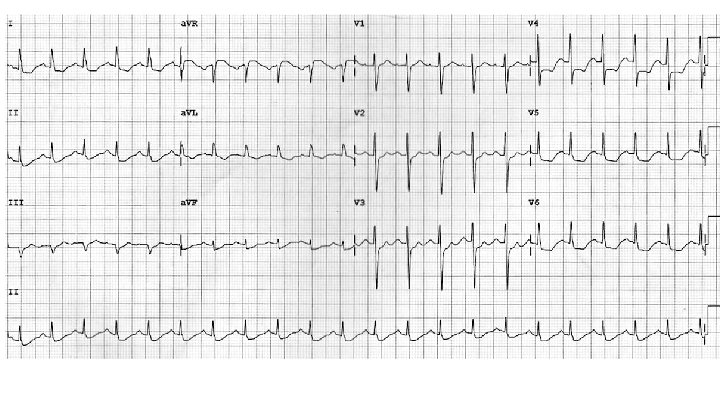
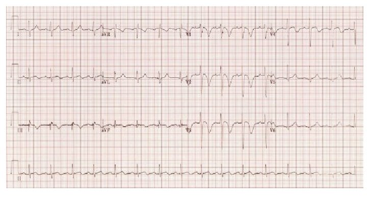
ST depression criteria -New horizontal or downsloping ST depression >= 0. 5 mm in two contiguous leads -Deeper ST depressions indicate higher likelihood of ACS and worse prognosis

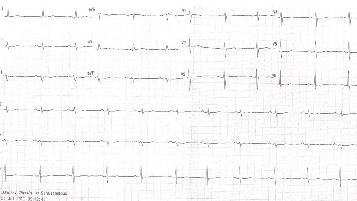
T wave inversions -Inversions deeper than 1 mm in two contiguous leads

Nonspecific ST-T changes -ST-T changes that don’t meet above criteria

Differential diagnosis for Nonspecific ST-T changes -Physiologic variant -Metabolic changes -Electrolyte abnormalities -CVA -Fever -Myocarditis -Acidosis/Alkalosis -Pericarditis -Endogenous catecholamines -Pulmonary emboli -Drugs -Myocardial ischemia -Acute abdominal process -Pulmonary processes
 in duration At least 1 mm")
Pathologic Q waves - 40 ms (1 mm) in duration At least 1 mm deep Greater than 25% of entire QRS amplitude Two contiguous leads
- Slides: 18