Inguinal Hernias Hydroceles and undescended testes Dr Ibrahim

Inguinal Hernias, Hydroceles and undescended testes Dr. Ibrahim Alsbou

Inguinal Hernias • A hernia may be defined as a protrusion of a portion or whole of an organ or tissue, through an abnormal opening. Groin Hernia Three types • Inguinal – Indirect (only common one in childhood) – Direct • Femoral
 indirect type (99%) •")
Epidemiology • 1– 5% of term males • Male (80%) indirect type (99%) • Right (60%), Left (30%), bilateral (10%) • ↑ Incidence in preterm infants (e. g. , 12% at 32 weeks) • ↑ Incidence in conditions of increased intra-abdominal pressure – Abnormal content (e. g. , ascites, peritoneal dialysis) – Abnormal abdominal wall configuration (e. g. , exomphalos, gastroschisis, bladder extrophy)

Embryology § The embryonic testis descends from the region of the deep inguinal ring through the anterior abdominal wall muscles to its final destination in the scrotum. It is preceded by a tongue of parietal peritoneum, which becomes the tunica and processus vaginalis. § The processus normally obliterates at ~35 weeks of gestation, but failure is common and results either in a hydrocele or an inguinal hernia depending on the degree of patency. § In girls, the canal is occupied by the round ligament, but sac formation from a putative processus may occur in the same way. § Contents of the sac may commonly include omentum, small and large bowel intestine, and lateral corner of bladder, and in girls the ovary. § Rare contents include the appendix (Amyand hernia), Meckel’s diverticulum (Littre hernia), or only part of the small bowel(Richter hernia).

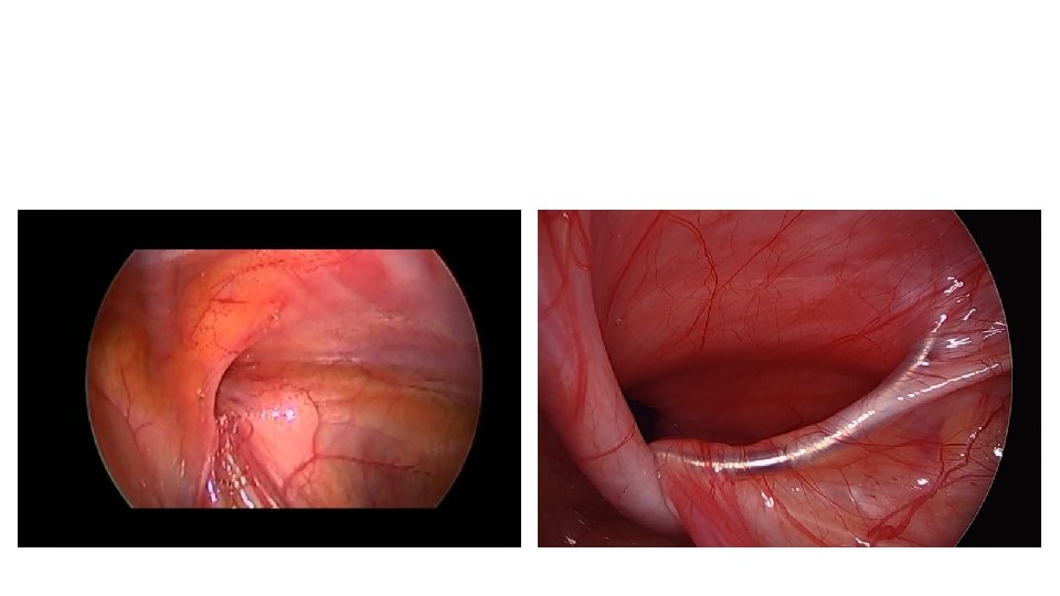
Anatomy of Inguinal Canal • Entry – deep inguinal ring (defect in transversalis fascia; medial border contains epigastric vessels). • Exit – superficial inguinal ring (triangular defect in external oblique: base is body of pubis. Cord structures can be felt on underlying bone. ) • Contents – spermatic cord has three layers (external and internal spermatic fascia, sandwiching a layer of muscle – cremaster), three nerves (ileo-inguinal, genital branch of genito-femoral, and sympathetics), and three other structures (vas, vessels, and lymphatics). • The processus envelopes the central structures (vas/vessels) anteriorly and is enveloped by the fascial layers. . During open surgery, the fascial layers are teased off the anterior aspect and processus/sac unrolled to expose the posterior aspect – the route to the vas/vessels.

Clinical Features • Intermittent swelling, noted during crying and descending toward the scrotum/labia. • This may reduce either spontaneously or on gentle, lateral pressure. • The bulge may be difficult to elicit in the outpatient setting, but examination should reveal unilateral thickening and slipperiness − the “silken cord sign. ” This, in conjunction with a good history, is adequate to proceed with surgery. • The irreducible hernia presents as a firm, tender mass occupying the inguinal canal and possibly extending to the scrotum. • Defining the testis as separate confirms the diagnosis.

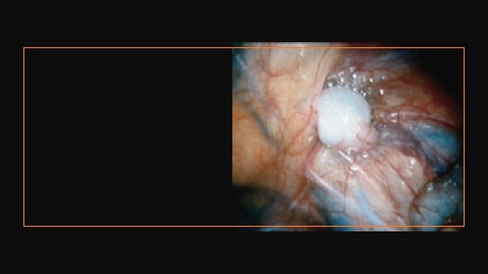
Management An acute obstructed hernia should be reduced manually. If this fails, then emergency surgery is warranted. Surgery: Inguinal Hernia Skin-crease groin incision 1. Incise external oblique (to open canal). 2. Identify sac and separate from vas and vessels. This is often difficult with a flimsy sac in infants, but it is important to keep proximal parts together if possible. 3. Ligation of the sac at the deep inguinal ring (herniotomy). Caveats 1. The bladder or the ovaries are the usual internal structures, which can form part of a sliding hernia. These need careful separation and probably a purse-string under vision to control neck of sac. 2. Sometimes, in small infants, there is a definite posterior wall weakness predisposing to recurrence. Perform Bassini repair (suture conjoint tendon down to inguinal ligament).

There has been much recent debate around the value of exploration of the contralateral side(to prevent a metachronous hernia becoming symptomatic). Thus, factors increasing the chances of this have included gender (female>male), laterality (right>left), and age (preterm>term). Some will insert a telescope via the hernial sac to assess the contralateral deep ring, although even this has problems. Despite this, policy remains that of leaving surgery to the affected side only

Outcome • Complication rate should be <1% – Risk of damage to vas or vessels, recurrence, atrophy, or iatrogenic ascent of the testis. • ↑↑ Risk following an episode of incarceration.

Hydrocele • A collection of fluid in the space surrounding the testicle between the layers of the tunica vaginalis.
 • Acquired")
Classification • Congenital – due to patency of processus vaginalis (usually children) • Acquired (usually adults) – Idiopathic – Secondary (e. g. , post-trauma, tumor, lymphatic infiltration (W. bancrofti, scrotal filariasis)) ØThe processus vaginalis should obliterate by the time of birth (some studies suggest 80% are actually patent). Failure results in ingress of fluid from peritoneal cavity to fill tunica vaginalis. ØAn abdominal-scrotal hydrocele is a variant where there is a large sac extending from scrotum to retroperitoneum – this tends not to have an obvious connection with the peritoneal cavity. ØA hydrocele of the cord is a fluid-filled cavity within the processus and palpable between superficial inguinal ring and upper scrotum. This may feel like a third testicle

Clinical Features • Hydroceles are usually seen as asymptomatic swellings with diurnal variation (absent in morning). They may also be present only intermittently. • The key sign on examination is the ability to get above (i. e. differentiates from hernia) and transilluminate (showing fluid nature). • Sometimes, if communicating, the swelling can be reduced slowly by sustained pressure.
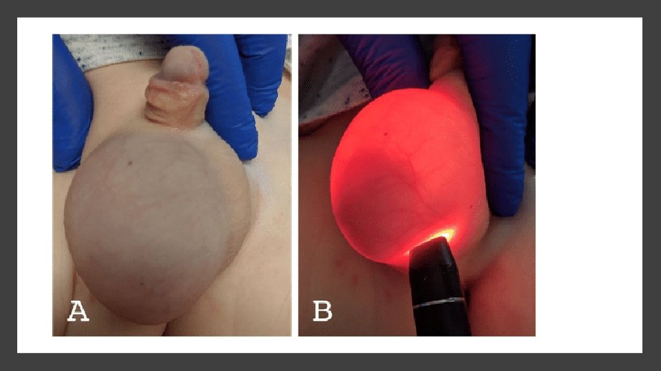

Management • Management is predominantly noninterventional, as most will resolve spontaneously by 1– 2 years of age. • Some giant examples do need surgery even in infancy, as they tend to bury the penis.

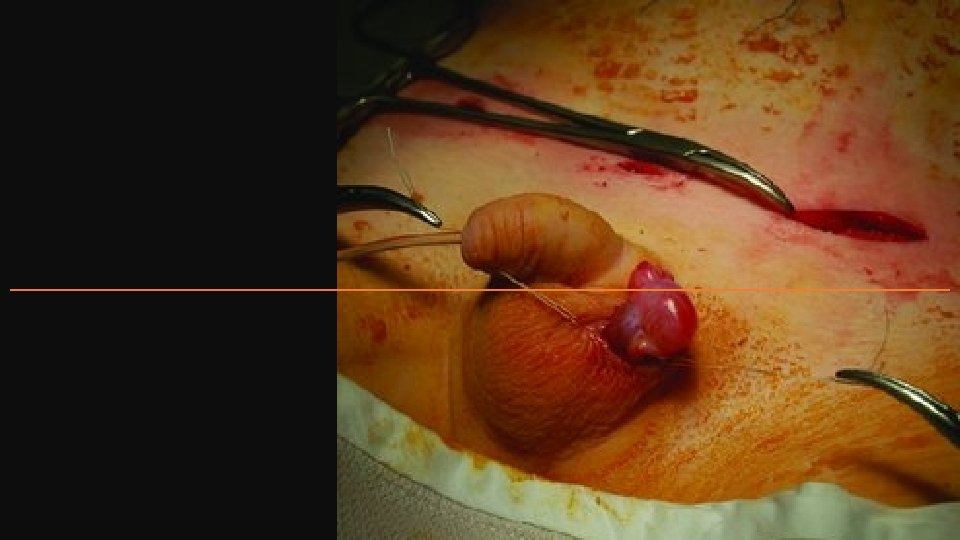
Surgery: Hydrocele Skin-crease groin incision 1. Canal usually left intact, with separation of cremaster fibers at the level of superficial inguinal ring to identify deeper processus vaginalis. 2. Separation from vas and vessels. 3. Ligation – usually combined with the opening of distal sac to drain all the fluid. Ødo not perform office aspiration of hydrocele either as diagnostic process or even as an attempt at treatment (unless postoperative). ØRecurrence (<5%) is uncommon, but its risk is increased if it is longstanding and its sac is thick-walled. If confident that previous surgery did indeed ligate the PPV, then redo surgery should aim to obliterate tunica vaginalis by excision or eversion (Jaboulay) or plication (Lord).

The Undescended Testes Failure of the testis is to descend along its normal pathway into the scrotum is the commonest endocrine disorder in the male.

Epidemiology Incidence • Preterm boys − ~30%. • Term 3– 5%. – Falling to <1% at 6 months • An undescended testis (UDT) is associated with 4– 5 -fold increased risk of cancer and a risk of infertility.

Associated Anomalies • Patent processus vaginalis, inguinal hernia, and abnormal epididymis • Hypospadias • Abdominal wall defects, e. g. , gastroschisis, exomphalos • Cerebral palsy, mental retardation • Prune belly syndrome • Wilms’ tumor
 and within the")
Embryology Testes develop from the gonadal ridge (antero-medial to the mesonephros) and within the coelomic cavity behind the peritoneum and below the developing kidneys. 1. 3– 5 weeks – undifferentiated gonad develops along gonadal ridge. 2. 6 weeks – primodial germ cells migrate from the yolk sac into the gonadal ridges and differentiate into gonocytes. Determination of a testis is controlled by the SRY gene (together with WT-1, SF-1, SOX 9). 3. 7– 8 weeks – Sertoli cells develops and start secreting MIS (Mullerian Inhibiting Substance) or antimullerian hormones, which causes regression of Mullerian derivatives. 4. 9 weeks – Leydig cells form and start secreting testosterone, which induces both the differentiation of the Wolffian ducts into male internal accessory reproductive organs and masculinization of the external genitalia. v. Testicular descent then occurs in two phases. 5. 10– 15 weeks – Transabdominal phase – controlled by the Leydig cell hormone (insulin-like hormone [INS]). By the third month, testes occupy a position immediately above the internal inguinal ring. 6. 24– 35 weeks – Inguino-scrotal phase – regulated by androgens and the genitofemoral nerve. Most (75%) infants born with UDT will have spontaneous descent by 4– 6 months postnatally (corrected for term), thought to be due to a postnatal surge of testosterone.

Etiology The precise cause is unknown but possible causes include: • Defects in testosterone, INS, and MIS. • HOXA 106 and HOXA 11 (developmental genes) may have some role. Hypogonadotrophic hypogonadism: Absence of gonadotrophin surge, which is normally seen at 60– 90 days after birth, is an important sign of abnormal testicular function. This surge is called mini-puberty. If this surge is absent, Leydig cells do not proliferate and germ cells do not mature, leading to infertility. • Differential body growth in relation to spermatic cord/gubernaculum. • ↓ Intra-abdominal pressure – UDT is seen in a number of conditions with this feature (e. g. , prune belly syndrome, exstrophy, exomphalos, gastroschisis).

Clinical Features • Unilateral ~75% right>left • Nonpalpable testes ~20% • About 10% of total UDT have blind-ending vessels above the internal ring Palpable testes may be found: • Above tubercle 10– 15% • At the pubic tubercle 35– 40% • Upper scrotum 15– 20% • Ectopic 5– 8%

Examination • Examine supine, and with crossed legs and if required in the upright position. • Asymmetry of the scrotum suggests unilateral UDT, whereas a bilateral hypoplastic scrotum suggests bilateral UDT. • If the testis is not obviously palpable, the fingers should be kept near the iliac crest and gently milked toward the scrotum. • The fingers of one hand feel the testis while the other hand keeps the testis in that position. • Even if it comes down to the bottom of the scrotum, it is important to assess whether it remains over there for some time after release to differentiate it from a retractile testis. • An enlarged contralateral testis suggests either ipsilateral atrophy or absence. Perineal, penile, and femoral regions should be examined for ectopic testes.

Investigations • For unilateral UDT without hypospadias, no further investigations are required. • For unilateral UDT with hypospadias or bilateral nonpalpable UDT, investigation to rule out a disorder of sex development (DSD) is necessary. – LH (lutinizing hormone) – FSH (follicle-stimulating hormone) – h. CG (human chorionic gonadotrophin) stimulation test A negative testosterone response after h. CG stimulation, in the presence of ↑LH and ↑FSH, is suggestive of bilateral absent testes (anorchia). (NB: MIS can also be assayed for the presence of sertoli cells. ) • US, CT, and MRI have high false-negative results, so should be done only in difficult situations. • Abnormalities of the upper renal tract may be ruled out by US if associated with hypospadias or bilateral UDT. • A genitogram is helpful if an intersex problem is suspected.
 treatment has been practiced")
Management Role of Hormone Therapy Hormonal (h. CG, Gn. RH) treatment has been practiced in Europe for about 40 years. On the basis of some recent studies, the “Nordic consensus, ” however, has recommended against the use of hormones, considering the poor immediate benefits and the possible long-term adverse effects on spermatogenesis (hormones induce increased apoptosis of the germcells). This recommendation remains controversial.

Orchidopexy As descent is uncommon after 6 months, according to the Nordic consensus on treatment of UDT, orchidopexy is recommended soon after 6 months of age (corrected for term) or upon diagnosis, if it occurs late. Operative intervention is not required for normally retractile testes.

Palpable UDT 1. Skin-crease groin incision 2. Mobilization of testis 3. Opening of inguinal canal 4. Hernial sac or processus vaginalis dissected free and transfixed at its neck 5. Mobilization of testis on vas and vessels up to and beyond the level of deep ring 6. Tension-free placement in dartos pouch

Nonpalpable Testis Studies have suggested that even abdominal testes are histologically normal up to 6 months, but then show a sharp decrease in spermatogonia with age. • EUA and diagnostic laparoscopy – if actually palpable convert to simple orchidopexy. • Two-stage Fowler–Stephens operation. This is indicated for high or intra-abdominal testis and can be performed open or laparoscopically (latter now more popular). Early decision is needed to avoid unnecessary devascularization of the blood supply coming along the vas deferens and cremaster muscles. 1. First stage – the testicular blood vessels are ligated or clipped, so that the testis develops its blood supply from vessels along the vas and cremaster muscle. 2. Second stage (>6 months after) – division of ablated testicular vessel pedicle and mobilization entirely on vas overlying peritoneum. All children should be followed up at 3 months and after 1 year to check the testes for viability, size, and location.

Complications • Testicular atrophy – 1– 5% cases after open orchidopexy – 20– 30% after FS orchidopexy • Vas injury-1– 5% • Recurrence, renascent – Up to 10% is usually due to inadequate mobilization and inadequate hernial sac dissection. Revision orchidopexy is indicated. • About half of acquired (previously not operated) UDT may come down on their own; hence, a conservative approach until puberty is indicated by some authors, although there is a risk to fertility, with lower sperm counts being reported in this group. Many authors would recommend orchidopexy in this situation.
. • Bilateral UDT – ↓↓ fertility (~25% have adequate")
Outcome Fertility (difficult to predict). • Bilateral UDT – ↓↓ fertility (~25% have adequate sperm counts). • Unilateral UDT: ↓ fertility (~ 90% with ~ 75% have normal sperm counts after surgery at a mean age of 10 years). • Subfertility – waiting time to pregnancy is longer in bilateral UDT. • Fertility according to the preoperative site of the testis is difficult to predict. – Bilateral – ↑ position of the testes ↓ lower the fertility. – Unilateral – preoperative site is not a major determinant.
- Slides: 32