Inger presents ABNORMAL ACTIVITY IN HYPOTHALAMUS AND AMYGDALA

Inger presents:

ABNORMAL ACTIVITY IN HYPOTHALAMUS AND AMYGDALA DURING HUMOR PROCESSING IN HUMAN NARCOLEPSY WITH CATAPLEXY Sophie Schwartz, Aurelie Ponz, Rositsa Poryazova, Esther Werth, Peter Boesiger, Ramin Khatami and Claudio L. Bassetti Presented by: Inger Appanaitis

Cataplexy: It’s no laughing matter! Sudden loss of muscle tone with preserved consciousness triggered by variety of emotions. Telling or hearing jokes is most common trigger of cataplectic attack.

Cataplexy Respiratory and eye muscles not affected Episodes last a few seconds → 30 minutes Sleep deprivation increases frequency and severity of attack Partial or complete Slackening of jaw → total collapse Affects 1 in 2000 individuals

Narcolepsy: Repeated, irresistible ‘sleep attacks’ Cataplexy Recurrent intrusions of REM sleep into the transition period between sleep and wakefulness. (N 1, N 2, N 3, N 2, REM)

Characteristics of Narcolepsy • Affects 0. 02 -0. 16% of adults – 70% of people with narcolepsy also suffer from cataplexy (NC) • Genetic inheritance: 5 -15% of first-degree relatives (DSM-IV) • (Siegel et al 2001: Suggest damage to hypocretin system) • Some individuals take naps in order to manage sleepiness – Average 2 -6 sleep episodes/day

Laughter

Anger

Embarrassment

NC caused by reduction or loss of hypocretin/orexin in hypothalamus 1998: De Lecea* and Sakurai De Lecea- Hypocretin: Hypothalmic location and sequence homology to secretin Treatment for obesity Sakurai- Orexin: Greek ‘orexis’=appetite 3 rd ventricle injections induced feeding in rats Hypocretin (HCRT)= Orexin
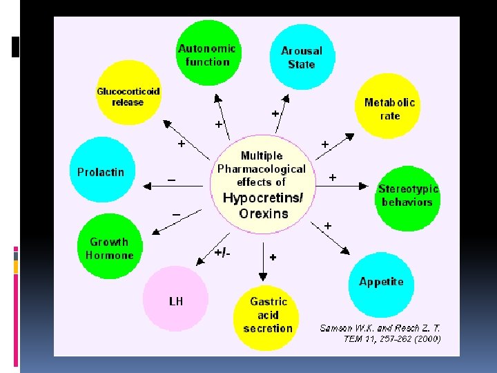

Hypocretin- First characterization • 2 distinct hypocretins – Hcrt 1 and Hcrt 2 • No difference between males and females • NT found in dorsal and lateral hypothalamus – Accumulation within axon terminals and vesicles suggests that they may have intercellular signaling activity – Hcrt 2 is neuroexcitatory • Increase in frequency of postsynaptic currents

Coincidence or destiny? Luis De Lecea and Thomas Kilduff Former director of narcoleptic dog colony at Stanford Sakurai group continued research w/ knock-out mice Hcrt-2 causing cataplexy Stanford group (canine narcolepsy)- systematic chromosome analysis Reported mutated, non-functional version of Hcrt-2 gene

Role played by Hcrt ← Inhibitory Feedback ← Direct Inhibition ← Mutually Inhibitory Circuit

‘Snorting a Brain Chemical Could Replace Sleep’- Siegel Can cross BBB Reduces sleepiness w/out causing edginess Orexin A reversed the effects of sleep deprivation in monkeys, allowing them to perform like well-rested monkeys on cognitive tests Brains looked ‘awake’ in PET Possibilities…

Schwartz’s Funny Study Suprapontine brain mechanisms- associated with the cataplectic effects of emotion in human NC- remain essentially unknown Assess brain activity in patients while observing humorous pictures Hypothalamic and amygdalar brain regions f. MRI

Why amygdala? • Dogs demonstrated changes of neuronal firing in the amygdala during cataplexy; • Amygdala found to be strongly activated during REM sleep in normal humans; • Involvement in emotional information processing in both animals and humans. • Hypothalamus represents a second main suprapontine brain site whose dysfunction might contribute to cataplexy in NC

Objective • To date, imaging studies failed to reveal consistent brain abnormalities in NC patients. Advanced neuroimaging techniques could not demonstrate any systematic structural or functional change in the hypothalamus and/or amygdala. • Hypothesis: Patients may show abnormal processing of external emotional inputs within limbic circuits or increased activation of efferent motor systems – Use f. MRI to monitor neural activity elicited by humorous versus neutral pictures in NC (patients) and healthy volunteers (controls)

Test Subjects:
 Stanford cataplexy questionnaire (>32.")
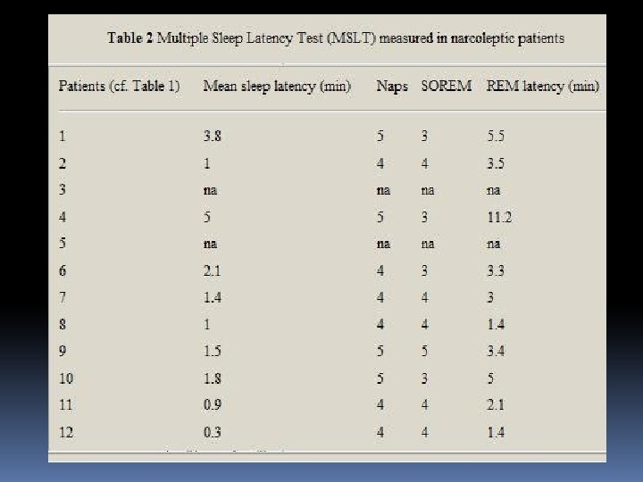
Sleep Tests • • • Epworth Sleepiness Scale (0 -24) Stanford cataplexy questionnaire (>32. 5%) Ullanlinna Narcolepsy Scale (>10) Swiss-Narcolepsy Scale (<00 Multiple Sleep Latency Test (MSLT) • Measures the time it takes from the start of a daytime nap period to the first signs of sleep, called sleep latency. The test is based on the idea that the sleepier people are, the faster they will fall asleep. • Can be used to test for narcolepsy, to distinguish between physical tiredness and true excessive daytime sleepiness.

Humor judgment paradigm Mini-sequences with neutral picture followed by same pic with either neutral or humorous element Humor intensity scale 0 -3 f. MRI-ed 39 funniest pics w/neutral counterparts Patients judged whether they found these funny or not

Neutral

Humorous

Results- Group Patients and controls did not differ in the proportion of images judged as humorous Patients had slower rxn times indicating excessive daytime sleepiness f. MRI revealed (+) in amygdala and insula and frontal regions known to be recruited by the affective content of humorous inputs and experiences
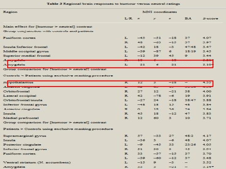

Results- Controls Tested for regions showing ↑f. MRI signal during humourous trials in controls but not in patients Controls showed a maximal activity difference in R - hypothalmus Consistent with hypothesis that patients would have decreased hypothalamic activity Controls also showed increased activity in other areas associated with emotional regulation

Increased response to humour in right hypothalamus, medial prefrontal and cingulate cortex for controls relative to NC patients Schwartz, S. et al. Brain 2008 131: 514 -522; doi: 10. 1093/brain/awm 292 Copyright restrictions may apply.

Results- NC patients showed ↑ response to humorous stimuli in the R-amygdala ↑ activity in R- Inferior parietal and in fusiform cortex May reflect impact of top-down influences from amydgala on sensory pathways

Increased amygdala response to humour in NC patients compared to controls Increased f. MRI signal in left insula and nucleus accumbens in NC-patients (but not in controls)

Conclusion • Narcolepsy is associated with increased amygdalar activity together with reduced medial prefrontal and hypothalamic activity during humor processing – Regions associated with emotional processing – Also found increased activity in L-Nucleus accumbens • Involved in humor processing • Findings provide evidence for an implication of amygdalar circuits in the pathophysiology of human narcolepsy and abnormal responses to positive emotions in patients

Cataplectic patient in Reiss study Dramatic reduction in hypothalamic activity Similar to sleep state Initial overdrive and compensatory shutdown of the hypothalamus resulting in catalplectic attack? Massive supression of hypothalamic activity may be an essential component of a cascade of neural events leading to muscle atonia [all trials after attack were not modeled]

Additional Conclusions by Reiss Patients rated significantly fewer humorous cartoons as funny compared to controls Funny cartoons rated as less funny Patients showed ↑activity in hypothamus* Cataplectic patient- ↓activity Patients had ↑ activation of R-inferior frontal gyrus Patients train themselves to suppress laughter and avoid humorous material

09 -23 -09 Dear Inger, Please find attached a ppt with a humor & a neutral mini-sequence. Good luck for your presentation! Sophie
- Slides: 35