Inflammatory Bowel Disease IBD The main objectives of
")
Inflammatory Bowel Disease (IBD)

The main objectives of this lecture was: 1. to learn the student what IBD means, what are the risk factors, and main presentation. 2. To know how differentiate between ulcerative colitis and crohns disease. 3. To know how mange these disorder and the main nutritional therapy and follow up to prevent the late complications

IBD represent two distinctive disorders of idiopathic chronic intestinal inflammation: Crohn disease and ulcerative colitis. Both disorders are characterized by unpredictable exacerbations and remissions. The onset is during adolescence and young adulthood. IBD is more common in urban areas than in rural areas.

Both genetic and environmental influences are involved in the pathogenesis of IBD. The risk in family members of an affected individual in the range of 7 -22%; a child whose parents both have IBD has a greater than 35% chance of acquiring the disorder. A perinuclear antineutrophil antibody (p. ANCA) is found in about 70% of individuals with ulcerative colitis compared with less than 20% of those with Crohn disease.

Cigarette smoking is a risk factor for Crohn disease but paradoxically protects against ulcerative colitis. In about 10% of individuals with chronic colitis, It is not possible to make a definitive diagnosis, this disorder is called indeterminate colitis. Extraintestinal manifestations occur slightly more commonly with Crohn disease than with ulcerative colitis.

Chronic Ulcerative Colitis Ulcerative colitis is localized to the colon and spares the upper gastrointestinal tract. Disease usually begins in the rectum and extends proximally for a variable distance. When it is localized to the rectum, the disease is ulcerative proctitis, whereas disease involving the entire colon is pancolitis.

About 30% of children with ulcerative proctitis experience proximal spread of the disease. Men are slightly more likely to acquire ulcerative colitis than are women; the reverse is true for Crohn disease.

Clinical Manifestations Blood in the stool and diarrhea are the typical presentation of ulcerative colitis. Constipation may be observed in those with proctitis. tenesmus, urgency, cramping abdominal pain, occurs in more extensive disease. The onset may range from insidious with gradual progression of symptoms to fulminant.

Fulminant colitis defined as Fever, severe anemia, hypoalbuminemia, leukocytosis, and greater than five bloody stools per day for 5 days. Anorexia, weight loss, and growth failure may be present. The clinical course of ulcerative colitis is marked by exacerbations. After initial symptoms, about 5% of children with ulcerative colitis have a prolonged remission (>3 yr).

The use of nonsteroidal anti-inflammatory drugs is predispose to exacerbation. The risk of colon cancer begins to increase after 8 -10 yr of disease and may then increase by 0. 5 -1% per yr. Because colon cancer is usually preceded by changes of mucosal dysplasia, it is recommended that patients who have ulcerative colitis for more than 10 yr be screened with colonoscopy and biopsy every 1 -2 yr.

Extraintestinal manifestations that tend to occur more commonly with ulcerative colitis than with Crohn disease include: 1. pyoderma gangrenosum, 2. sclerosing cholangitis & chronic active hepatitis 3. ankylosing spondylitis. 4. Iron deficiency may result from chronic blood loss as well as decreased intake.

5. Folate deficiency is unusual but may be accentuated in children treated with sulfasalazine, which interferes with folate absorption. 6. Anemia of chronic disease. 7. Secondary amenorrhea is common during periods of active disease in older girls.

Differential Diagnosis 1. Infectious colitis: Bacterial Campylobacter jejuni, Clostridium difficile, Escherichia coli, Shigella, Yersinia. Parasite Entamoeba histolytica , viral colitis in immunocompromised individuals. 2. Dietary protein intolerance 3. Systemic vasculitis (SLE, dermatomyositis) 4. Henoch-Schönlein purpura

5. Hemolytic-uremic syndrome 6. Crohn colitis. Diagnosis 1. Typical presentation in the absence of an identifiable specific cause 2. Laboratory studies CBP: anemia (either iron deficiency or the anemia of chronic disease), WBC ↑ with more severe colitis.

ESR : elevated, it may be normal even with fulminant colitis. Hypoalbuminemia. 3. Plain radiographs of the abdomen: may demonstrate loss of haustral markings in an air-filled colon or marked dilatation with toxic megacolon. 4. Barium enema.

Small ulcerations are distributed uniformly about the colonic circumference and continuously from the rectum to the proximal transverse colon

late changes: The colon is featureless, reduced in caliber, and shortened.

5. Sigmoidoscopy & colonoscopy with biopsy : Classically, disease starts in the rectum with a gross appearance characterized by erythema, edema, loss of vascular pattern, granularity, and friability. The endoscopic findings of ulcerative colitis result from microulcers, which give the appearance of a diffuse abnormality. With very severe chronic colitis, pseudopolyps may be seen.

colonoscopy should not be performed when fulminant colitis is suspected because of the risk of provoking toxic megacolon or causing a perforation during the procedure. Biopsy of involved bowel demonstrates evidence of acute and chronic mucosal inflammation. Typical findings are cryptitis, & crypt abscesses.

Treatment A medical cure for ulcerative colitis is not available; treatment is aimed at controlling symptoms and reducing the risk of recurrence. 1. In mild colitis : * Aminosalicylate 1. Sulfasalazine: is composed of a sulfur moiety linked to the active ingredient 5 -aminosalicylate.

This linkage prevents the premature absorption of the medication in the upper gastrointestinal tract, allowing it to reach the colon, where the two components are separated by bacterial cleavage. The dose is 50 -75 mg/kg/24 hr (divided into 2 -4 doses) PO . Onset of action may take several weeks. Sulfasalazine treats colitis. Hypersensitivity to the sulfa component is the major side effect of sulfasalazine.
 to treat")
2. Other less allergenic preparations of 5 -aminosalicylate (mesalamine, 40 -60 mg/kg/day) to treat ulcerative colitis and prevent recurrences. Aminosalicylate may also be given in enema form and is especially useful for proctitis. Hydrocortisone enemas (100 mg) are used to treat proctitis , once a day (usually bedtime) for 2 -3 wk.

2. I n moderate to severe pancolitis or colitis that is unresponsive to 5 -aminosalicylate therapy treated with oral corticosteroids (prednisone). The dose is 1 -2 mg/kg/24 hr, taper to an alternate-day dose within 1 -3 mo. Children who requiring frequent corticosteroid therapy are started on immunomodulators such as azathioprine (1. 5 -2. 5 mg/kg/day) or 6 -mercaptopurine (1 -1. 5 mg/kg/day).
: Performed for intractable")
Infliximab, a monoclonal antibody may use in fulminant colitis. Surgical treatment(Colectomy): Performed for intractable disease, complications of therapy, and fulminant disease that is unresponsive to medical management.
 Involves any region of the alimentary")
Crohn Disease (Regional Enteritis, Regional Ileitis, Granulomatous Colitis) Involves any region of the alimentary tract from the mouth to the anus. Gastrointestinal involvement in Crohn disease is transmural.
 but may involve the")
The initial presentation most commonly involves ileum and colon (ileocolitis) but may involve the small bowel alone in about 30% (70% of these patients have terminal ileitis alone) or colon alone in 10%-15%. Upper gastrointestinal involvement (esophagus, stomach, duodenum) is seen in up to 30% of children.

Clinical Manifestations Children with ileocolitis typically have cramping, abdominal pain, and diarrhea, sometimes with blood. Ileitis may present as right lower quadrant abdominal pain alone. Crohn colitis may be associated with bloody diarrhea, tenesmus, and urgency.

Systemic signs and symptoms are more common in Crohn disease than in ulcerative colitis and include: Fever, malaise, and easy fatigability are common Growth failure with delayed bone maturation and delayed sexual development may precede other symptoms by 1 or 2 yr. Children may present with growth failure as the only manifestation of Crohn disease. Primary or secondary amenorrhea.
. Gastric or duodenal involvement may be associated")
Perianal disease is common (tags, fistula, abscess). Gastric or duodenal involvement may be associated with recurrent vomiting and epigastric pain. Partial small bowel obstruction from inflammation or stricture, may cause cramping abdominal pain and intermittent abdominal distention. Enteroenteric or enterocolonic fistulas→ malabsorption.
→ signs of urinary infection, or fecaluria. Perianal")
Enterovesical fistulas (between bowel and urinary bladder)→ signs of urinary infection, or fecaluria. Perianal fistulas Intra-abdominal abscess, Hepatic or splenic abscess , Anorectal abscesses, Perianal abscess. Extraintestinal manifestations that are especially associated with Crohn disease include oral aphthous ulcers, peripheral arthritis, erythema nodosum, digital clubbing, episcleritis, renal stones (uric acid, oxalate), and gallstones.

Differential Diagnosis 1. Infectious enteropathies: Yersinia, Giardia, tuberculosis 2. Small bowel lymphoma 3. Periappendiceal abscess 4. Growth hormone deficiency or gluten-sensitive enteropathy (celiac disease) 5. juvenile rheumatoid arthritis.
, diarrhea,")
Diagnosis 1. History : any combination of abdominal pain (especially right lower quadrant), diarrhea, vomiting, anorexia, weight loss, growth retardation, and extraintestinal manifestations. 2. physical examination: Children often appear chronically ill, pale, have weight loss and malnourished, Digital clubbing.
, WBC normal or")
3. laboratory studies: *CBP→ anemia, elevated platelet count (>600, 000/mm 3), WBC normal or mildly elevated. *Low serum albumin level *ESR → elevated or normal *Stool α 1 -antitrypsin level may be elevated *Anti-Saccharomyces antibodies are identified in 55% of children with Crohn disease but in only 5% of children with ulcerative colitis.

4. Endoscopic and radiologic findings *Small bowel follow-through: show aphthous ulceration and thickened, narrowing of the lumen anywhere in the gastrointestinal tract. Linear ulcers may give a cobblestone appearance to the mucosal surface. Other manifestations on radiographic studies that suggest more severe Crohn disease are fistulas between bowel (enteroenteric or enterocolonic), sinus tracts, and strictures.

Double-contrast barium enema examination in Crohn's colitis demonstrates numerous aphthous ulcers (the tiny spots on the lining of the intestine).

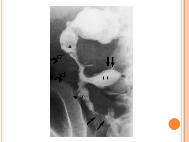
Stricture, terminal ileum - colonoscopy

Severe stenosis of the terminal ileum. Inflammatory effacement of the mucosal folds and small ulcerations characterize the proximal nonstenotic segment.

*Ultrasonography and contrast CT : identifying intraabdominal abscess. *MRI: localize areas of active bowel disease. It is useful during pregnancy. *Colonoscopy with biopsy: Findings on colonoscopy include patchy, nonspecific inflammatory changes, aphthous ulcers, linear ulcers, and strictures. Findings on biopsy → noncaseating granulomas.

Treatment 1. For mild terminal ileal disease or mild Crohn disease of the colon: mesalamine (40 -60 mg/kg/day). Sulfasalazine may be effective for mild Crohn colitis but will not be helpful for small bowel disease. 2. For more extensive or severe small bowel or colonic disease:
. Tapering can begin by 3 -4 wk and continue")
Corticosteroids (prednisone, 1 -2 mg/kg/day). Tapering can begin by 3 -4 wk and continue over several months. Steroid enemas → used for distal colon disease. Children who become refractory to corticosteroid therapy or become dependent on daily dosing→ Immunomodulators such as azathioprine (1. 5 -2. 5 mg/kg/day) or 6 -mercaptopurine (1. 0 -1. 5 mg/kg/day). A beneficial effect of these drugs may be delayed for 3 -6 mo after starting therapy.
 will be associated with marked symptom improvement in 5070%")
Infliximab (5 mg/kg given intravenously) will be associated with marked symptom improvement in 5070% of patients & serve as a bridge until the immunomodulators take effect. 3. To treat perirectal fistula→ Metronidazole (10 -20 mg/d. L/day), Azathioprine and 6 -mercaptopurine. 4. For children with growth failure → Nutritional therapy: enteral nutritional approach. High-calorie oral supplements. 5. Psychologic counseling & Social support.

Surgical therapy Indictions: *localized disease of small bowel or colon that is unresponsive to medical treatment *bowel perforation *fibrosed stricture with symptomatic partial small bowel obstruction * intractable bleeding.

Intra-abdominal or liver abscess treated by ultrasonographic or CT-guided catheter drainage and concomitant intravenous antibiotic treatment. severely symptomatic perianal fistula may require fistulotomy.


- Slides: 46