INFLAMMATORY BOWEL DISEASE Describe the different types of

INFLAMMATORY BOWEL DISEASE

Describe the different types of IBD. Discuss the management of ulcerative colitis. Discuss etiology, clinical features and management of ulcerative colitis. What are the ulcerative lesions of the colon. Discuss the management of ulcerative colitis.

4. A patient gives chronic history of diarrhoea and blood in stool presents with multiple fistulae in the perineum and multiple stricture in small intestine. The diagnosis is – a) b) c) d) Crohn’s disease Radiation enteritis Ulcerative Colitis Ischemic bowel disease

5. Pt. with recurrent diarrhoea, pseudopolyp, lead pipe appearance on Ba enema has – a) b) c) d) Ulcerative colitis Crohn’s disease Irritable bowel syndrome Short bowel syndrome
 b) c) d)")
6. Pyoderma – gangrenosum is most commonly associated with – a) b) c) d) Ulcerative colitis Crohns disease Amoebic colitis Ischemic colitis
")
7. Most common cause of death in Crohn’s disease is due to – a) b) c) d) Sepsis Thromboembolic complication Electrolyte disturbance Malignancy

8. Two 34 year old men have a stoma – one a colostomy in the left iliac fossa after rectal surgery, and other an ileostomy in the right iliac fossa after a panproctocolectomy for ulcerative colitis. Contents of the colostomy bag in the left iliac fossa are relatively small in volume and consist of well-formed stools. Contents of the ileostomy bag in the right iliac fossa are relatively copious non-smelly fluid. What is the best explanation for the difference? A. B. C. D. E. More than 75% of fluid absorption from the entire gut takes place in the colon Random variation The caecum absorbs more fluid than the small intestine There is net secretion of fluid in the left (descending) colon The colon absorbs a small but significant proportion of ingested plus secreted fluid in the gut

Ulcerative colitis

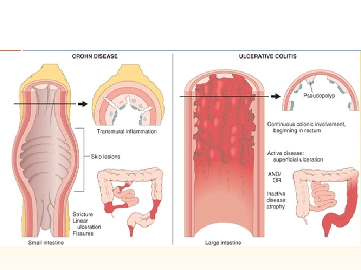
UC is one of the 2 major types of IBD. Characteristically involves only the large bowel. The exact etiology is unknown. HLA–B 27 is identified in most patients with ulcerative colitis. It is a lifelong illness that has a profound emotional and social impact on the affected patients. Spreads proximally from the anal verge in an uninterrupted pattern to involve part or the entire colon. UC occasionally involves the terminal ileum, as a result of an incompetent ileocecal valve.

ETIOLOGY Unknown. Factors contributing to UC include: genetic factors, immune system reactions, environmental factors, NSAID use, low levels of antioxidants, psychological stress factors, a smoking history, and consumption of milk products.

Genetics Genetically susceptible individuals have abnormalities of humoral and cell-mediated immunity. Enhanced reactivity against commensal intestinal bacteria and this dysregulated mucosal immune response predisposes to colonic inflammation. A family history of ulcerative colitis (observed in 1 in 6 relatives) is associated with a higher risk for developing the disease.

Immune reactions Autoantibodies against intestinal epithelial cells may be involved. The presence of antineutrophil cytoplasmic antibodies (ANCA) and anti– Saccharomyces cerevisiae antibodies (ASCA) is a well-known feature of inflammatory bowel disease.

Other etiologic factors Vitamins A and E are found in low levels in patients with ulcerative colitis. Psychological and psychosocial stress can precipitate exacerbations. Smoking is negatively associated with ulcerative colitis. This relationship is reversed in Crohn disease. Milk consumption may exacerbate the disease.

Pathology The rectum is involved in all circumstances. It is a diffuse inflammatory disease, primarily affecting the mucosa and superficial submucosa. There are multiple minute ulcers. When the disease is chronic, inflammatory polyps (pseudopolyps) occur. Microscopic ally, there is an increase in inflammatory cells in the lamina propria, the walls of crypts are infiltrated by inflammatory cells and there are crypt abscesses. There is depletion of goblet cell mucin. With time precancerous changes can develop (= severe dysplasia or carcinoma in situ).

History Patients predominantly complain of rectal bleeding. Frequent stools and mucous discharge from the rectum. Tenesmus. Severe dehydration, especially in the elderly population.

Extracolonic manifestations Uveitis, pyoderma gangrenosum, pleuritis, erythema nodosum, ankylosing spondylitis, primary sclerosing cholangitis and spondyloarthropathies. Uveitis is the most common.

Physical Examination Physical findings are normal in patients with mild disease. Patients with severe disease can have signs of volume depletion and toxicity, including the following: Fever Tachycardia Significant abdominal tenderness Weight loss
 a severe initial attack,")
In general, a poor prognosis is indicated by (1) a severe initial attack, (2) disease involving the whole colon and (3) increasing age, especially after 60 years.

Grading mild – rectal bleeding or diarrhoea with four or fewer motions per day and the absence of systemic signs of disease; moderate – more than four motions per day but no systemic signs of illness; severe – more than four motions a day together with one or more signs of systemic illness: fever over 37. 5°C, tachycardia more than 90 min, hypoalbuminaemia less than 30 g l, weight loss more than 3 kg.

Ulcerative Colitis Crohn Disease Only colon involved Panintestinal Continuous inflammation extending proximally from rectum Skip-lesions with intervening normal mucosa Inflammation in mucosa and submucosa only Transmural inflammation No granulomas Noncaseating granulomas Perinuclear ANCA (p. ANCA) positive ASCA positive Bleeding (common) Bleeding (uncommon) Fistulae (rare) Fistulae (common)

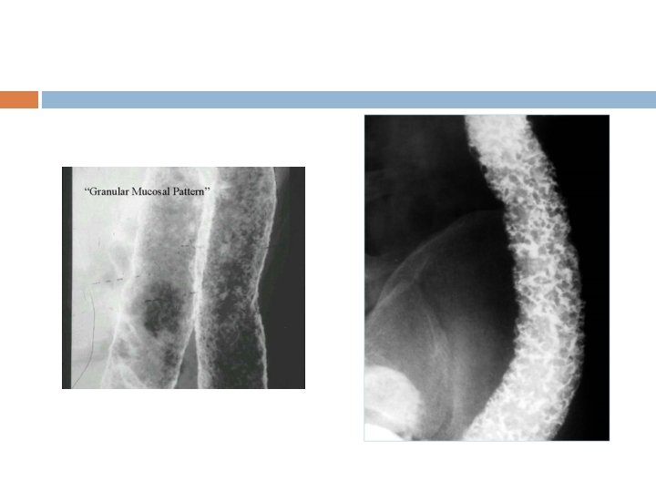
WORKUP Best made with endoscopy. Double-contrast barium enema examination. ANCA and ASCA. Anemia. Thrombocytosis. Hypoalbuminemia. Hypokalemia. Hypomagnesemia. Elevation of ESR and CRP. Stool studies to exclude other causes.

Barium enema The principal signs are: • loss of haustration, especially in the distal colon; • mucosal changes caused by granularity; • pseudopolyps; • in chronic cases, a narrow contracted colon.

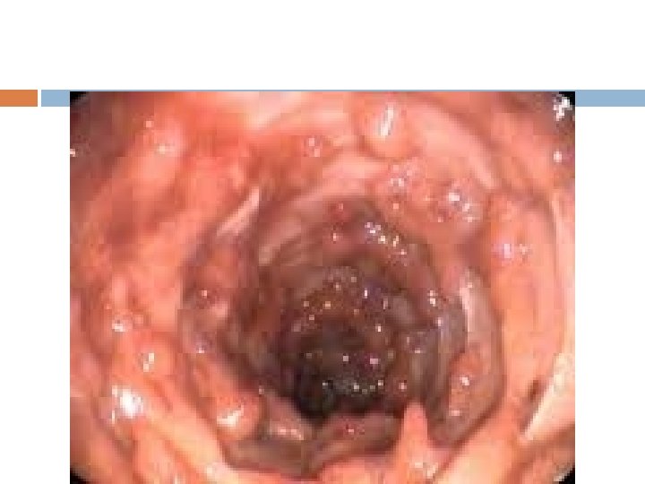
Colonoscopy and biopsy The mucosa is hyperaemic and bleeds on touch, There may be a pus-like exudate. Pseudopolyps. Ulcers are seen that appear to coalesce. Colonoscopic biopsy is done: 1. to establish the extent of inflammation; 2. to distinguish between UC and Crohn’s colitis; 3. to monitor response to treatment; 4. to assess longstanding cases for malignant change.

Colonoscopy and biopsy Extensive disease - Evidence of ulcerative colitis proximal to the splenic flexure Left-sided disease - Ulcerative colitis present in the descending colon up to, but not proximal to, the splenic flexure Proctosigmoiditis - Disease limited to the rectum with or without sigmoid involvement

Complications ACUTE: Toxic megacolon Perforation Haemorrhage CHRONIC: Cancer Extra-intestinal manifestations

TREATMENT Medical management with corticosteroids and anti-inflammatory agents. Symptomatic treatment with antidiarrheal agents and rehydration. Surgery is contemplated when medical treatment fails or when a surgical emergency (eg, perforation of the colon) occurs.

5 -aminosalicylic Acid Derivatives 1. 2. 3. : Sulfasalazine is useful in treating mild-to-moderate ulcerative colitis and maintaining remission. Sulfasalazine is 5 aminosalicylate (5 -ASA) coupled to a sulfapyridine. It is poorly absorbed in the proximal bowel, and the bacteria in the colon uncouple the 5 -ASA from the sulfa moiety, allowing 5 -ASA to exert its anti-inflammatory effect on the colonic mucosa by inhibiting prostaglandin synthesis. It acts locally in the colon to reduce the inflammatory response and systemically inhibits prostaglandin synthesis. Balsalazide is a prodrug that is converted into 5 -aminosalicylic acid through bacterial azo reduction. Metabolites of drug may decrease inflammation by blocking the production of arachidonic acid metabolites in colon mucosa. Mesalamine is the drug of choice for maintaining remission. It is useful for the treatment of mild-to-moderate ulcerative colitis. It is better tolerated and has less adverse effects than sulfasalazine. Enema and suppository forms are typically used in patients with distal colitis.

Tumor Necrosis Factor Inhibitor These agents prevent the endogenous cytokine from binding to the cell surface receptor and exerting biological activity. These agents adversely affect normal immune responses and allow development of superinfections; reactivation of latent TB has been reported in patients with previous exposure to TB. Infliximab, Adalimumab, Golimumab.

Immunosuppressant Agents These agents regulate key factors of the immune system. Effective in bringing steroidresistant disease under control. Azathioprine, Cyclosporine, 6 -Mercaptopurine, Tacrolimus.

Corticosteroids Decrease inflammation by suppressing the migration of polymorphonuclear leukocytes and reversing increased capillary permeability. They are used for induction of remission in moderate-to-severe active ulcerative colitis. They have no role in maintaining remission; long-term use can cause adverse effects.

Alpha 4 Integrin Inhibitors Emerging as option for moderate-to-severe active IBD in patients who have had an inadequate response with, lost response to, or were intolerant to a TNF blocker or immunomodulator; or had an inadequate response with, were intolerant to, or demonstrated dependence on corticosteroids. Vedolizumab.

Antimicrobials They prolong GI transit time and decrease secretion via peripheral mu-opioid receptors. Loperamide,

Treatment of mild disease In disease confined to the rectum, topical mesalazine given by suppository is the preferred therapy. Left-sided colonic disease is best treated with a combination of mesalazine suppository and an oral aminosalicylate.

Treatment of Acute, Severe disease Acute, severe ulcerative colitis ie, >6 bloody bowel movements/d, with one of the following: fever >38°C, hemoglobin level < 10. 5 g/d. L, heart rate >90 bpm, erythrocyte sedimentation rate >30 mm/h, or C-reactive protein level >30 requires hospitalization and treatment with intravenous high-dose corticosteroids (hydrocortisone 400 mg/d or methylprednisolone 60 mg/d).

Alternative induction medications like Cyclosporine, tacrolimus, infliximab, adalimumab, and golimumab are used in steroid-resistant disease.

Maintenance therapy Once remission has been achieved, maintenance therapy is recommended for all patients to prevent relapse. Oral aminosalicylates are indicated for disease that responded to ASA or steroids. Patients who are unable to maintain remission or are intolerant to 5 -ASA, Azathioprine and 6 mercaptopurine are alternatives. Extended-release oral formulation of budesonide provides the benefit of a powerful anti-inflammatory drug delivered locally while avoiding many of the systemic side effects associated with systemic steroids. Probiotics.

Indications for surgery It is the definitive therapy for ulcerative colitis. Failure of medical management is the most common indication for surgery. Indications for urgent surgery in patients with ulcerative colitis include (1) toxic megacolon refractory to medical management, (2) fulminant attack refractory to medical management, and (3) uncontrolled colonic bleeding. Indications for elective surgery in ulcerative colitis include (1) long-term steroid dependence, (2) dysplasia or adenocarcinoma found on screening biopsy, (3) and disease present for 7 -10 years.

Types of Surgery Total abdominal colectomy with ileostomy: done in emergency setting as a “first aid procedure”. The only problem is that because the rectal stump is left behind chances of haemorrhage are always there. Proctocolectomy and ileostomy: has the lowest complication rate. Indicated in patients who are not candidates for restoration. The patient is left with a permanent ileostomy.

Restorative proctocolectomy with an ileoanal pouch: In this operation, a pouch is made out of ileum as a substitute for the rectum and sewn or stapled to the anal canal. This avoids a permanent stoma. Complications are pelvic sepsis , poor function and pouchitis or inflammation of the pouch , women may suffer from reduced fertility.

Colectomy and ileorectal anastomosis: used if there is minimal rectal inflammation. Annual rectal inspection is required Has the advantage of stoma avoidance and minimal risk to sexual function.
: rarely used. Complications include early")
Ileostomy with a continent intra-abdominal pouch (Kock’s procedure): rarely used. Complications include early leak with formation of fistulae and late subluxation of the valve. Pouch survival at 10 years was 87% in one study.

CROHN’S DISEASE
 b) c) d) e) In Crohn’s disease all are seen except: Hyperplastic")
1. a) b) c) d) e) In Crohn’s disease all are seen except: Hyperplastic polyps Diverticulosis Fissuring ulcer Epitheloid granuloma Crypt abscess
 Stomach not involved b) No")
2. Crohn’s disease is associated with following – a) Stomach not involved b) No granulomatous + Transmucosal fissures c) Continuous involvement d) Through and trough involvement of thickness of bowel wall
 Regional")
3. Transmural inflammation with skip lesions is colon are characteristic of – a) Regional ileitis (Crohn’s disease) b) Ischemic colitis c) Ulcerative colitis d) Non specific colitis

Crohn disease is an idiopathic, chronic inflammatory process that can affect any part of the gastrointestinal tract from the mouth to the anus. Occurs as a result of an imbalance between proinflammatory and anti-inflammatory mediators. Approximately 30% of Crohn disease cases involve the small bowel, particularly the terminal ileum, another 20% involve only the colon, and 45% involve both the small bowel and colon. The characteristic presentation is abdominal pain and diarrhea, which may be complicated by intestinal fistulization or obstruction.

Etiology Genetic, Microbial, Immunologic, Environmental, Dietary, Vascular, Psychosocial factors Smoking and the use of OCPs and NSAIDs.

PATHOPHYSIOLOGY Chronic inflammation from T-cell activation leading to tissue injury is implicated in the pathogenesis of Crohn’s disease. After activation by antigen presentation, unrestrained responses of type 1 T helper (Th 1) cells predominate as a consequence of defective regulation. Th 1 cytokines such as interleukin (IL)-12 and TNF-α stimulate the inflammatory response. Inflammatory cells recruited by these cytokines release nonspecific inflammatory substances, including arachidonic acid metabolites, proteases, platelet activating factor, and free radicals, which result in direct injury to the intestine.

Microscopically, the initial lesion starts as a focal inflammatory infiltrate around the crypts, followed by ulceration of superficial mucosa. Later, inflammatory cells invade the deep mucosal layers and, in that process, begin to organize into noncaseating granulomas. The granulomas extend through all layers of the intestinal wall and into the mesentery and the regional lymph nodes.

Macroscopically, the initial abnormality consists of hyperemia and edema of the involved mucosa. Later, discrete superficial ulcers form over lymphoid aggregates and are seen as red spots or mucosal depressions (see the image below). These can become deep, serpiginous ulcers located transversely and longitudinally over an inflamed mucosa, giving the mucosa a cobblestone appearance. The lesions are often segmental, being separated by healthy areas, and are referred to as skip lesions

HISTORY Low-grade fever. Prolonged diarrhea with abdominal pain. Weight loss. Generalized fatigability. Crampy or steady right lower quadrant or periumbilical pain. The pain precedes and may be partially relieved by defecation. Diarrhea is not grossly bloody and is often intermittent. Colonic Crohn disease may be clinically indistinguishable from ulcerative colitis, with symptoms of bloody mucopurulent diarrhea, cramping abdominal pain, and urgency to defecate. Those with perianal disease may have debilitating perirectal pain, malodorous discharge from the fistula, and disfiguring scars from active disease or previous surgery.

Ho Recurrent attacks of SAIOAIO. Enterovesical fistula may present as recurrent urinary tract infections and pneumaturia. Enterovaginal fistulae may present as as feculent vaginal discharge. Enterocutaneous fistulae may present as feculent soiling of the skin.

Physical examination Chronic intermittent fever Diffuse or localized abdominal tenderness. Fullness or a discrete mass may be appreciated, typically in the right lower quadrant of the abdomen. Inspection of the perianal region can reveal skin tags, fistulae, ulcers, abscesses, and scarring. Mucocutaneous or aphthous ulcers, erythema nodosum and pyoderma gangrenosum. Eye examination may reveal episcleritis. Arthritis and arthralgia.

Complications Intra-abdominal abscesses. Fistulae sinus tracts. Bowel strictures and adhesions. Perforation. Colonic malignancy.

Disease activity and scoring systems Montreal classification system is based on the following 3 variables: Age at diagnosis Disease distribution/location Disease behavior
 has 3 categories, as follows : A 1 –")
Age at diagnosis (A) has 3 categories, as follows : A 1 – ≤ 16 years A 2 – 17 -40 years A 3 – > 40 years Disease distribution/location (L) has the following 4 categories, 1 of which is a modifier for upper GI involvement : L 1 – Ileal L 2 – Colonic L 3 – Ileocolonic L 4 – Isolated upper GI disease; L 4 is a modifier that can be added to L 1 -L 3 when there is concomitant upper GI involvement Disease behavior (B) has 1 interim category (B 1) and 2 specified categories, with an additional modifier for perianal diseases (p), as follows : B 1 – Nonstricturing, nonpenetrating; B 1 p: nonstricturing, nonpenetrating with perianal involvement B 2 – Stricturing; B 2 p: stricturing with perianal involvement B 3 – Penetrating; B 3 p: penetrating with perianal involvement

WORKUP Complete blood cell count Hypoalbuminemia Deficiencies in iron and micronutrients Liv Acute inflammatory markers, such as CRP level or ESR, may correlate with disease activity. Stool samples should be tested for the presence of WBCs, occult blood, routine pathogens, ova, parasites, and Clostridium difficile toxin. Fecal calprotectin has been proposed as a noninvasive surrogate marker of intestinal inflammation in IBD.

WORKUP Plain radiography or CT of the abdomen can be used to assess for bowel obstruction or intra-abdominal abscesses. Barium contrast studies. CT enterography or MR enterography. MRI of the pelvis or transrectal ultrasonography can identify perianal fistulae and detect pelvic and perianal abscesses. Endoscopy and biopsy.

TREATMENT-Medical Steroids are the mainstay of treatment. 5 -ASA compounds. Those who have symptoms and signs of a mass or an abscess are also treated with antibiotics. Azathioprine is now standard maintenance therapy. Anti-TNF therapy. Nutritional support.

TREATMENT-Surgical Indications for surgery Surgical resection will not cure CD. Surgery is therefore focused on the complications of the disease. • recurrent intestinal obstruction; • bleeding; • perforation; • failure of medical therapy; • intestinal fistula; • fulminant colitis; • malignant change; • perianal disease.

1. Ileocaecal resection. 2. Segmental resection. 3. Colectomy and ileorectal anastomosis. 4. Emergency colectomy. 5. Laparoscopic surgery 6. Temporary loop ileostomy. 7. Proctocolectomy. 8. Strictureplasty. 9. Anal disease is usually treated conservatively.
- Slides: 64