Inflammatory Arthritis Rheumatoid Arthritis Dr R B Kalia

Inflammatory Arthritis. Rheumatoid Arthritis Dr R B Kalia, Additional Professor , Department of Orthopaedics

Leaning Objective • Clinical Features of RA • Investigations • Diagnosis • Indications for Surgery in Arthritis • Various procedures possible • Rational choice in treatment

Clinical Features • Chronic multisystem disease of unknown cause. • persistent inflammatory synovitis • Peripheral joints in a symmetric distribution • synovial inflammation causes cartilage destruction and bone erosions and subsequent changes in joint integrity
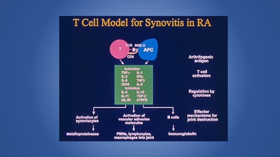

Effects of IL-6 • B cell maturation • Ig, • rheumatoid factor, • hypergammaglobulemia • Hepatocyte stimulus • acute phase proteins (high ESR) • decreased albumin synthesis

Course of RA • Quite variable mild oligoarticular illness of brief duration with minimal joint damagea relentless progressive polyarthritis with marked functional impairment

Epidemiology • RA occurs in 0. 5‐ 1. 0% of the population • Women affected three times more often than men • Prevalence increases with age • Onset most frequent in fourth and fifth decades.

Articular Manifestations • Typically a symmetric polyarthritis • Peripheral joints with pain, tender‐ ness, and swelling • Morning stiffness is common • PIP and MCP joints frequently involved • Joint deformities may develop after persistent inflammation.

Systemic • Fever • Decreased appetite • Muscle wasting

Extraarticular Manifestations • Cutaneous‐rheumatoid nodules, vasculitis • Pulmonary‐nodules, interstitial disease. • Ocular‐keratoconjunctivitis sicca, episcleritis, scleritis • Hematologic‐anemia, Felty's syndrome (splenomegaly and neutropenia) • Cardiac‐pericarditis, myocarditis • Neurologic‐myelopathies secondary to cervical spine disease, entrapment, vasculitis

EVALUATION • Hx and physical exam with careful examination of all joints. • Rheumatoid factor (RF) is present in >66% of pts; its presence correlates with severe disease, nodules, extraarticular features. • Antibodies to cyclic citrullinated protein {anti‐CCP) have similar sensitivity but higher specificity than RF • may be most useful in early RA • Presence most common in pts with aggressive disease with a tendency for developing bone erosions.

Other laboratory data • CBC, ESR. • Synovial fluid analysis‐useful to rule out crystalline disease, infection. • Radiographs‐juxta‐articular osteopenia, joint space narrowing, marginal erosions. • Chest x‐ray should be obtained.
 • SEROLOGY (0‐")
2010 ACR/EULAR Classification Criteria for RA • JOINT DISTRIBUTION (0‐ 5) • SEROLOGY (0‐ 3) • SYMPTOM DURATION (0‐ 1) • ACUTE PHASE REACTANTS (0‐ 1) > 6 – Definitely RA

JOINT DISTRIBUTION • 1 large joint • 2‐ 10 large joints • 1‐ 3 small joints (large joints not counted) • 4‐ 10 small joints (large joints not counted). • >10 joints (at least one small joint) 0 1 2 3 5

SEROLOGY • Negative RF AND negative ACPA • Low positive RF OR low positive ACPA • High positive RF OR high positive ACPA 0 2 3

SYMPTOM DURATION /ACUTE PHASE REACTANTS • < weeks ‐ 0 • > 6 weeks ‐ 1 • Normal CRP AND normal ESR 0 • Abnormal CRP OR abnormal ESR 1

SURGERY FOR RHEUMATOID ARTHRITIS Indicated when the disease has progressed to such a stage • Pain is unrelieved by medication • Mechanically unstable joint • • Arthroscopic synovectomy/ open synovectomy Proximal tibial osteotomy Arthrodesis Total joint arthroplasty

Goals • Relieve pain • Prevent destruction of cartilage or tendon • Improve function of joints by • Increasing or decreasing motion a) Correcting deformity b) Increasing stability c) Improving effective muscle forces

Functional Impairment Class I ‐ Can carry out all usual activities without handicap Class II ‐ Can perform normal activities despite the handicap of discomfort or limited motion at one or more joints Class III ‐Are limited to few of the duties of their usual occupation or self‐care Class IV ‐Are largely or completely incapacitated, are bedridden or confined to a wheelchair, and are limited to little or no self‐care.

SYNOVECTOMY- Rheumatoid Arthritis The procedure consists of • Removing the diseased synovium • Decreasing the inflammatory mediators and protecting the cartilage. Indicated in patients with • minimal structural damage to the joint • Refractory to pharmacological agents. • Open synovectomy • Arthroscopic synovectomy.

Synovial villi with nodular lymphocytosis , marked increase in plasma cells with synovial cell hyperplasia and hypertrophy

Synovectomy • Removing the superficial layers of the synovium with a shaver • Down to a defining plane between the synovium and subsynovial tissues. • Smooth shiny fibers of the capsule can be seen

TOTAL JOINT ARTHROPLASTY • Moderate to severe destruction of cartilage and subchondral bone • Relieve pain and improve function in most joints

Case 1 • 36 years old • Seropositive Rheumatoid Arthritis • CRP 5 • ESR 34 mm • Unable to walk more than a dozen steps • Severe restriction of movement • Received DMARDS for 15 years
 b) c) d) Arthroscopic synovectomy Tibio‐femoral Fusion")
What is the appropriate further Management? a) b) c) d) Arthroscopic synovectomy Tibio‐femoral Fusion Total knee replacement Unicondylar knee replacement

Bl TKR

Total Knee Replacement Complications may be more frequent in patients with rheumatoid arthritis than in those with osteoarthrosis because of • Poor healing of tissue • Deep wound infections • Severe flexion contracture • Severe joint laxity or osteopenia • Involvement of multiple other joints limiting rehabilitation.

Pre op radiographs

Post Op after knee replacement

Rheumatoid Arthritis Hip The following procedures have proved useful • Synovectomy, • Arthrodesis • Total hip arthroplasty • Resection of the femoral head and neck

SYNOVECTOMY • Indicated early in the course of juvenile rheumatoid arthritis when joint destruction is minimal. • Temporary symptomatic relief and improved function can often be achieved in carefully selected patients.

RESECTION ARTHROPLASTY • Severe rheumatoid arthritis of long duration and contractures of multiple joints are not candidates for hip arthroplasty. • Rare functional class IV patients ‐there is no hope for rehabilitation to an ambulatory status. • When there is increasing pain and when deformities interfere with perineal hygiene‐ Girdlestone resection or neck resection have been useful

Case 2 • 42 years old • Rheumatoid arthritis for 20 years • Increasing pain and stiffness right hip for 4 years • Severe restriction of function and ADA affected • Flexion deformity 20 degrees adduction deformity 20 degrees

Radiographs
 Shentons")
MCQ 2 • Which of the following radiological feature is not present? a) Shentons arch is broken b) Reduced joint space c) Protrusio acetabuli d) Medialization of head

Cemented THR

UPPER EXTREMITY • Shoulder‐ Adduction and internal rotation deformity • Elbow ‐ flexion deformity of the, limitation of pronation and supination • Flexion deformity of the wrist • Ulnar deviation of the hand, and flexion and ulnar deviation of the fingers Treating the affected part with rest usually relieves pain Loss of function often follows.

Total shoulder Arthroplasty

Elbow • Involved in 20% to 50% of patients with rheumatoid arthritis. • The function of the joint may deteriorate • Compromising activities of daily living and independence.

Surgical procedures for rheumatoid arthritis elbow • Synovectomy (most often combined with radial head excision) • Total elbow arthroplasty. • Often requires a release of the collateral ligaments and complete capsulotomy to optimize movement after surgery. • Combined with a resection of proximal radial head to improve pronation and supination.

Radiographic - Lateral view

TER
 b) c) d) Arthroscopic")
MC Q 1 What is the appropriate further Management? a) b) c) d) Arthroscopic synovectomy Tibio‐femoral Fusion Total knee replacement Unicondylar knee replacement
 Shenton’s")
MCQ 2 • Which of the following radiological feature is not present? a) Shenton’s arch is broken b) Reduced joint space c) Protrusio acetabuli d) Medialization of head

Conclusions • Rh Arth is a multisystem disease • If not diagnosed early – Significant damage to joints • Classical presentation may/May not be present • Each diagnostic test needs to be understood • Joint replacement – End stage disease

Thank You
- Slides: 48