Inflammation Lecture 2 Dr Bushra Altarawneh Morphologic patterns

Inflammation Lecture 2 Dr. Bushra Al-tarawneh

Morphologic patterns of acute inflammation Serous Inflammation. Fibrinous Inflammation. Suppurative Inflammation. Membranous Inflammation.

Serous inflammation � Characterized by the outpouring of a watery, relatively protein-poor fluid that, depending on the site of injury, derives either from the plasma or from the secretions of mesothelial cells lining the peritoneal, pleural, and pericardial cavities. � Fluid in a serous cavity is called an effusion. � End with respiratory or cardiac impairment. � The skin blister resulting from a burn or viral infection is a good example of the accumulation of a serous effusion either within or immediately beneath the epidermis of the skin.

Skin blister showing the epidermis separated from the dermis by a focal collection of serous effusion. Serous pleural inflammation (Ascites)

Fibrinous inflammation Occurs in sever injuries. There will be increase vascular permeability lead to exudation of large molecules (such as fibrinogen) passing the endothelial barrier. � Histologically, the accumulated extravascular fibrin appears as an eosinophilic meshwork of threads. � Exudates ► Resolution: Degraded by fibrinolysis and the accumulated debris is removed by macrophages. � Exudates ► Organization ► Scarring. Failure of removing fibrin completely ► ingrowths of fibroblasts and blood vessels.

Organization of fibrinous pericardial exudate forms dense fibrous scar tissue that bridges or obliterates the pericardial space and restricts myocardial function.
 inflammation � These are manifested by the collection of large amounts of")
Suppurative (purulent) inflammation � These are manifested by the collection of large amounts of purulent exudate (pus) consisting of neutrophils, necrotic cells, and edema fluid. � Certain organisms (e. g. , staphylococci) are more likely to induce such localized suppuration and are therefore referred to as pyogenic (pus-forming). � Presented as abscess or ulcer.

Abscess : Focal collection of pus caused by pyogenic organisms or by secondary infections of necrotic foc � Composed of: Large amounts of purulent exudate (pus) consisting of neutrophils, necrotic cells and edema fluid. � �

� Ulcer: It is local defect, or excavation, of the surface epithelium produced by sloughing (shedding) of inflammatory necrotic tissue. � Ulcers are most commonly encountered in: (1) The gastrointestinalor genitourinarytract (2) The subcutaneous tissue.

Pseudomembranous inflammation of mucous membranes Ø Severe injury with extensive epithelial necrosis → sloughing → large shallow ulcers covered by false or pseudomembrane (white or creamy color layer). � Composed of: Fibrin, dead epithelium, neutrophils, red blood cells and bacteria. � Ex. : Diphtheria and pseudomembranous colitis.

Pseudomembrane in Diphtheria Pseudomembranous colitis : Inflammation of mucous membranes of colon.

Categories of Chronic Inflammation Ø Chronic inflammation is inflammation of prolonged duration (weeks to years) in which continuing inflammation, tissue injury, and healing, often by fibrosis, proceed simultaneously. � It is either: 1 - Progression of acute inflammation to chronic. Occurs when the acute response cannot be resolved due to: A. Persistence of the injurious agent. B. Interference of normal healing process. 2 - Chronic inflammation from the onset. (e. g. Some viral infections)

Causative agents of chronic inflammation 1 - Low-grade, persistent irritant agents that are unable to penetrate deeply or spread rapidly. 2 - Moderate to low virulent microbes like viruses, certain bacteria, fungi, and larger parasites that are difficult to eradicate like: Mycobacteria Tuberculosis Treponema pallidum Syphilis Actinomyces Actinomycosis 3 - Foreign bodies such as talc, silica, asbestos, and surgical suture materials.

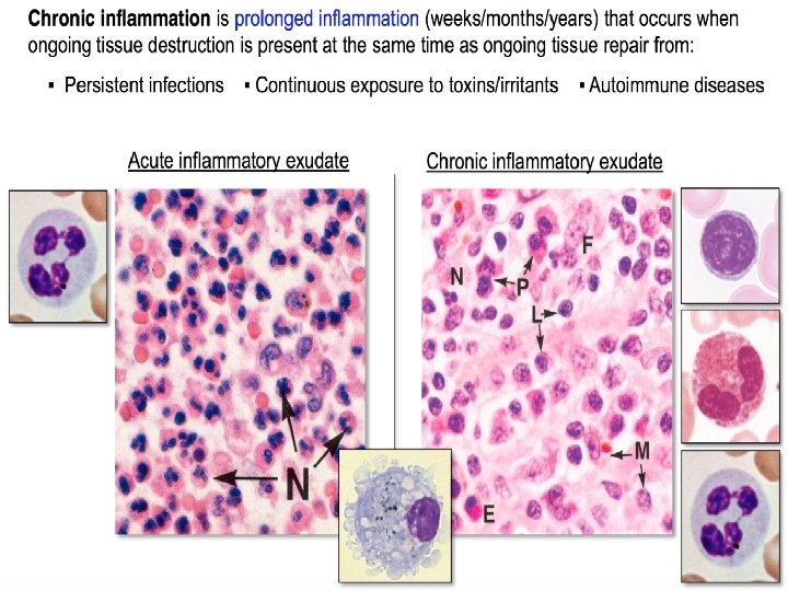
Features of chronic inflammation 1. Long duration: Persists for weeks, months or years. 2. Tissue destruction: Greater than in acute inflammation (Induced by products of the inflammatory cells). 3. The inflammatory infiltrate is a mixture of mononuclear cells: Macrophages, lymphocytes and plasma cells. 4. Productive rather than exudative: Production of fibrous tissue through formation of granulation tissue. 5. Healing will occur by proliferation of large of fibroblasts end with fibrosis. (Scar) number

Microscopical features of chronic inflammation: 1. Infiltration by mononuclear cells: Ø Lymphocytes: B-lymphocytes plasma cell Ab. T-lymphocytes sensitized cytokines. Ø Plasma cells : Antibody-producing cells. Ø Macrophages: Blood monocytes when extravasated it became activated macrophages. Ø Eosinophiles and mast cell: Specially presents in allergic reaction. 2. Tissue destruction induced by infl. Cells. 3. Healing by granulation tissue composed of macrophages, new blood vessels & fibroblasts.

Classification of chronic inflammation 1 - Nonspecific chronic nflammation. 2 - Granulomatous inflammation.

1. Nonspecific Chronic Inflammation Infection Chemotaxis. Macrophages & lymphocytes infiltration Fibroblast proliferation Scar formation (Fibrosis)

2. Granulomatous inflammation � Granulomatous inflammation is a distinctive pattern of chronic inflammation characterized by aggregates of activated macrophages with scattered lymphocytes. These macrophages will be activated and modified to be similar to epithelial cells so called: Epithelioid cells. q Many epithelioid cell united together forming: Giant cell. q A dense membrane of connective tissue encapsulates the lesion and isolates it. q

Diagram of typical TB granuloma Multinucleated GC Epithelioid cells Caseation necrosis Lymphocytes Firoblasts
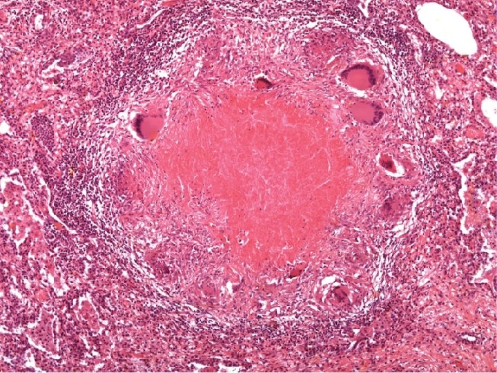

Giant cells The epithelioid cells coalesce, forming a large, multinucleated giant cell that attempts to surround the foreign Agent. .

Examples of granulomatous inflammation:

Chronic Inflammatory Cells Chronic inflammatory cells: v Macrophage. v Lymphocytes: T & B lymphocytes v Eosinophils cells. v Mast cells.

Macrophages It is the dominant cells of chronic inflammation derived from circulating blood monocyte after their emigration into the tissue forming tissue macrophage that scattered in all connective tissues. 1 - Liver: Kupffer cells 2 - Spleen and lymph nodes: Sinus histiocytes 3 - Central nervous system: Microglial cells 4 - Lungs : Alveolar macrophages

In all tissues macrophages act as: 1. Filters for microbes. 2. Senescent cells to alert the immune system to injurious stimuli. (T and B lymphocytes). Activation of Macrophage: Macrophages are activated by: 1 - Nonimmunologic stimuli like bacterial endotoxin. 2 - Cytokines from immune-activated lymphocytes.

Differences between Activated macrophage and blood monocyte: � Increased cell size. � Increased content of lysosomal enzymes. � More active metabolism. � Greater ability to kill ingested organisms. � Longer half-lives. � Greater capacity for phagocytosis than monocytes.

Macrophage Activation

Macrophage Lymphocytes interaction Lymphocytes & macrophages interact in a bidirectional way resulting in a cycle of cellular reactions that play an important role in chronic inflammation. Activated Macrophages produce cytokines (IL-12) activate T-lymph. promoting more antigen presentation and cytokine secretion. T-lymph. activate macrophages Produce cytokines (IFN -γ)

Lymphocyte During chronic inflammation both T and B lymphocytes migrate into inflammatory sites Activation of T and B lymphocytes Activated T-Lymphocyte Activated B-Lymphocyte Cytotoxic T-Lymphocyte Natural Killer cells Plasma cells Immunoglobulins

Plasma cells � Plasma cells produce antibodies directed against: 1 - Persistent antigens in the inflammatory site. 2 - Altered tissue components. � In some strong chronic inflammatory reactions the accumulation of : Lymphocytes Macrophages Plasma cells � Have morphologic features of lymphoid organs, so appear as lymph nodes.

Eosinophils are found in: 1 - Inflammatory sites around parasitic infections. 2 - Part of immune reactions mediated by Ig. E, associated with allergies. Eosinophil granules contain major basic protein, a highly charged cationic protein that is toxic to parasites but also causes epithelial cell necrosis.

Mast cells Widely distributed in the connective tissues throughout the body. Seen in acute & chronic inflammation. � In individuals prone to allergic reactions: � Mast cells are bounded with Ig. E antibody specific for certain antigens Ig. E-armed mast cells Release infl. Mediators Early vascular changes of inflammation. � Ig. E-armed mast cells are central players in allergic reactions, including anaphylactic shock.

Ig. E

The components of acute and chronic inflammation

Acute chronic Thank You
- Slides: 36