Infectious Bursal Disease Gumboro Defination Infectious bursal disease
")
Infectious Bursal Disease (Gumboro)
 is an acute and highly contagious viral infection")
Defination • Infectious bursal disease (IBD) is an acute and highly contagious viral infection of immature chickens. • IBD is characterized by destruction of lymphocytes in the bursa of Fabricius (BF) and to a lesser extent in other lymphoid organs. • Affected chickens have reduced antibody response to vaccinations, strong post vaccinal reactions, and increased susceptibility to concurrent or secondary infections.

Susceptibility • Only chickens develop IBD after infection by serotype 1 viruses. • Turkeys may be asymptomatic carriers of serotype 2 • The Pekin duck can also be an asymptomatic carrier of serotype 1 viruses

• The age of maximum susceptibility is between 3 and 6 weeks corresponding to the period of maximum bursa development, during which the acute clinical signs are observed. • Infections before 3 weeks are generally subclinical and immunosuppressive. • Clinical cases may be observed up to the age of fifteen to twenty weeks
 m")
Etiology • IBD is caused by a birnavirus (infectious bursal disease virus; IBDV) m non enveloped doublestranded (RNA) • Two serotypes of IBDV have been identified. • The serotype 1 viruses cause disease in chickens • Serotype 2 strains infect chickens and turkeys but have not caused clinical disease or immunosuppression in these hosts
, classical virulent, variant, or hypervirulent")
• IBDV may be considered apathogenic, attenuated (vaccines), classical virulent, variant, or hypervirulent (vv. IBDV). • Very stable hardy virus. • Able to withstand a wide p. H range (p. H 2 -12). • Heat stable (still viable after 30 minutes at 60°C). • High level of resistance to most commonly used disinfectants. • Survives in the poultry house environment for

Transmission of IBD Virus • Only horizontal transmission by the oral or respiratory pathway. • Infected subjects excrete the virus in faeces as early as 48 h after infection, and may transmit the disease by contact over a sixteen-day period • The disease is transmitted by direct contact with excreting subjects, or by indirect contact with any inanimate or animate (farm staff, animals) contaminated vectors

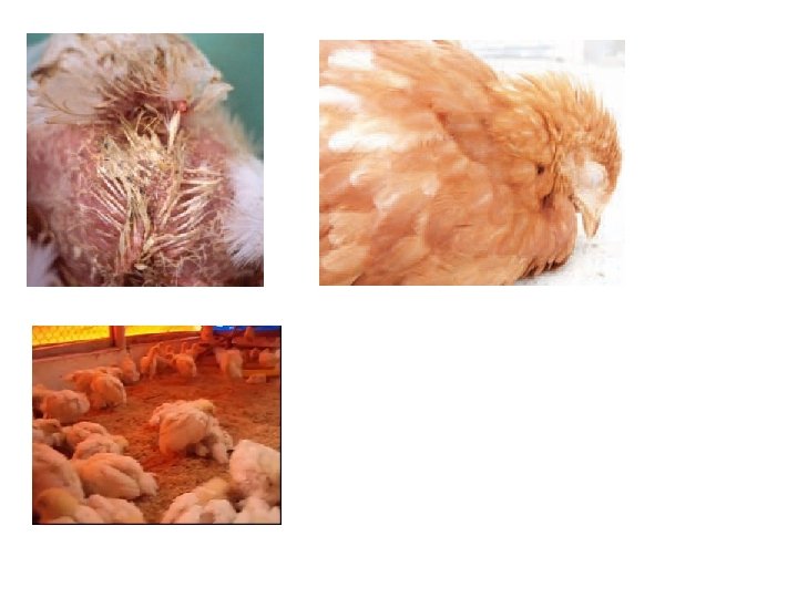
Signs • incubation period of 2 -3 days. • Morbidity is high with a mortality usually 0 - 20% but sometimes up to 60% • Depression. • Inappetance. • Unsteady gait. • Huddling under equipment. • Vent pecking. • Diarrhoea with urates in mucus.

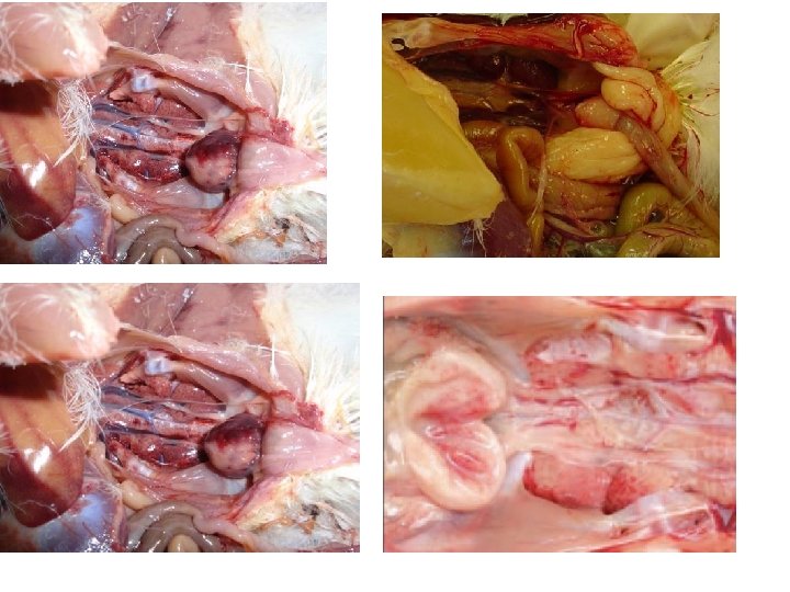
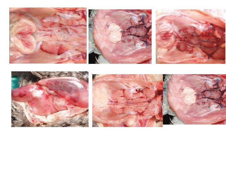
Post-mortem lesions • Oedematous bursa (may be slightly enlarged, normal size or reduced in size depending on the stage), may have haemorrhages, rapidly proceeds to atrophy. • Hemorrhage may be present in the thigh and pectoral muscles • Dehydration. • Swollen kidneys with urates.
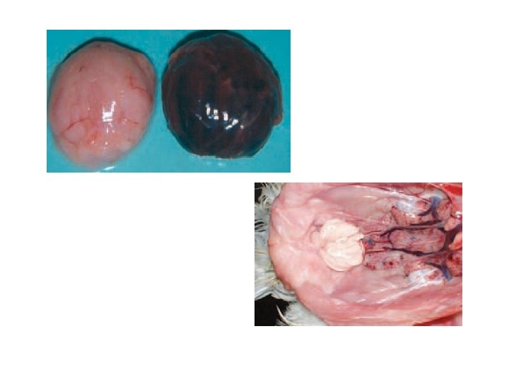

Diagnosis - flocks’ history, and of the clinical signs and lesions - Confirmation of a diagnosis of clinical IBD can be made at necropsy by examining the BF during the early stages of disease for characteristic gross lesions - A filtered homogenate of the bursa of Fabricius is inoculated 9 -11 day embryonated eggs originating from hens free of anti-IBDV antibodies.

• Isolation and identification of the agent provide the most certain diagnosis of IBD, and Identification of the agent by using - Identification by the agar gel immunodiffusion test - immunofluorescence , (AC-ELISA) and by molecular techniques

• Isolation of virus in cell culture or embryos Inoculate 0. 2 ml of sample into the yolk sac of five 6– 8 -day-old specific antibody negative (SAN) chicken embryos and on to the chorioallantoic membrane of five 9– 11 -dayold chicken embryos • Isolation of virus in chickens 2. Serological tests

• Agar gel immunodiffusion test or Enzymelinked immunosorbent assay

Prevention and Control of IBD An effective IBD prevention and control program must involve an • effective breeder vaccination program, • effective biosecurity program, and • effective broiler vaccination program

• Immunization of breeders is an important part of the IBD control program. Antibodies produced by the hen are passed through the egg to the broiler chick. These maternal antibodies, if present in adequate levels, protect the chicks against subclinical IBD.

• An example of a comprehensive breeder vaccination program where subclinical IBD is a problem might have a vaccine schedule such as this: at 12 to 15 days of age -- IBD live; at 30 to 33 days of age -- IBD live; at 85 days of age -- IBD live or inactivated; and at 120 days of age --IBD inactivated. Revaccinate at 38 to 42 weeks of age with an inactivated IBD vaccine if breeder titers are low or of poor uniformity. Routinely monitor breeder IBD antibody titers to ensure vaccines are administered properly and that the chickens respond appropriately.

• Effective control of IBD in commercial broilers requires that field virus exposure be reduced by proper clean-up and disinfection between flocks, and that traffic (people, equipment and vehicles) onto the farm be controlled. The development and enforcement of a comprehensive biosecurity program is the most important factor in limiting losses due to

• A third factor to consider in the IBD prevention and control program is vaccination of the broilers to prevent clinical IBD. • Three categories of vaccines, based on their pathogenicity, have been described: 1) mild, 2) intermediate, and 3) virulent. The intermediate type IBD vaccines are most commonly used. • The intermediate can stimulate the broiler to produce antibodies earlier than the mild-type vaccines, without significant damage to the BF as may occur with the virulent type vaccines

Nutritional Deficiencies A nutritional deficiency can arise simply due to: - • A nutrient being omitted from the diet, or • interaction between nutrients or between nutrients and antinutritional factors. • The composition of individual ingredients in a diet is variable; some nutrients are comparatively unstable,

• Stress due to bacterial, parasitic, or viral infections; high or low temperatures; low humidity; or drugs may either interfere with absorption of a nutrient or increase the quantity required.

Rickets or. Osteomalacia • Rickets are nutritional diseases seen in chickens, turkeys and ducks that results in soft bones, with the leg bones often becoming bowed and hampering the bird’s ability to stand walk

Causes • Deficiency or imbalance of circulating calcium, vitamin D 3 or phosphorous. • Imbalanced or deficient diet, some medications and also some mould toxins. • mycotoxins, can interfering with the absorption of nutrients.

Signs and Lesions: • Rickets most commonly occurs in young meat birds. • The primary pathologic change is inadequate bone mineralization • Young broilers and poults exhibit lameness, usually around 10 -14 days of age.

• Their bones are rubbery, and the rib cage is flattened and beaded at the attachment of the vertebrae • There is often an enlargement of the ends of the long bones, with a widening of the epiphyseal plate

Prevention and treatment • ensuring that poultry diets have an appropriate level and balance of calcium and phosphorous, they must be adequate in vitamin D 3.

• For normal bone calcification, calcium and phosphorous need to be supplied not only in adequate amounts but in a ratio to each other of about 2: 1. Excess of either calcium or phosphorous can cause rickets. Vitamin D 3 plays a critical role in regulating the absorption and metabolism of calcium.

• Rickets caused through the presence of dietary mycotoxins can be treated by replacing the toxin-contaminated feed and by supplementing vitamin D 3 to three or fourfold of the usual levels.

Cage Layer Fatigue • High-producing laying hens maintained in cages sometimes show paralysis around the time of peak egg production due to a fracture of the vertebrae that subsequently affects the spinal cord.

• The fracture is caused by an impaired calcium flux related to the high output of calcium in the eggshell. • Because medullary bone reserves become depleted, the bird uses cortical bone as a source of calcium for the eggshell. • The condition is rarely seen in floor-housed birds, suggesting that reduced activity or exercise is a predisposing factor.

Signs • Affected birds are invariably found on their sides in the back of the cage. • Death occurs from starvation or dehydration because the birds cannot reach feed or water.

Treatment: • A high incidence of cage layer fatigue can be prevented by ensuring the normal weight-forage of pullets at sexual maturity and by giving pullets a high-calcium diet (minimum 3. 5% calcium) for at least 14 days prior to first oviposition.

• Diets must provide adequate quantities of calcium and phosphorus to prevent deficiencies. • Affected birds will recover if moved to the floor.
 • Perosis, which occurs in young chicks, is characterized by enlargement")
Manganese Deficiency (perosis) • Perosis, which occurs in young chicks, is characterized by enlargement and malformation of the tibiometatarsal joint, twisting and bending of the distal end of the tibia and the proximal end of the tarsometatarsus, thickening and shortening of the leg bones, and slippage of the gastrocnemius or Achilles tendon from its condyles.

cause • Perosis or chondrodystrophy is encountered in young birds whose diet is deficient in manganese (Mn) or some of the following vitamins: choline, nicotic acid, pyridoxine, biotin or folic acid.
- Slides: 38