INFECTIOUS BURSAL DISEASE Dr SANJIV KUMAR ASSTT PROFESSOR

INFECTIOUS BURSAL DISEASE Dr. SANJIV KUMAR ASSTT. PROFESSOR, DEPTT. OF PATHOLOGY, BVC, BASU, PATNA

INTRODUCTION SYNONYMS: Infectious nephrosis Acute B- Gumboro disease, bursitis and Avian highly contagious infection of chickens Lymphocytes are the primary target cells Bursa, First lymphoid organ, is severely affected report in Gumboro (Delware District of USA) Economically significant, because heavy mortality in 3 – 6 wks old chickens and severe prolonged immunosuppression of chickens infected at an early age.
 - Birna = two Serotype 1 IBDV Variation")
Etiology Birna virus ( ds RNA) - Birna = two Serotype 1 IBDV Variation in virulence - from apathogenic to highly virulent strains Serotype 2 IBDV Non pathogenic or Immunosuppressive

Transmission Mainly oro faecal route but may be by conjuctiva or respiratory route Affected birds excrete the virus in faeces for 10 -14 days Virus is very stable Remains highly infectious for many months (up to 122 days) in the poultry environment Role of mechanical vectors (Human, wild birds, insects) No vertical transmission and carriers

PATHOGENESIS After ingestion, the virus multiplies in macropahges of small intestine (Duodenum, jejeunum and caecum). Thereafter, reaches liver (kupffer cells). Comes in circulation, reach Bursa (multiplies in B lymphocytes). Second massive viremia and finally destroys the lymphoid follicles in the bursa of Fabricius as well as the circulating B-cells in the secondary lymphoid tissues such as GALT (gut-associated lymphoid tissue), CALT (conjunctiva), BALT (Bronchial) caecal tonsils, Harderian gland, etc.
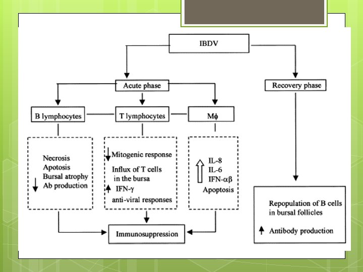
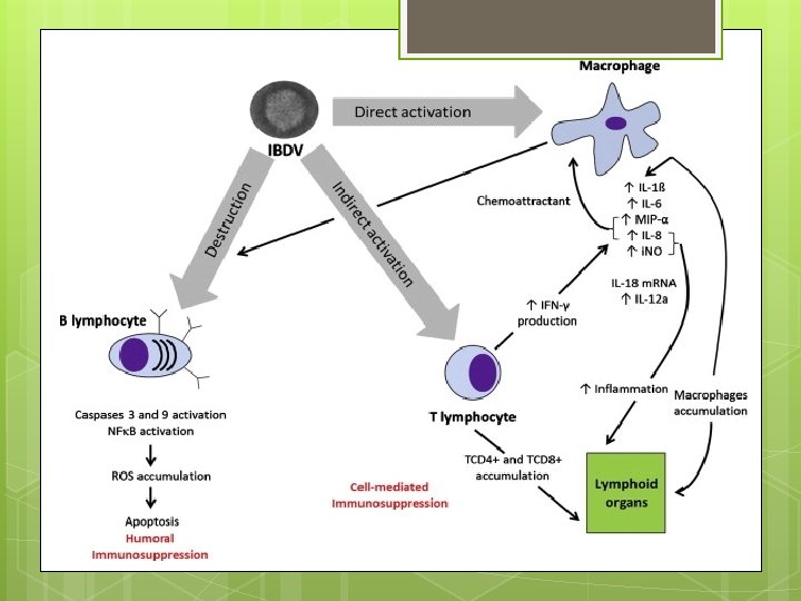

Susceptible age 3 -6 wks of age B - cells and their precursors are the main target cells T- Lymphocytes are relatively unaffected Renal pathology (swollen with urate deposits and cell debris) are due to severely swollen bursa Mechanism for muscular haemorrhage is not known (may be due to interference of virus with the normal blood clotting mechanism) Acute disease and death is due to the necrotizing effect of these viruses on the host tissues. Bursal infection in early life can result in impaired immune responses. Kidney failure is a common cause of mortality.

Consequences Lowered resistance to diseases Negative interference with effective vaccination. Subclinical infection in younger than 3 wks —> immunosuppression Chemical bursectomy (using cyclophosphamide) in 3 day old chicks and subsequent challenge at 4 wks —> Resistant to disease Similar results in surgically bursectamised 4 wks old chickens ( with mild necrosis of lymphatic tissue and 1000 times less viral production) but 100% mortality in control non-bursectamised birds

CLINICAL SIGNS Severity depends upon age, breed, and MDA level of the chick as well as the virulence of virus. Incubation period: 2 - 3 days. 3 -6 wks old chicks are affected. Depression White watery diarrhoea Soiled vent Anorexia Ruffled feathers Reluctance to move Closed eyes and death Morbidity - 10 – 100% Mortality: 0 - 20% (Normally), 90 – 100% (VVIBDV) Milder form - Little or No signs Suboptimal growth Course of the disease Short, leading to death or recovery (in individual bird) Mortality reaches a peak 3 -5 days after infection
, some times at the")
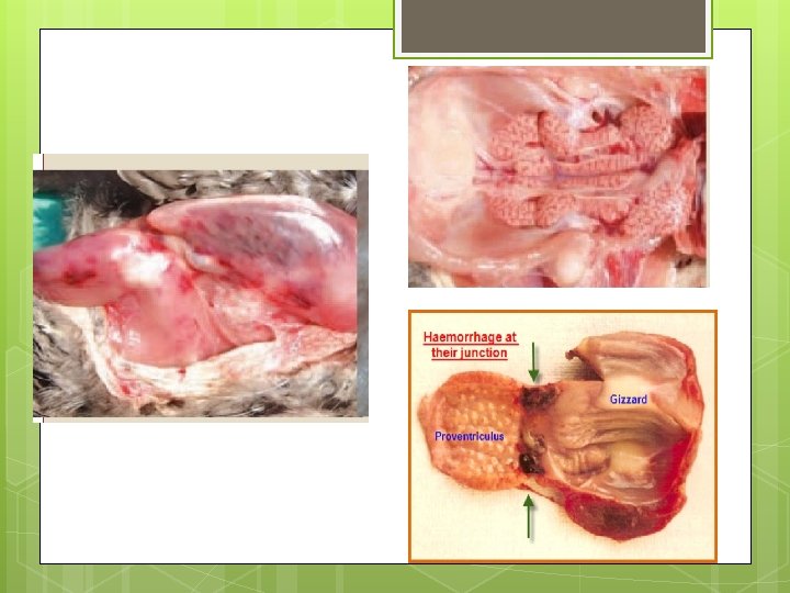
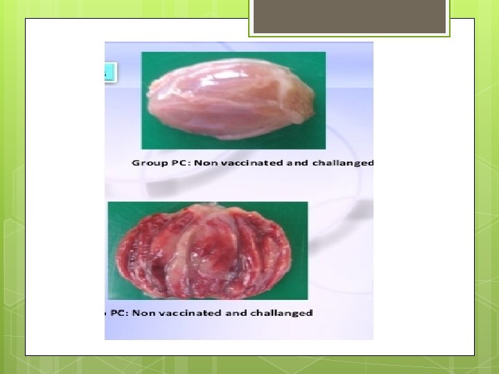
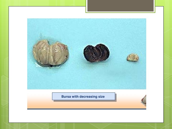
GROSS PATHOLOGY Dehydration of carcass Muscular haemorrhage (thigh and pectoral), some times at the junction of proventriculus and gizzard. Haemorrhages of pectoral leg muscles are typical of IBD Intestine Liver- with excess mucus Hepatomegaly and peripheral infarcts Spleen- Splenomegaly Kidneys- Swelling and white appearance, dilatation of tubules with urates ( cell debris, occasionally).
 Atrophy (after 3 – 8 days)")
Bursa Enlarged, inflamed, edematous and cream coloured (early) Atrophy (after 3 – 8 days) Haemorrhage on the internal and serosal surfaces Caseous core within the lumen from sloughed epithelium
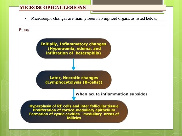

Spleen - Moderate lymphoid cell necrosis Thymus and caecal tonsil - Lymphoid cellular reaction (early stage), but less extensive damage Harderian Kidneys Liver gland - Depletion of plasma cells - Non – specific, degenerative changes - Mild perivascular infiltration of monocytes.

DIAGNOSIS Based on history, clinical signs and gross lesions Serological test AGPT (using macerated bursa) ELISA To identify the presence of antigen Immunoperoxidase staining Immunofluorescence (in frozen bursal sections or smears) Virus isolation (rarely) - Time consuming process Inoculation of suspected bursa into 10 – 11 days old embryonated eggs Some strains grow on Chick embryo fibroblast, vero cells or certain lymphoblastoid cell cultures Abs may develop after infection (detected by NT, ELISA, Precipitation test). It is useful when MDA declines below detectable levels) Nucleic acid probe, Ag-capture ELISA (using MCAbs. , ), RT-PCR

DIFFERENTIAL DIAGNOSIS Coccidiosis Ranikhet disease Haemorrhagic syndrome of muscles and other haemorrhages Avitaminosis A FLKS Water deprivation with swollen kidneys Excess renal urates.
- Slides: 19