
Infark Miokard Akut IMA Mohammad Saifur Rohman dr






- Slides: 119
Infark Miokard Akut (IMA) Mohammad Saifur Rohman, dr. Sp. JP, Ph. D. FICA
Outlines Anatomy of coronary artery l Coronary artery disease : atherosclerosis l Progression of CAD IMA l Diagnosing IMA l IMA Treatment l
Major components Muscle; contractile apparatus l Nerve; conducting system and innervation l Vessel; Supporting oxygen from the blood l Extra cellular matrix l Endocardium; valves l Pericardium l Etc. l
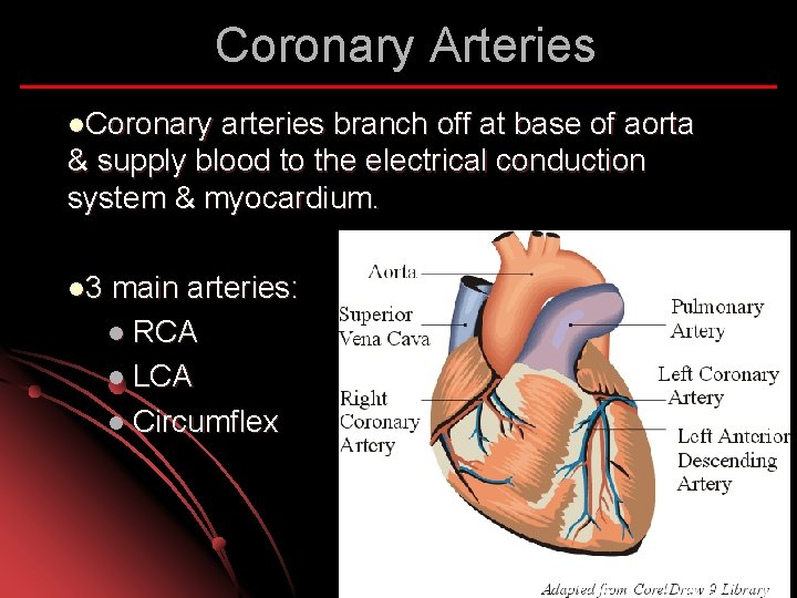
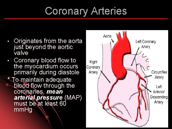
Coronary Arteries l. Coronary arteries branch off at base of aorta & supply blood to the electrical conduction system & myocardium. l 3 main arteries: l RCA l LCA l Circumflex
Coronary Arteries Originates from the aorta just beyond the aortic valve • Coronary blood flow to the myocardium occurs primarily during diastole * To maintain adequate blood flow through the coronaries, mean arterial pressure (MAP) must be at least 60 mm. Hg •
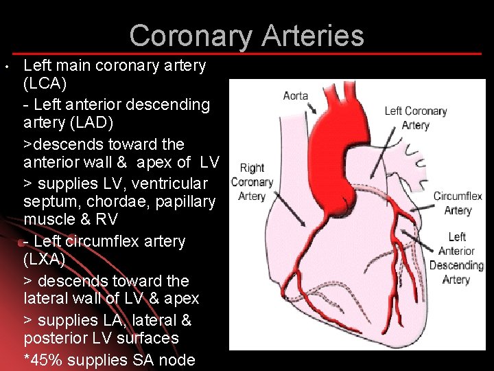
Coronary Arteries • Left main coronary artery (LCA) - Left anterior descending artery (LAD) >descends toward the anterior wall & apex of LV > supplies LV, ventricular septum, chordae, papillary muscle & RV - Left circumflex artery (LXA) > descends toward the lateral wall of LV & apex > supplies LA, lateral & posterior LV surfaces *45% supplies SA node
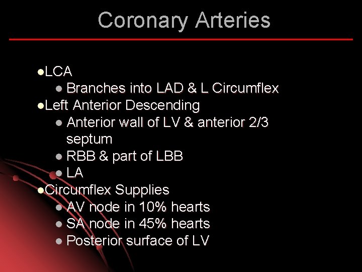
Coronary Arteries l. LCA l Branches into LAD & L Circumflex l. Left Anterior Descending l Anterior wall of LV & anterior 2/3 septum l RBB & part of LBB l LA l. Circumflex Supplies l AV node in 10% hearts l SA node in 45% hearts l Posterior surface of LV
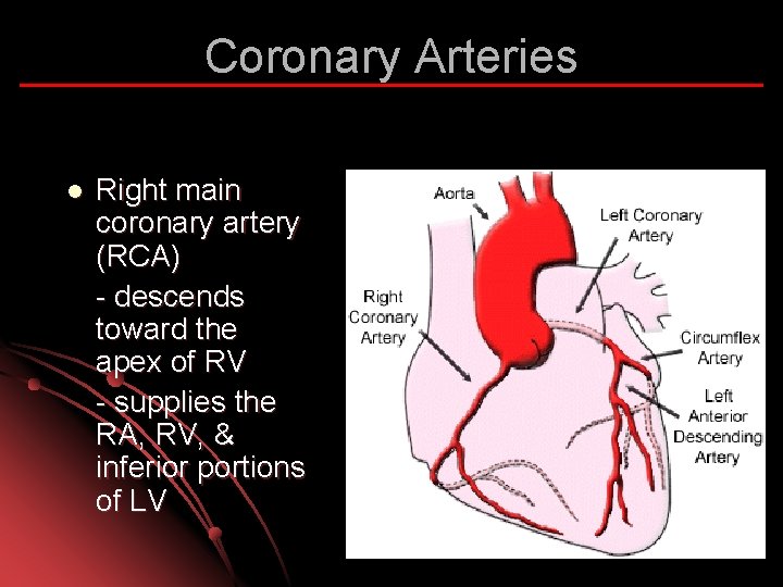
Coronary Arteries l Right main coronary artery (RCA) - descends toward the apex of RV - supplies the RA, RV, & inferior portions of LV
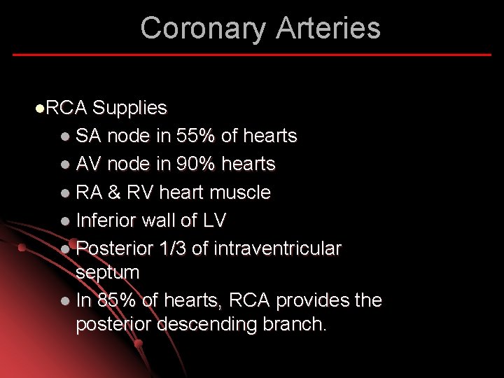
Coronary Arteries l. RCA Supplies l SA node in 55% of hearts l AV node in 90% hearts l RA & RV heart muscle l Inferior wall of LV l Posterior 1/3 of intraventricular septum l In 85% of hearts, RCA provides the posterior descending branch.
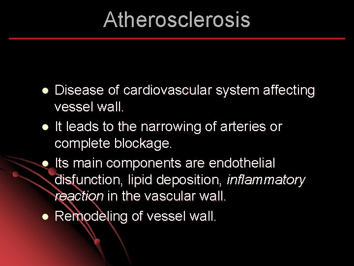
Atherosclerosis l l Disease of cardiovascular system affecting vessel wall. It leads to the narrowing of arteries or complete blockage. Its main components are endothelial disfunction, lipid deposition, inflammatory reaction in the vascular wall. Remodeling of vessel wall.
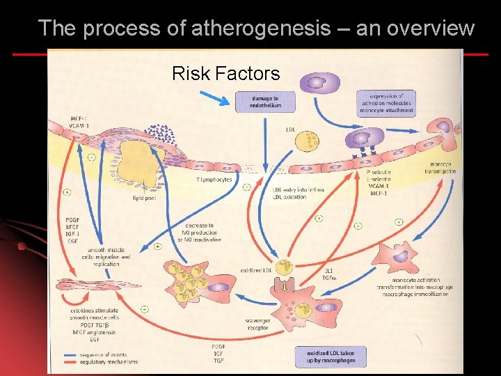
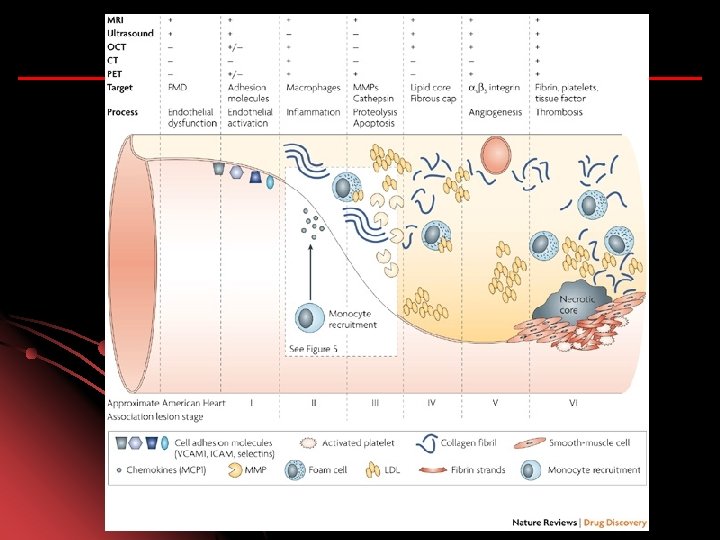
The process of atherogenesis – an overview Risk Factors
Smoking
Hypertension
Obesity
DIABETES
Family history: Man < 55 yo Women < 65 yo
CV Prevention
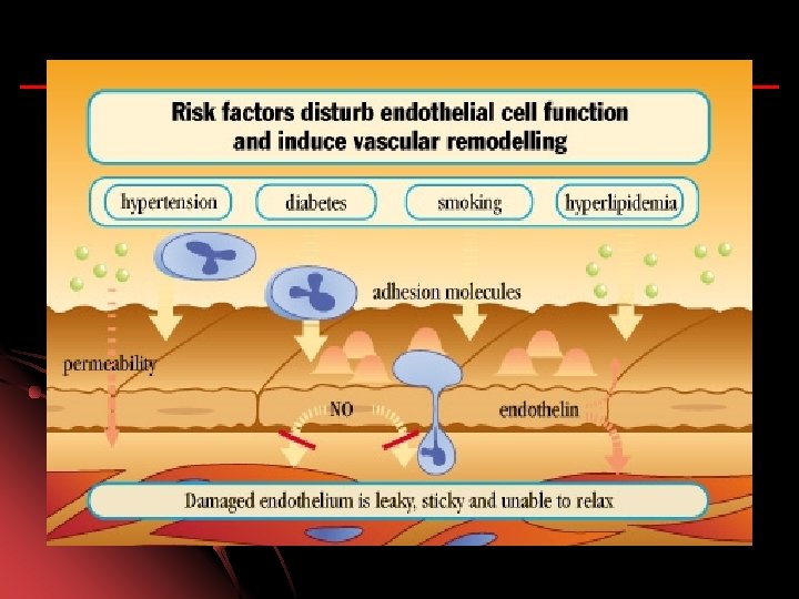
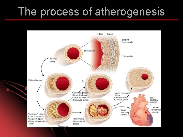
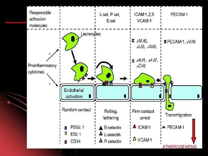
The development of atherosclerosis l l l The key event – damage to the endothelium caused by excess of lipoproteins, hypertension, diabetes, components of cigarette smoke. Endothelium becomes more permeable to lipoproteins. Lipoproteins move below the endothelial layer (to intima). Endothelium loses its cell-repelent quality. Inflammatory cells move into the vascular wall.
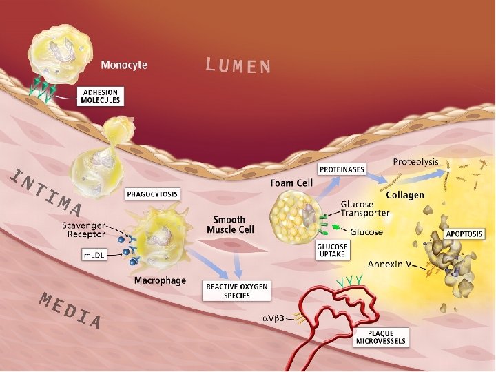
Triggers for inflammation in atherosclerosis l l l LDL retained in the intima (in part by binding to proteoglycan) undergoes oxidative modification. Lipid hydroperoxides, lysophospholipides, carbonyl compounds localize in the lipid fraction. Oxygen free-radicals inactivate NO rapidly. NO + superoxide (O 2. -) peroxynitrite (ONOO-). NO has no longer vasoprotective function.
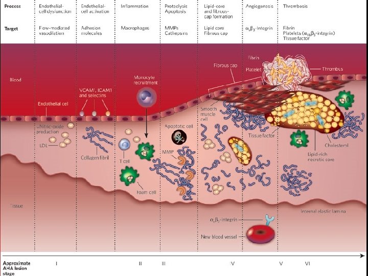
The process of atherogenesis l l l Lipid entry into the arterial wall is a key process in atherogenesis. Hypercholesterolemia – factor for VCAM-1 and MCP-1 induction. LDL and VLDL are most atherogenic, enter vascular wall more easily. LDL – in plasma are protected against oxidation by vit. E, ubiquinon, plasma antioxidants (b-carotene, vit. C). Out of plasma, LDL phospholipides and fatty acids oxidize.
The process of atherogenesis l l l Activated macrophages produce enzymes – lipoxygenases, myeloperoxidase, NADPH oxidase ROS Oxidized LDL are cytotoxic to endothelial cells, mitogenic for macrophages. Oxidized LDL apolipoprotein apo. B 100 bind to the scavenger receptor. Scavenger receptors are not subjected to regulation by intracellular cholesterol level. Macrophages take up oxidized LDL, overload with lipids.
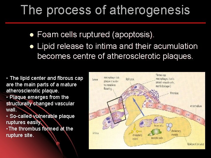
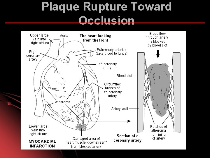
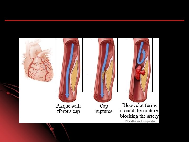
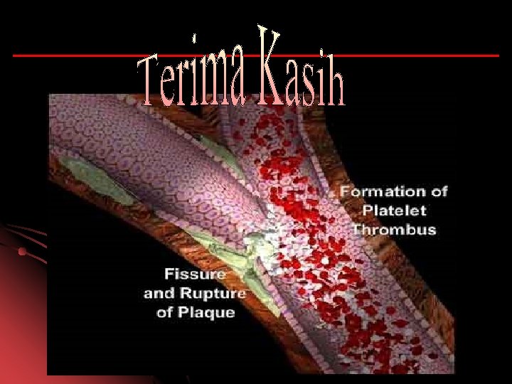
The process of atherogenesis l l Foam cells ruptured (apoptosis). Lipid release to intima and their acumulation becomes centre of atherosclerotic plaques. • The lipid center and fibrous cap are the main parts of a mature atherosclerotic plaque. • Plaque emerges from the structurally changed vascular wall. • So-called vulnerable plaque ruptures easily. • The thrombus formed at the rupture site.
The process of atherogenesis
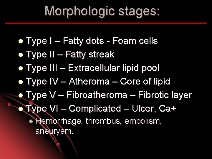
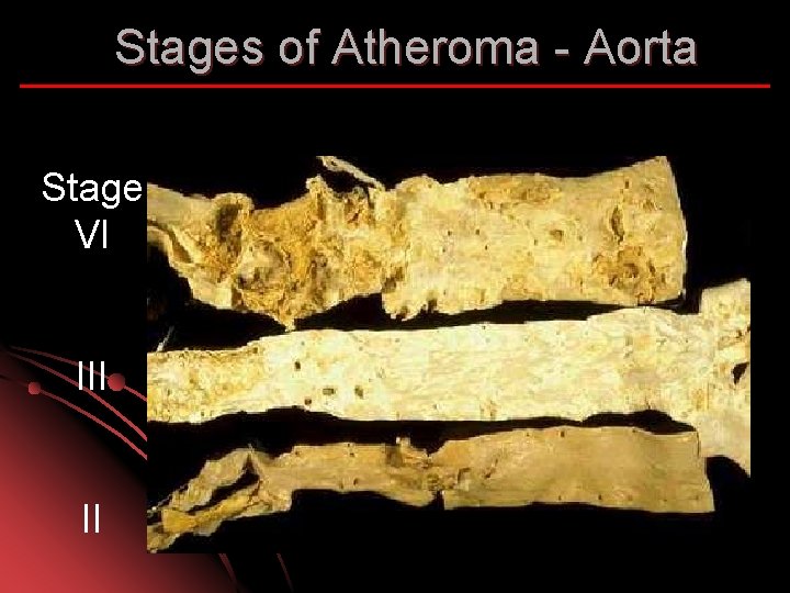
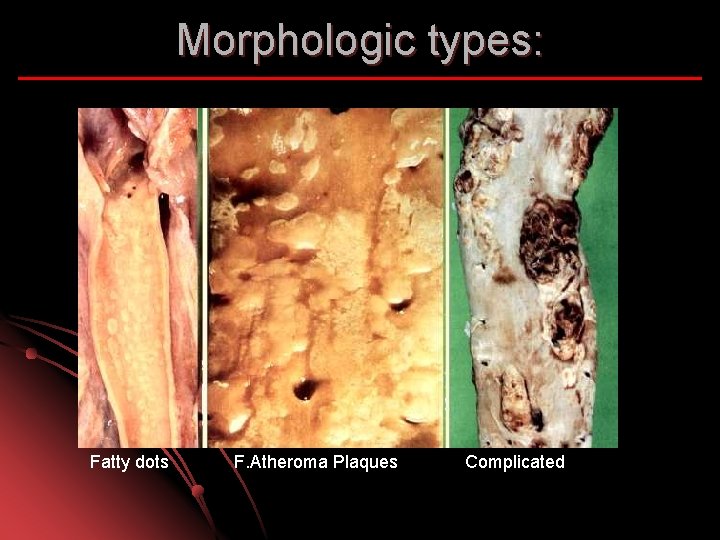
Morphologic stages: Type I – Fatty dots - Foam cells l Type II – Fatty streak l Type III – Extracellular lipid pool l Type IV – Atheroma – Core of lipid l Type V – Fibroatheroma – Fibrotic layer l Type VI – Complicated – Ulcer, Ca+ l l Hemorrhage, aneurysm. thrombus, embolism,
Stages of Atheroma - Aorta Stage VI II
Morphologic types: Fatty dots F. Atheroma Plaques Complicated
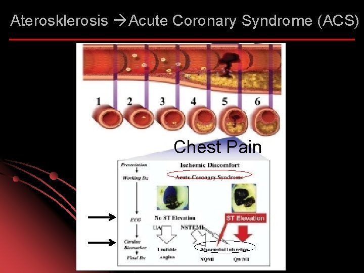
Aterosklerosis Acute Coronary Syndrome (ACS) Chest Pain
Plaque Rupture Toward Occlusion
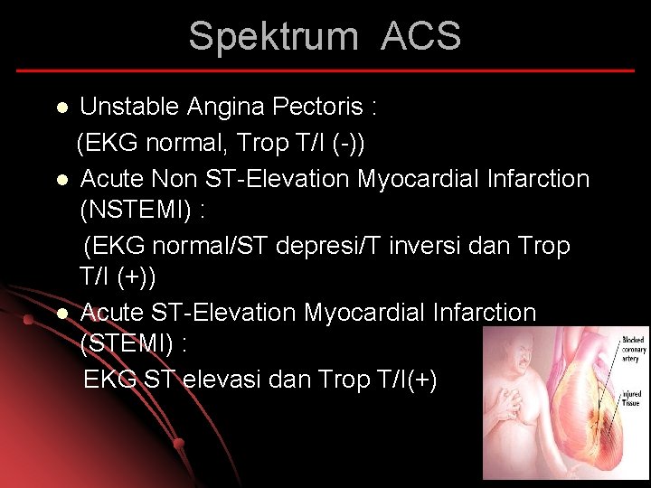
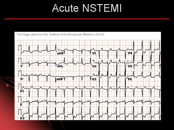
Spektrum ACS Unstable Angina Pectoris : (EKG normal, Trop T/I (-)) l Acute Non ST-Elevation Myocardial Infarction (NSTEMI) : (EKG normal/ST depresi/T inversi dan Trop T/I (+)) l Acute ST-Elevation Myocardial Infarction (STEMI) : EKG ST elevasi dan Trop T/I(+) l
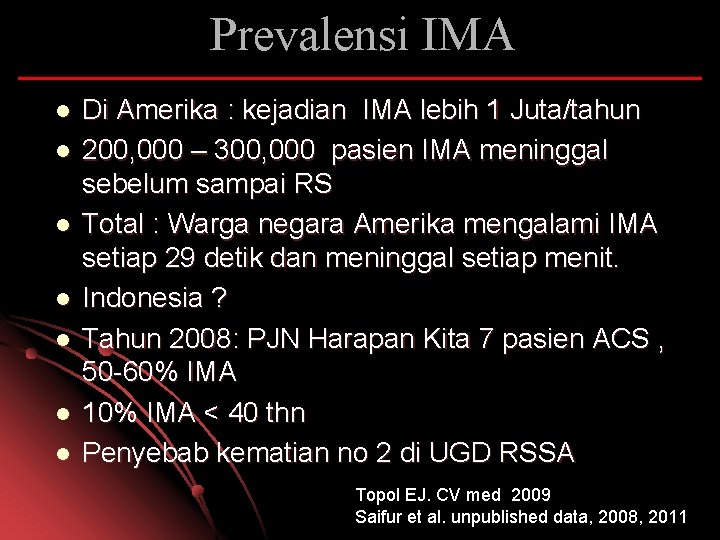
Prevalensi IMA l l l l Di Amerika : kejadian IMA lebih 1 Juta/tahun 200, 000 – 300, 000 pasien IMA meninggal sebelum sampai RS Total : Warga negara Amerika mengalami IMA setiap 29 detik dan meninggal setiap menit. Indonesia ? Tahun 2008: PJN Harapan Kita 7 pasien ACS , 50 -60% IMA 10% IMA < 40 thn Penyebab kematian no 2 di UGD RSSA Topol EJ. CV med 2009 Saifur et al. unpublished data, 2008, 2011
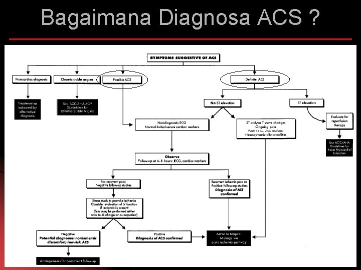
Bagaimana Diagnosa ACS ?
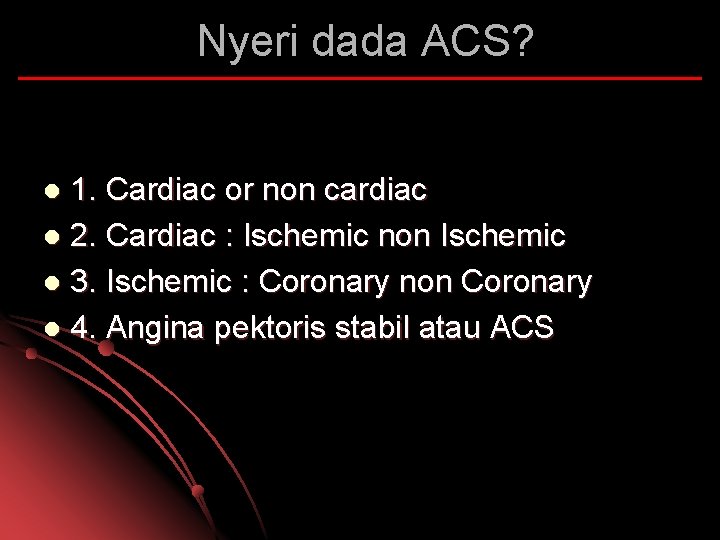
Nyeri dada ACS? 1. Cardiac or non cardiac l 2. Cardiac : Ischemic non Ischemic l 3. Ischemic : Coronary non Coronary l 4. Angina pektoris stabil atau ACS l
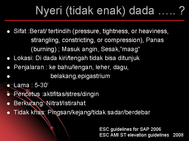
Nyeri (tidak enak) dada …. . ? l l l l Sifat : Berat/ tertindih (pressure, tightness, or heaviness, strangling, constricting, or compression), Panas (burning) ; Masuk angin, Sesak, ”maag” Lokasi: Di dada kiri/tengah tidak bisa ditunjuk Penjalaran : ke bahu/lengan, leher, dagu, belakang, epigastrium Lama : 5 -30’ Pencetus : aktifitas/stres/dingin Berkurang: Nitrat/Istirahat Tidak khas: Pingsan/kejang/tidak sadar/berdebar ESC guidelines for SAP 2006 ESC AMI ST elevation guidelines 2008
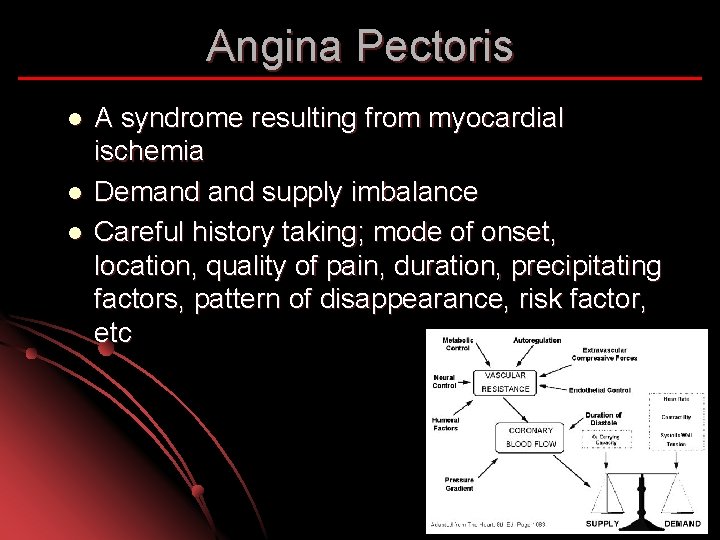
Angina Pectoris l l l A syndrome resulting from myocardial ischemia Demand supply imbalance Careful history taking; mode of onset, location, quality of pain, duration, precipitating factors, pattern of disappearance, risk factor, etc
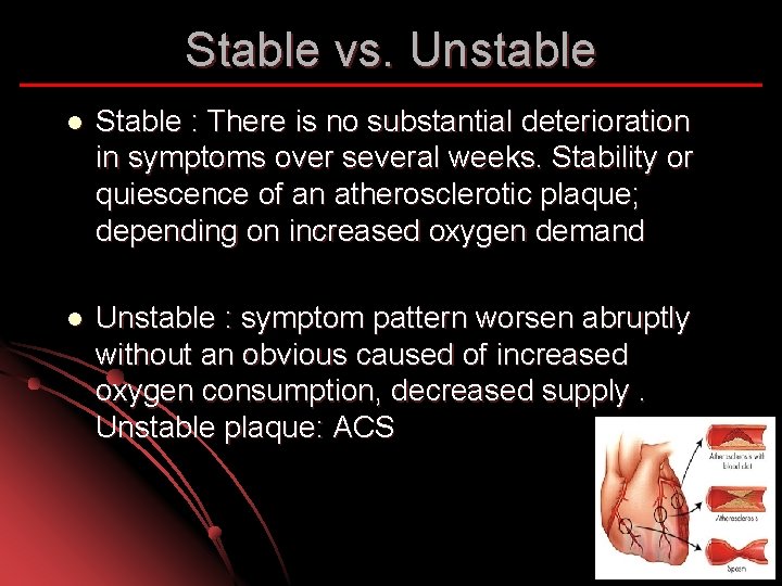
Stable vs. Unstable l Stable : There is no substantial deterioration in symptoms over several weeks. Stability or quiescence of an atherosclerotic plaque; depending on increased oxygen demand l Unstable : symptom pattern worsen abruptly without an obvious caused of increased oxygen consumption, decreased supply. Unstable plaque: ACS
Stable Angina Pectoris Canadian Class : l I Ordinary physical activity does not cause angina l II Slight limitation of ordinary activity l III Marked limitation of ordinary activity l IV Inability to carry on any activity without discomfort
Nyeri dada khas ACS Angina awitan baru derajat 3 menurut klasifikasi kanada kardiovaskuler group ( nyeri dada timbul pada aktifitas ringan sehari-hari) l Angina saat istirahat > 20 menit l Perburukan derajat angina menjadi derajat 3 dalam beberapa hari – 1 bln terakhir (Crescendo angina) l Atypical l
Hati-hati : Angina Equivalent Indigestion, belching, dyspnea l DM, wanita, manula (post operative) l Didapatkan 5% dari ACS l 2% dipulangkan ternyata ACS l Braunwalds Heart Disease 8 th Ed 2008
DD Chest pain Ischemic l Stenosis Aorta l Regurgitasi Aorta l Hypertrophic Cardiomyopathy l Angina pada Hypertensi l Hipertensi pulmonal berat 11 th ed Hurst’s the heart 2005
DD Chest Pain Non Ischemic l Diseksi Aorta l Pericarditis l Mitral valve prolaps 11 th ed Hurst’s the heart 2005
DD Chest Pain Gastro intestinal l Esophageal spasm/reflux/rupture l Peptic Ulcer Neuromusculoskeletal l Costochondritis l Herpes zoster l Chest wall pain dan tenderness etc 11 th ed Hurst’s the heart 2005
DD chest pain Pulmonary l Pulmonary emboli l Pneumothorax l Penumonia with pleural involvement Pleurisy Psychogenic l Axiety/depression/cardiac psychosis etc 11 th ed Hurst’s the heart 2005
Non Angina Pain Hanya terasa pada sebagian kecil dada kiri/kanan (bisa di tunjuk) l Berkahir berjam sampai berhari. l Biasanya tidak berkurang dengan nitrogliserin l Mungkin dicetuskan oleh debaran. l ESC guidelines for SAP 2006 ESC AMI ST elevation guidelines 2008
EKG Secepat mungkin – 10’ setelah pasien tiba l Diulang apabila meragukan adanya kenaikan segmen ST (ST televasi) l Bandingkan denga EKG sebelumnya l Pasang monitor EKG l
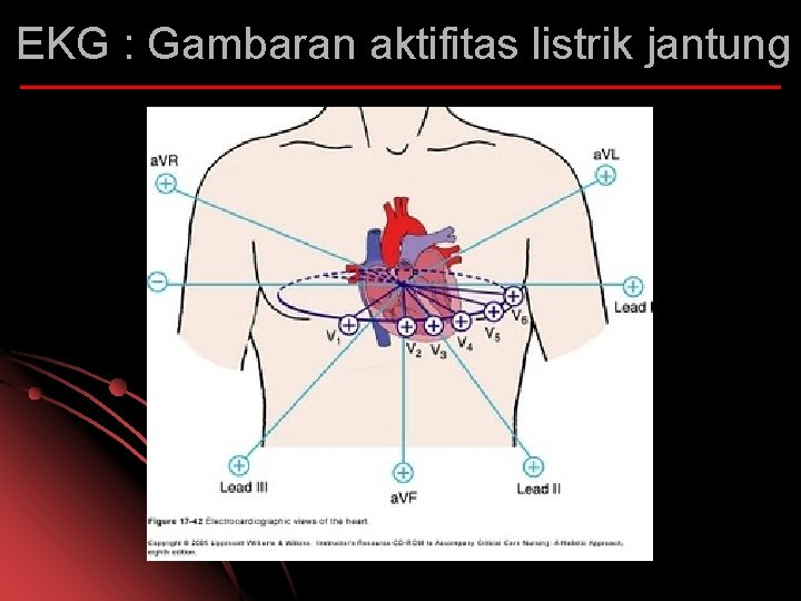
EKG : Gambaran aktifitas listrik jantung
EKG pada ACS EKG dapat menentukan adanya: l Old/Recent/Acute infarction l Pericarditis l Arrhythmias l Pembesaran jantung
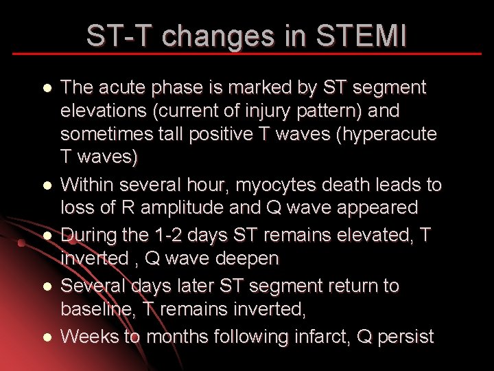
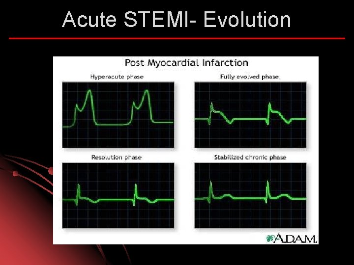
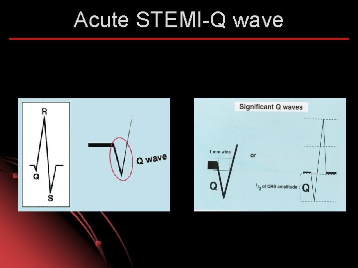
ST-T changes in STEMI l l l The acute phase is marked by ST segment elevations (current of injury pattern) and sometimes tall positive T waves (hyperacute T waves) Within several hour, myocytes death leads to loss of R amplitude and Q wave appeared During the 1 -2 days ST remains elevated, T inverted , Q wave deepen Several days later ST segment return to baseline, T remains inverted, Weeks to months following infarct, Q persist
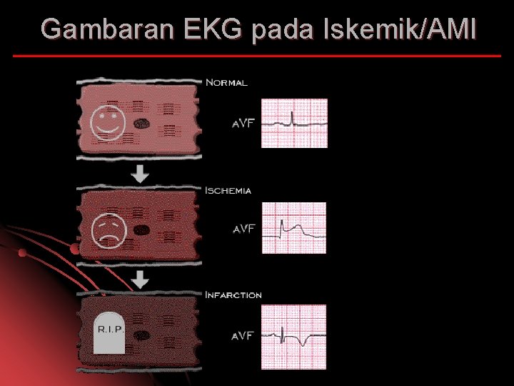
Gambaran EKG pada Iskemik/AMI
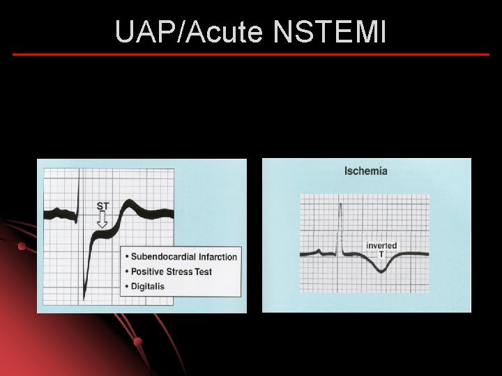
UAP/Acute NSTEMI
Acute NSTEMI
Acute STEMI- Evolution
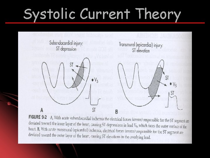
Systolic Current Theory
Diastolic current Theory Leaky K+ l Never fully repolarize l Relatively more negative than normal l Current directed away from infarct area l Baseline shift downward l
Acute STEMI-Q wave
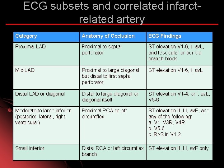
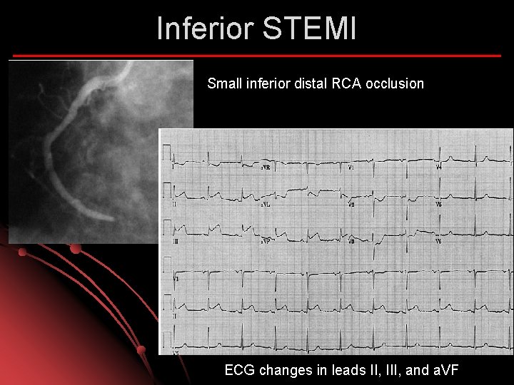
ECG subsets and correlated infarctrelated artery Category Anatomy of Occlusion ECG Findings Proximal LAD Proximal to septal perforator ST elevation V 1 -6, I, av. L, and fascicular or bundle branch block Mid LAD Proximal to large diagonal but distal to first septal perforator ST elevation V 1 -6, I, av. L Distal LAD or diagonal Distal to large diagonal or diagonal itself ST elevation V 1 -4, or I, av. L, V 5 -6 Moderate to large inferior (posterior, lateral, right ventricular) Proximal RCA or left circumflex ST elevation II, III, av. F, and any of the following: a. V 1, V 3 R, V 4 R b. V 5 -6 c. R>S in V 1 -2 Small inferior Distal RCA or left circumflex ST elevation II, III, av. F only branch
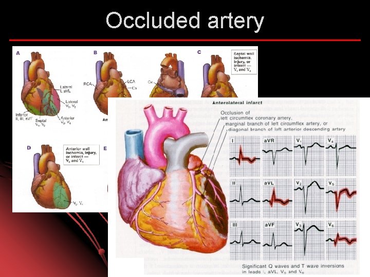
Occluded artery
Anterior STEMI ECG demonstrates large anterior infarction
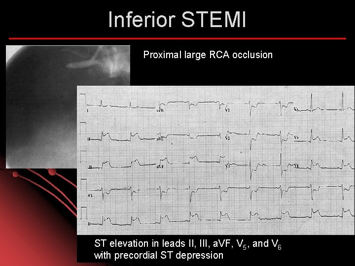
Inferior STEMI Proximal large RCA occlusion ST elevation in leads II, III, a. VF, V 5, and V 6 with precordial ST depression
Inferior STEMI Small inferior distal RCA occlusion ECG changes in leads II, III, and a. VF
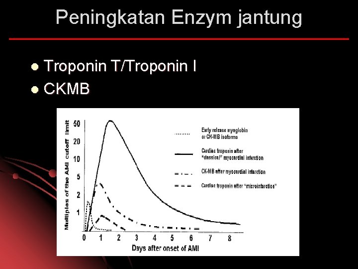
Peningkatan Enzym jantung Troponin T/Troponin I l CKMB l
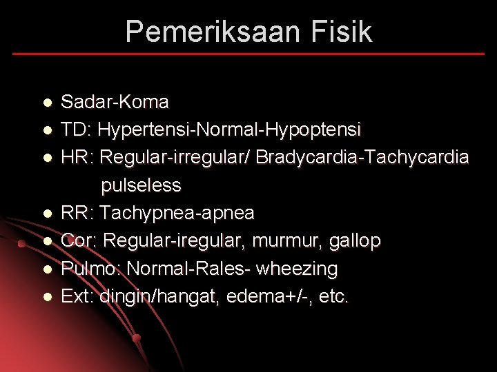
Pemeriksaan Fisik l l l l Sadar-Koma TD: Hypertensi-Normal-Hypoptensi HR: Regular-irregular/ Bradycardia-Tachycardia pulseless RR: Tachypnea-apnea Cor: Regular-iregular, murmur, gallop Pulmo: Normal-Rales- wheezing Ext: dingin/hangat, edema+/-, etc.
Komplikasi MI Mechanical l Electrical l Ischemia l Embolic l Inflammation l
Komplikasi Mekanik Ventricular Septal Rupture l Mitral Regurgitation l Cardiac free wall rupture l Large ventricular aneurysms l LV pump failure and cardiogenic shock l Dynamic LVOT obstruction l RV failure l
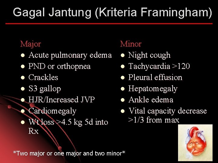
Gagal Jantung (Kriteria Framingham) Major Minor l Acute pulmonary edema l Night cough l PND or orthopnea l Tachycardia >120 l Crackles l Pleural effusion l S 3 gallop l Hepatomegaly l HJR/Increased JVP l Ankle edema l Cardiomegaly l Vital capacity decrease >1/3 from max l Wt loss >4. 5 kg 5 d into Rx *Two major or one major and two minor*
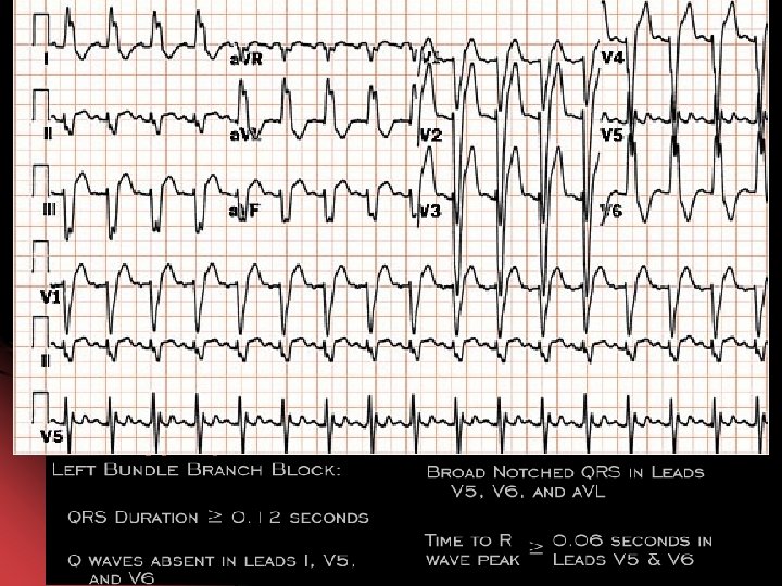
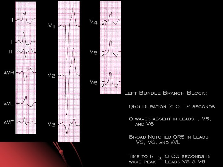
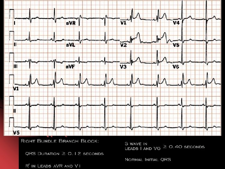
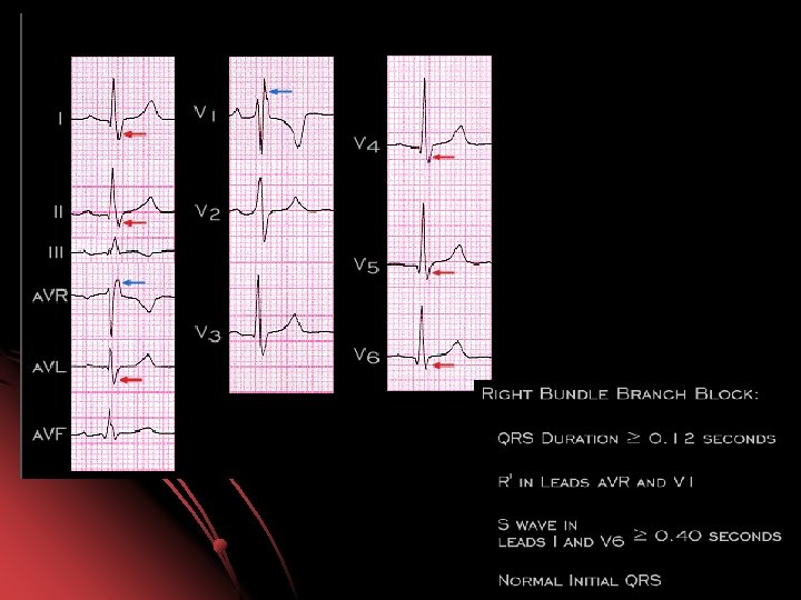
Komplikasi Elektrik SA Dysfunction l Atrial Fibrillation l First-Second degree AV block l Total AV Block l Left Bundle Branch Block l Right Bundle Branch Block l Ventricular Tachycardia l Ventricular Fibrillation l
Komplikasi Ischemik Perluasan Infark l Angina Post-infark l
Komplikasi Emboli l Systemic embolism ; stroke, limb ischemia, renal infarction, intestinal ischemia
Inflammation Complication Early Pericarditis l Late Pericarditis (Dresslers syndrome) l
Universal Definition of Myocardial Infarction Diagnosa AMI ditegakkan apabila min memenuhi 2 dari kriteria: l Gejala Ischemic l Perubahan EKG l Kenaikan/penurunan Troponin T/I
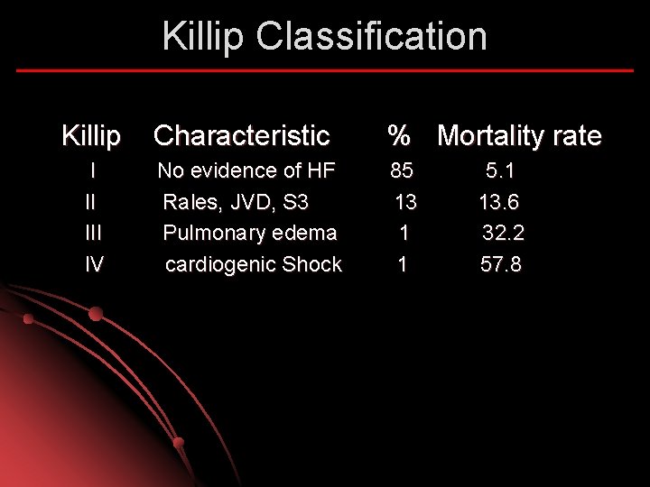
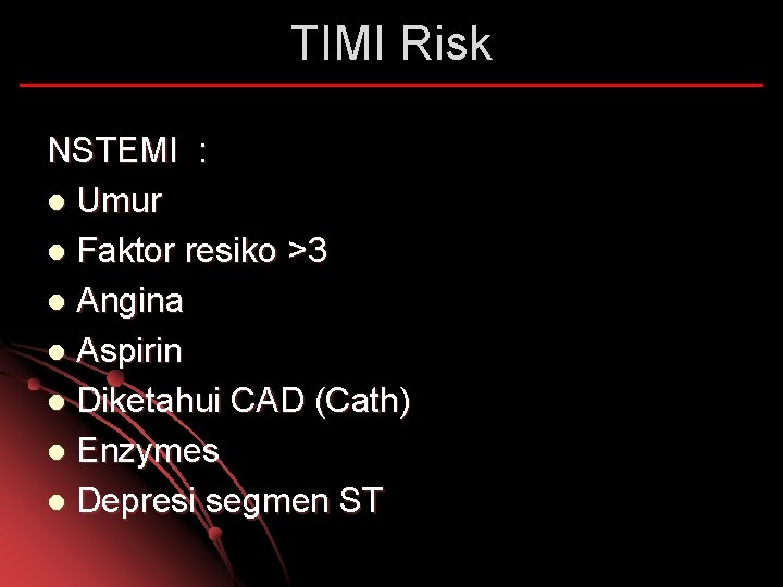
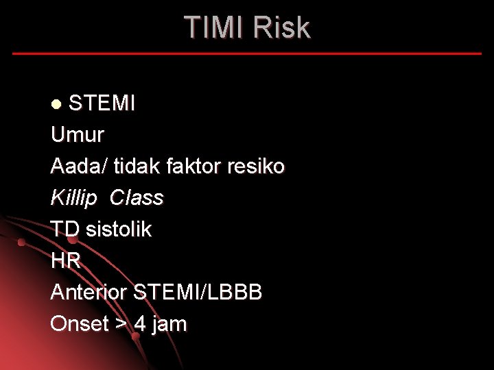
Diagnosis NSTEMI timi risk. . /7…. l Acute ant STEMI timi risk… /14 Killip… onset j…am l Cepat tegakkan diagnosa dan cepat bertindak (dalam hitungan menit) l Semakin tinggi resiko semakin agresif terapi yang diberikan l
Killip Classification Killip I II IV Characteristic % Mortality rate No evidence of HF Rales, JVD, S 3 Pulmonary edema cardiogenic Shock 85 13 1 1 5. 1 13. 6 32. 2 57. 8
TIMI Risk NSTEMI : l Umur l Faktor resiko >3 l Angina l Aspirin l Diketahui CAD (Cath) l Enzymes l Depresi segmen ST
TIMI Risk STEMI Umur Aada/ tidak faktor resiko Killip Class TD sistolik HR Anterior STEMI/LBBB Onset > 4 jam l
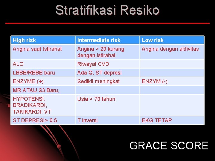
Stratifikasi Resiko High risk Intermediate risk Low risk Angina saat Istirahat Angina > 20 kurang dengan istirahat Angina dengan aktivitas ALO Riwayat CVD LBBB/RBBB baru Ada Q, ST depresi ENZYME (+) Sedikit meningkat ENZYM (-) MR ATAU S 3 Baru, HYPOTENSI, BRADIKARDI, TAKIKARDI. VT Usia > 70 tahun ST DEPRESI> 0. 5 T inversi EKG TETAP GRACE SCORE
Prinsip Terapi Cepat (time responsif), obati penyebab buka sumbatan l Terlambat: Fatal l Monitor ketat tanda vital sejak awal l Antisipasi dini tanda perburukan dan komplikasi l
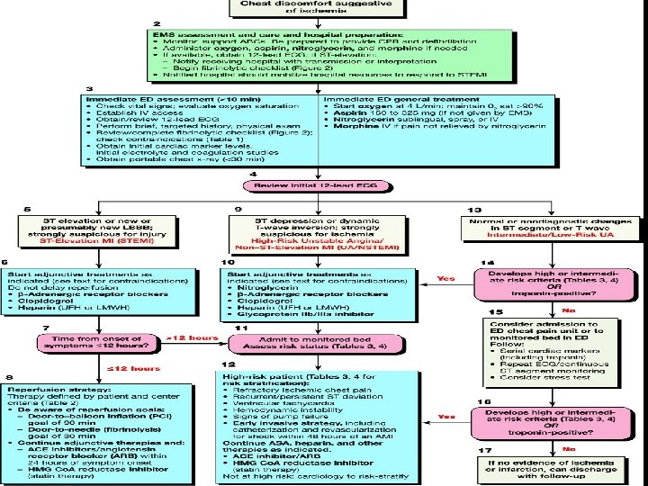
Terapi Awal ACS Atasi keadaan kegawat daruratan : asistol, apneu, syock, lung edema, gagal jantung dll. l Terapi reperfusi : PCI, Fibrinolitik, heparin l Antiischemic l Turunkan oksigen demand : Bed rest total, pendekatan psikologis, dll l Terapi komorbid; hipertensi, DM, dll l
Pentingnya Reperfusi Sumbatan total 15 -30 menit tanpa kolateral IMA l Reperfusion selamatkan miorkard l Kematian 1 bulan: 25 -30% 4 -6% dengan reperfusi (PCI, fibrinolytic, antithombotic) l ESC AMI ST elevation guidelines 2008
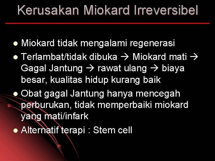
Kerusakan Miokard Irreversibel Miokard tidak mengalami regenerasi l Terlambat/tidak dibuka Miokard mati Gagal Jantung rawat ulang biaya besar, kualitas hidup kurang baik l Obat gagal Jantung hanya mencegah perburukan, tidak memperbaiki miokard yang mati/infark l Alternatif terapi : Stem cell l
The time is muscle
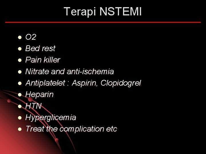
Terapi NSTEMI l l l l l O 2 Bed rest Pain killer Nitrate and anti-ischemia Antiplatelet : Aspirin, Clopidogrel Heparin HTN Hyperglicemia Treat the complication etc
Tips l l l Obat anti ischemik atau anti nyeri segera di berikan Anti platelet dan heparin dimasukkan secepatnya setelah diagnosis ACS-NSTEMI ditegakkan, jangan di tunda Turunkan kebutuhan/kerja jantung dengan berikan rasa nyaman dan aman pasien dan bed rest total Setengah duduk pada pasien dengan gagal jantung Pikirkan immediate/urgent PCI pada pasien resiko tinggi/hemodinamik tidak stabil/nyeri berkepanjangn/aritmia maligna dll
Alat diagnosis dan monitoring Perawat l l Keluhan berhubungan dengan fungsi dan beban jantung, deteksi dini komplikasi Monitor tanda vital, saturasi, perfusi, EKG, intake - output, balance. dll Kalau perlu ukur CVP, arteri line dll Laboratorium : Enzyme jantung, analisa gas darah, fungsi ginjal, elektrolit, komorbid (infeksi, dm, dll)
Intervensi keperawatan l l Turunkan kebutuhan dan bebang jantung misalnya istirahat fisik dan mental, kondisi hangat, tenang, rasa aman, pemilihan diet : NGT atau bantuan makanan, dilarang mengejan, atasi febris, rasa sakit atau sesak dll Bantu fungsi jantung: Inotropic, IABP, anti iskemik, dll Atasi komorbid/komplikasi: hipertensi, DM, infeksi, gagal jantung, gangguan ginjal, dll Atasi kekurangan atau kelebihan cairan, kalori, oksigen, PH, elektrolit, dll
Terapi STEMI l l l l l O 2 Bed rest Pain killer Nitrate and anti-ischemia Antiplatelet : Aspirin, Clopidogrel Fibrinolytic time to neddle : 30 m/PCI HTN Hyperglicemia Treat the complication etc
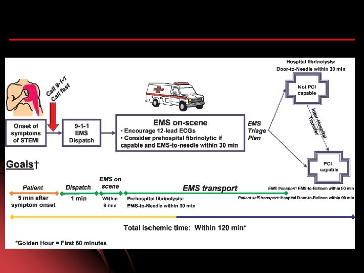
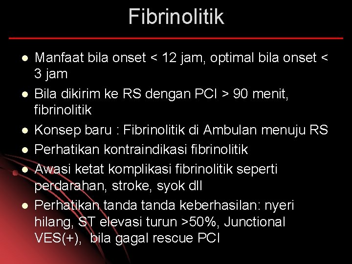
Fibrinolitik l l l Manfaat bila onset < 12 jam, optimal bila onset < 3 jam Bila dikirim ke RS dengan PCI > 90 menit, fibrinolitik Konsep baru : Fibrinolitik di Ambulan menuju RS Perhatikan kontraindikasi fibrinolitik Awasi ketat komplikasi fibrinolitik seperti perdarahan, stroke, syok dll Perhatikan tanda keberhasilan: nyeri hilang, ST elevasi turun >50%, Junctional VES(+), bila gagal rescue PCI
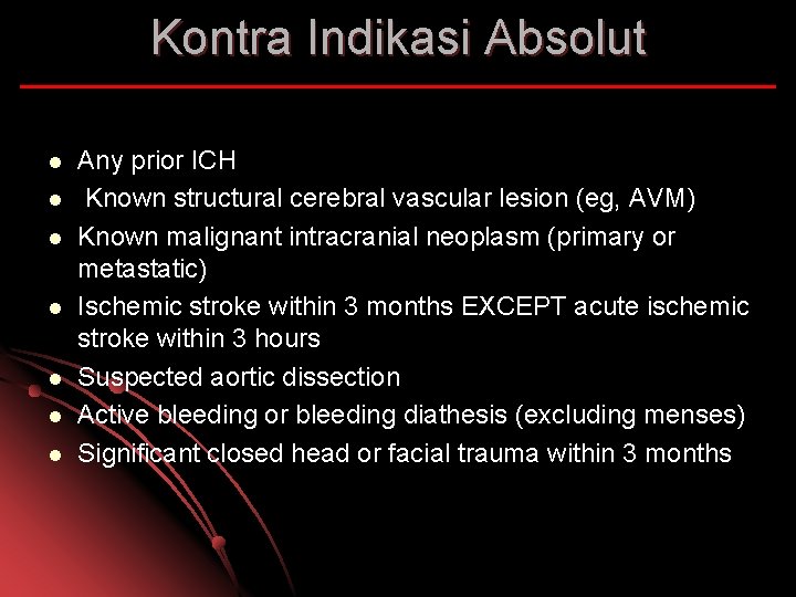
Kontra Indikasi Absolut l l l l Any prior ICH Known structural cerebral vascular lesion (eg, AVM) Known malignant intracranial neoplasm (primary or metastatic) Ischemic stroke within 3 months EXCEPT acute ischemic stroke within 3 hours Suspected aortic dissection Active bleeding or bleeding diathesis (excluding menses) Significant closed head or facial trauma within 3 months
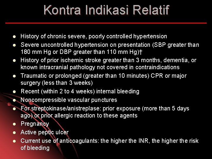
Kontra Indikasi Relatif l l l l l History of chronic severe, poorly controlled hypertension Severe uncontrolled hypertension on presentation (SBP greater than 180 mm Hg or DBP greater than 110 mm Hg)† History of prior ischemic stroke greater than 3 months, dementia, or known intracranial pathology not covered in contraindications Traumatic or prolonged (greater than 10 minutes) CPR or major surgery (less than 3 weeks) Recent (within 2 to 4 weeks) internal bleeding Noncompressible vascular punctures For streptokinase/anistreplase: prior exposure (more than 5 days ago) or prior allergic reaction to these agents Pregnancy Active peptic ulcer Current use of anticoagulants: the higher the INR, the higher the risk of bleeding
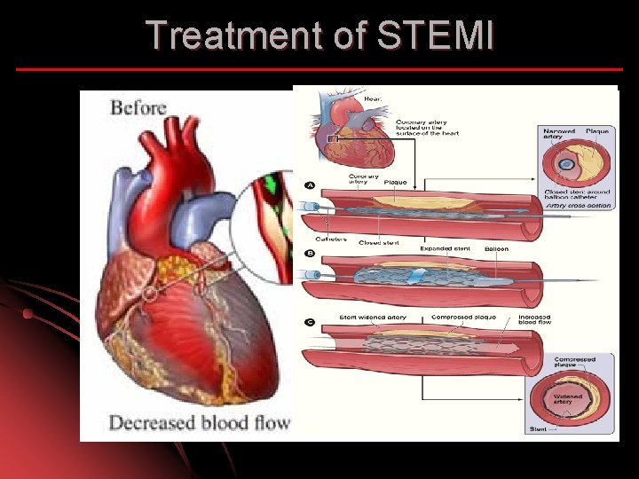
Treatment of STEMI
Percutaneous Coronary Intervention • Primary PCI : Pasien langsung di lakukan tindakan reperfusi dengan membuka sumbatan di arteri koroner tanpa dilakukan fibrinilotik terlebih dahulu • Rescue PCI : Dilakukan PCI setelah gagal dengan terapi fibrinolitik • Facilitated PCI : Pasien dilakukan fibrinolitik terlebih dahulu meskipun sudah ada rencana PCI • Urgent PCI: As soon as possible • Early PCI : Dalam waktu 24 jam pertama Early/urgent PCI: Resiko tinggi, hemodinamik tidak stabil, aritmia maligna, angina (+) dgn terapi, EF <40%, Gagal jantung, Riwayat PCI, CABG dl 6 bl
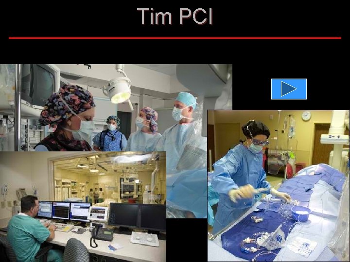
Tim PCI
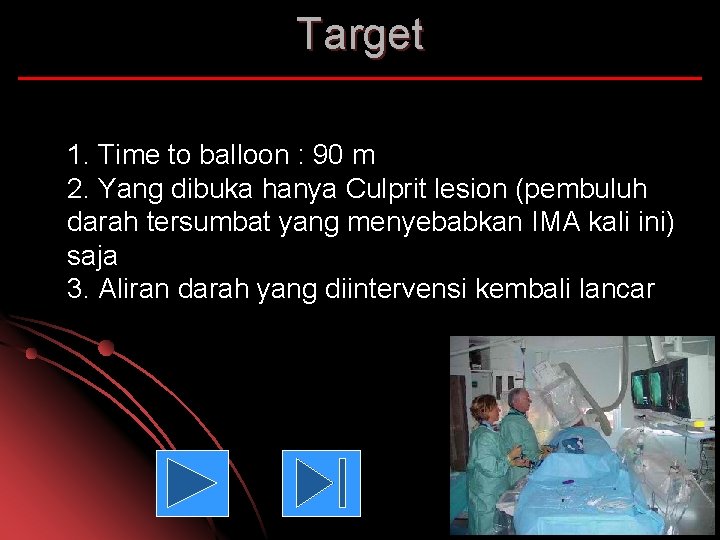
Target 1. Time to balloon : 90 m 2. Yang dibuka hanya Culprit lesion (pembuluh darah tersumbat yang menyebabkan IMA kali ini) saja 3. Aliran darah yang diintervensi kembali lancar
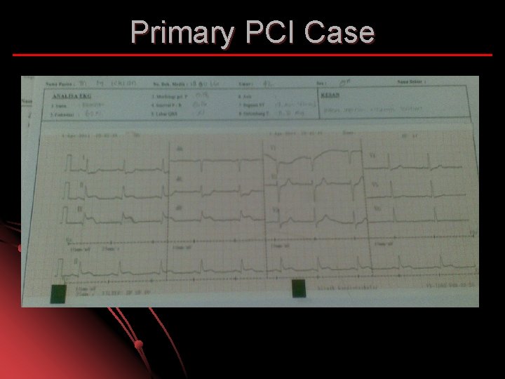
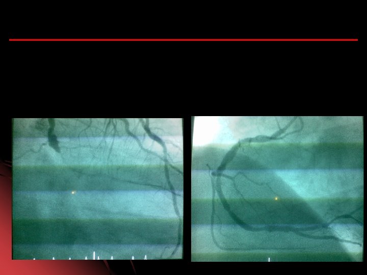
Primary PCI Case
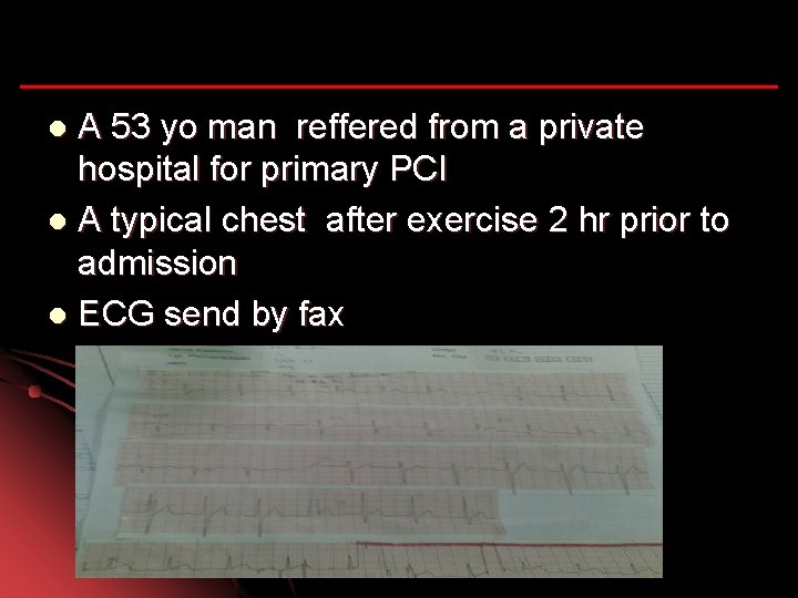
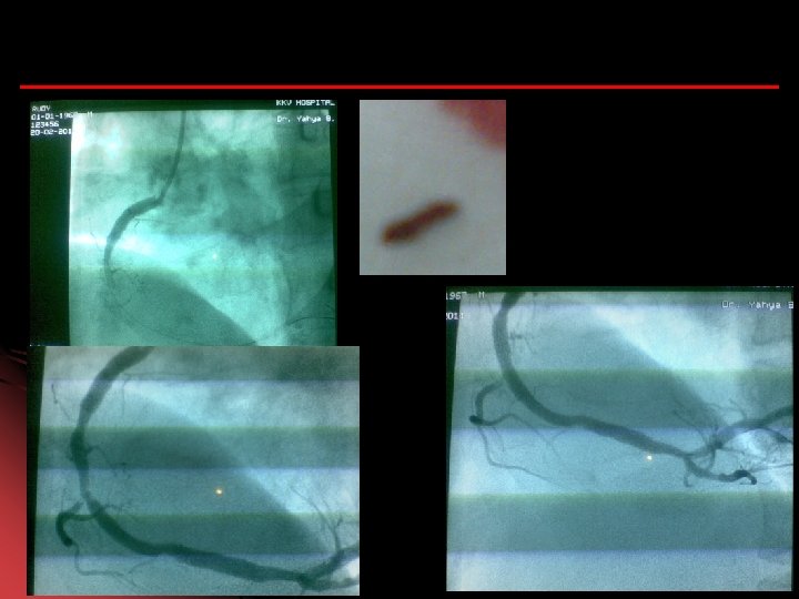
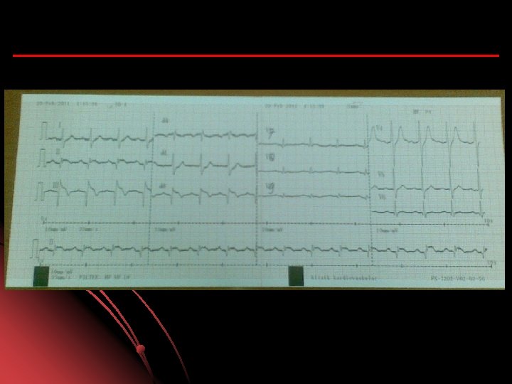
A 53 yo man reffered from a private hospital for primary PCI l A typical chest after exercise 2 hr prior to admission l ECG send by fax l
PCI appointment via phone l Patient directly transfer red to cath lab. l
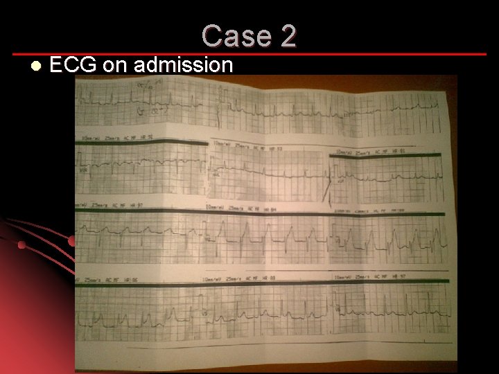
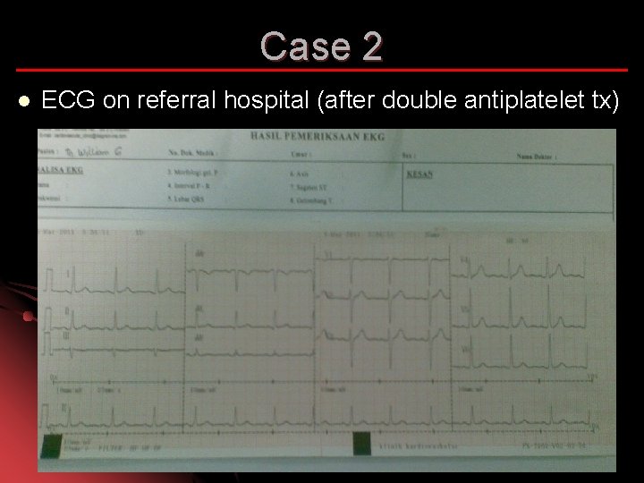
Case 2 Mr W. 28 yo, smoker and family history l Admit to Nearest private hospital with chief complaint of Heaviness while he was driving a car 30 min prior to admission l
Case 2 l ECG on admission
Case 2 l ECG on referral hospital (after double antiplatelet tx)
CABG l l l Failed PCI with persistent pain or hemodynamic instability in patients with coronary anatomy suitable for surgery. Persistent or recurrent ischemia refractory to medical therapy in patients who have coronary anatomy suitable for surgery, have a significant area of myocardium at risk, and are not candidates for PCI or fibrinolytic therapy. At the time of surgical repair of postinfarction ventricular septal rupture (VSR) or mitral valve insufficiency.
CABG l Cardiogenic shock in patients less than 75 years old with ST elevation, LBBB, or posterior MI who develop shock within 36 hours of STEMI, have severe multivessel or left main disease, and are suitable for revascularization that can be performed within 18 hours of shock l Life-threatening ventricular arrhythmias in the presence of greater than or equal to 50% left mainstenosis and/or triple-vessel disease.
Yang sering di lupakan…. . Edukasi pasien mengenai : l Mengapa bisa sampai sakit…. . Pola hidup l Kepatuhan untuk merubah pola hidup l Faktor resiko di kendalikan, rokok, HT, dll l Kepatuhan minum obat l Mencegah serangan jantung berikutnya dengan…. . merubah pola hidup, atur pola makan, olah raga teratur dan terukur l Reperfusi adalah awal dimulainya hidup baru …agar koroner tetap terbuka
Simpulan Tegakkan diagnosa dengan cepat dan tepat l Terapi dengan cepat dan tepat : Reperfusi l Monitor ketat l Cegah komplikasi l Edukasi untuk prevensi dan rehabilitasi l …. . ……. Kerja keras di awal……