In vivo Evaluation Animal Models Biodistribution Metabolism Studies

In vivo Evaluation Animal Models, Biodistribution, Metabolism Studies Lurdes Gano C 2 TN, Instituto Superior Técnico, Universidade de Lisboa Summer School: Development and Pre-clinical Evaluation of Radiopharmaceuticals – 4 -8 June 2018

Radiolabelled Biomolecules/Compounds with suitable Radiochemical and In Vitro Biological Profile In Vivo Biological Evaluation

In Vivo Biological Evaluation Ø Biodistribution in Animal Model Ø Pharmacokinetics in Animal Model Ø In Vivo Stability / Metabolic Studies Ø Molecular Imaging Ø Assessment of Therapeutic Potential (e. g. Tumor regression) Usefulness for clinical application as molecular imaging or radiotherapeutic agent

Molecular Imaging is the visualization, characterization and measurement of biological processes at the molecular and cellular levels in humans and other living systems Measure physiological parameters: Ø Receptor, antigen, enzyme concentration Ø Organ function Ø Metabolic processes How these parameters change during disease? Screening of therapeutic responsiveness

Molecular imaging techniques Ø Highly Specific Ø Indispensable in Diagnostics Ø Visualize specific molecular events Ø Enable earlier diagnosis Ø Monitor therapeutic responses (Radionuclide Therapy; Therapeutic Drug Development)

Molecular Imaging Modalities Radionuclide Imaging the in vivo biodistribution of radiotracers Quantitative SPECT PET • Resolution 1 mm • Resolution 2 mm • 80 -250 ke. V energies • 511 ke. V energy • Discriminate ≠ energies • More sensitive • Higher doses of radioactivity • Lower levels of radioactivity • Longer imaging times • Shorter imaging times
 Bioluminiscen Reflection of high- Measure Anatomical")
Molecular Imaging Modalities Optical Imaging Ultrasound Imaging 1) Bioluminiscen Reflection of high- Measure Anatomical frequency sound concentration and information/ waves in the body rates of relaxation Absortion of X-rays of H atoms in Combined with PET magnetic field and SPECT ce 2) Fluorescence MRI CT High Sensitivity; Low Sensitivity; Rapid; Resolution - 5 mm; Resolution 50 Very low μm; Sensitivity; Operator Rapid; Resolution 50 µm dependent; Qualitative Resolution 25 μm; High degree of scatter and absorption by tissues

• Design highly sensitive and specific imaging probes Various imaging modalities (radionuclides, fluorophores, nanoparticles) + Targeting ligands (Abs, Proteins, Peptides, Cells) • High affinity and specificity for target (Nanomolar concentration) • Targeting selectivity

Specific Interaction with biochemical and physiological processes Radiolabelled Targeting Biomolecules Molecular Imaging / Targeted Radionuclide Therapy / Theranostics Selective Binding to target Antigens, cellular membrane or nuclear Receptors Ø Increased Specificity – essential to minimize the unnecessary radiation exposure Ø Increased Accuracy Ø Potential ability to eradicate primary tumor and disseminated metastasis

Targeting Biomolecules: Ø Monoclonal Antibodies; Ø Antibody Fragments; Ø Small domain Antibodies Ø Nanobodies; Ø Peptides; Ø Small molecules; Ø …… Determines the fate of radiopharmaceuticals after administration

Animal Models

Animal Models Why use animal models in research? • To try and model human diseases • Understand molecular aspects of disease process • Essential for the development of clinically useful (radio)pharmaceuticals • Validation and quality control of (radio)pharmaceuticals • Mechanisms of localisation of compounds • Unique pharmacological and toxicological data • Predict biosafety and clinical efficacy

Animal Models 3 -Rs Principle Ø Replacement In vitro techniques; microorganisms; computer modelling Ø Reduction Study design – minimum animal number; Improve statistics; Use “lower” vs “higher” animals Ø Refinement Reduce pain and stress Non-invasive techniques Improve conditions

Animal Models Principles for animal experiments • Essential for significant relevant information • Obligation to treat animals with respect • Investigator has ultimate responsability • Balance between effects on animals and benefit for health • Appropriate species • Bred in captivity • Scientifically valid using minimum number • Well trained and competent staff • Brief experiments • No unnecessary repeats

Animal Models Normal Animals • Small rodents Rat; mice • Many physiological similarities Preserved basic layout and function of most organs • Provide useful information Biodistribution; In vivo stability; Interaction with molecular target in biological environment; Neuropeptide receptors widely expressed in mice (e. g. somatostatin analogues) usually interact as efficiently as human receptors; Most monoclonal antibodies towards human targets do not bind to their rodent equivalent.

Animal Models Disease Animal Models Ø Infection/ Inflamation Animal Models Ø Tumour-Bearing Animals Ø Transgenic Animals Biomolecules specifically bind in vivo to infection sites, antigens, overexpressed receptors, ….

Animal Models Tumour-Bearing Animals To predict the likely behaviour of the radiolabelled biomolecule in a cancer patient Depends on: Tumour source; Imunocompetence of the animal; Genetic manipulation Syngeneic Model – animals bearing tumours of their own species Spontaneous or carcinogen-induced Transplanted by administration of tumor cells (unnatural location; changes in the intratumoral signaling) Orthotopic Model - transplant of the tumor to the site as its origin (e. g. mamary gland, eye, bone marrow)

Animal Models Tumour-Bearing Animals Syngeneic Model • Well characterized cell lines • Immunocompetent hosts • Reproducible tumors • Low cost • Poor representation of human disease (diferent receptor subtypes, expression level, …) • Lack of target molecule homology between species

Animal Models Tumour-Bearing Animals Xenogeneic Model – animals bearing tumours of human origin Animals with imunodeficient system: Genetically modified 1. 2. Nude strains of mice or rats – lack of thymus; do not generate mature T cells SCID Mice (severe combined immunodeficient) - have a mutation, complete loss of humoral and cellular immune system

Animal Models Tumour-Bearing Animals Xenogeneic Model • Well characterized cells • Simple to implement • Expression of human homolog of the target • Homogeneity in tumor • Reproducible

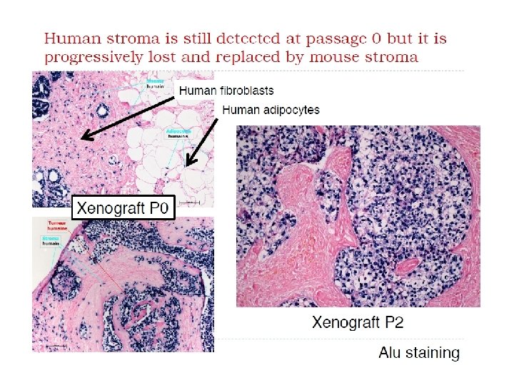
Animal Models Tumour-Bearing Animals Xenogeneic Model • Immunosupressed non human hosts • Tumor cells of human origin • Murine peritumoral milieu (blood vessels, stromal cells) • Imune environment of tumor • Different human tumor histology • More expensive • Require microbe-free animal housing

Animal Models Tumour-Bearing Animals Orthotopic Model • Best mimicking human carcinogenesis and metastatic patterns • Limited number of hosts • Surgical skills • Complex logistics • Non-homogeneity/ non reproducibility in tumor growth

Animal Models Induction of Xenotransplant Administration routes – subcutaneous administration of tumor cell suspension Tumor cells of human origin Murine stromal cells and blood vessels Abnormal imune environment of tumor

Animal Models Transgenic model Genetically modified animals to alter expression of target molecule Models of human disease 1. Administration of transfected cells (1 receptor subtype; 2. 3. different levels of expression) Reporter-gene imaging (expression of target molecule controled by a particular gene) Transgenic mice (incorporation of human gene, random, transient, relatively inefficient) 4. Gene targeting 5. “Knockout” mice (disruption of function of a selected gene)

Animal Models Gene targeting Genetically modified animals Introduction of human DNA homologous to the target mouse gene into embryonic stem cells Selected cells implanted in foster mothers Birth of 2 types of mice (only human gene or only mouse gene)

Animal Models Transgenic model • Controlled cancer progression in selected organs; • Resemble human carcinogenesis; • Immunocompetent host; • Limited availability; • Expensive; • Restricted experience; • Variations in tumor growth rates; • Demanding statistics
 Model Emerging platform for Translational Cancer Research; Observation:")
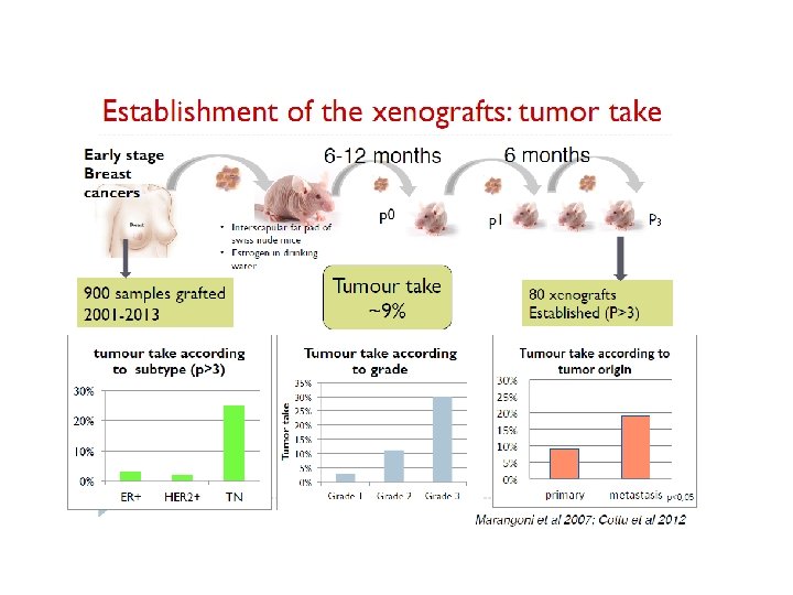
Animal Models Patient Derived Xenograft (PDX) Model Emerging platform for Translational Cancer Research; Observation: High failure rate of new molecules in late clinical development in oncology; Lack of efficacy J. Arrowsmith, P. Miller, Nat Rev Drug Discovery 2013

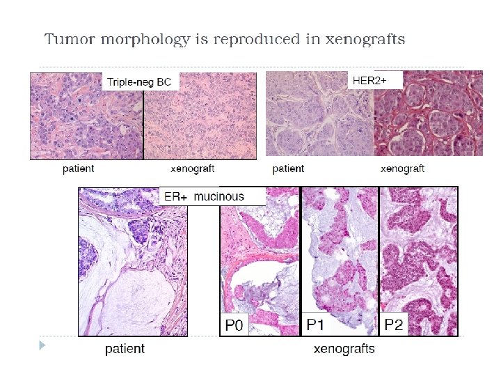
Animal Models PDX Model Way to increase the predictibility of preclinical studies Ø Use PDX animal model that more closely reproduce the heterogeneity of human cancers Ø Perform studies for genotype/response correlation Ø Maintain high correlation with the original tumor from patients Ø Still complementary to other models

Animal Models PDX Model

Animal Models PDX Model
 can be obtained")
Animal Models PDX Model PDX of Triple Negative Breast Cancer (TNBC) can be obtained with relative high rates compared to ER+ or Her 2+ since these TNBC xenografts are highly proliferating
 BUT There")
Animal Models PDX Model Gene expression is stable over time (tumor passage) BUT There are gene differences when comparing the xenograft with the patient Expression of tumor supressors is lost in 60 -70% xenografts

Radiopharmaceuticals Efficacy Ø Biodistribution and Pharmacokinetics Ø Rapid uptake in target tissue Ø High affinity and selectivity Ø Rapid clearance from blood and non-target organs Ø Residence time in target tissue long enough Ø Predominant kidney excretion with no tubular reabsorption Ø Specific Activity Ø in vitro/ in vivo stability
 • The")
Factors affecting performance of radiolabelled biomolecules • Affinity for the target (receptor) • The target density • The target accessibility (membrane or nuclear receptor) • Non-target expression of the receptor • The in vivo stability of biomolecule • The choice of radionuclide • The stability of the radiolabelled biomolecule complex • The physicochemical properties of the radiolabelled biomolecule (size, charge, lipophilicity)

Prerequisite for effective in vivo tumor targeting • In vivo metabolic stability in the biological milieu (metal chelate; enzymatic peptide chain) • Radiolabelled antibodies must retain imunoreactivity • Radiolabelled peptides must retain receptor binding ability • High radiochemical purity • High specific activity (at least 1 Ci/umol peptide) Unlabeled peptide bioconjugate would occupy saturable receptor sites

Prerequisite for effective in vivo tumor targeting • High target-to-background ratio • Rapid clearance from non-target organs (high contrast images – diagnosis Minimize radiotoxicity – therapy) • Rapid excretion into urine • Minimal hepatobiliar excretion • Rapid clearance of radioactivity from kidneys improve accuracy of diagnostic and minimize nefrotoxicity during therapy

Pharmakokinetic Aspects of Radiolabelled Antibodies vs Peptides Radiolabelled Antibodies • Slow blood clearance (MW; circulating antigens) Radiolabelled Peptides • Rapid blood clearance • More favourable pharmacokinetics
 •")
Preclinical screening of radiolabelled biomolecules • Determine biodistribution overtime (depends on the application) • Determine % Radioactivity Excretion; • Determine % I. A. per organ; % I. A. per gram; Dissection and counting Quantification by PET or SPECT camera Autoradiography • Target-to-non target ratio • Clearance of radiolabelled biomolecule and its radioactive metabolites

Fate of Radiolabelled Biomolecules in the Body • Absorption • Distribution X Reversible pass Vascular Comp → Tissues/ Organs • Elimination (Excretion + Metabolism) Pharmacokinetics parameters Clearance = Rate of elimination / Plasma concentration Mean residence time = 1 / K k= elimination rate constant

Biodistribution Studies Ø Distribution of radiolabelled biomolecule in main organs Ø Uptake and retention time in receptor-negative tissues vs receptor –positive tissues Ø Blocking experiments by co-administration of unlabelled biomolecule Ø Rate of blood clearance Ø Rate and route of excretion Ø In vivo stability of radiolabeled biomolecules

Biodistribution Studies Female MCF-7 xenografts Balb/C nude mice Ø Fast blood clearance Ø High kidney uptake Ø Uptake in ER rich organs (ovaries, uterus) and xenografts

Biodistribution Studies Species Variation Major variations between species: Uptake by specific organs Clearance Mouse heart beats much faster than the human; Mice breath much faster • Shorter tissue perfusion times • Shorter gastrointestinal transit time • More rapid pharmacokinetics

Biodistribution Studies There are some limitations in extrapolating data from animal models due to: § Different genotypes between mice and men § Size difference – specially dosimetric calculations § Faster sequestring and metabolizing

Biodistribution Studies Experimental Procedure • Administration of radiolabelled molecules (i. v. ; i. p. ); • Measure I. A. ; • Sacrifice , weight, whole body radioactivity measrument; • Organ dissection, weight and counting • Determination- % Excreted activity; %I. A. /g ; % I. A. /total organ

Bombesin antagonis radioligands SPECT/CT 99 m. Tc 111 In PET 68 Ga 64 Cu Agonist radioligands have been preferred since they internalize after receptor binding promoting intracellular accumulation of radionuclide Antagonists can bind more receptors - High receptor occupancy Biomolecules labelled with different radionuclides – Different biodistribution patterns

In vivo Stability Studies HPLC analysis of samples of blood serum; collected urine and organ homogenates (liver, kidney, brain, …) Treatment of biological samples (protein precipitation) Chromatographic analysis (HPLC)

In vivo Stability Studies HPLC analysis of samples of urine collected at sacrifice time J Nucl Med 2011 52: 1970

Biodistribution Studies PC-3 xenograft bearing mice Blockade with co-injection of peptide excess 100 ug Tyr-BBN J Med Chem 48: 100 Uptake decrease in receptor rich organs

Peptide Radionuclide Receptor Therapy • Specific • Improve patients quality of life • Rapid tumor uptake • Pain relief • Long residence time into tumor • Tumor regression • Rapid clearance from non target • Decrease level of tumor markers organs Clinical success of therapy Nefrotoxicity

Mechanisms of Urinary Excretion • Glomerular Filtration • Tubular Resorption – Active process (proximal tubule) Passive process (distal tubule) • Tubular Secretion – active process (proximal tubule)

Peptide Radionuclide Receptor Therapy Nefrotoxicity – limits the administration dose Neuropeptides • Predominant renal excretion • Glomerular filtration • Resorption in proximal tubule • Retention in lisossomes High radioactivity concentration in the kidney

Peptide Radionuclide Receptor Therapy Strategies to reduce nefrotoxicity Reduce kidney uptake • Co-administration of positive charged aminoacids solution (lysine and arginine) - 33 a 40% • Gelofusine Increases excretion of megalin ligands • Colchicine Blocks microtubules function – essential to endocitosis • Co-administration of albumin fragments (3 -50 k. Da) Interfers megalin mediated resorption

Biodistribution in mice

Strategies to reduce nefrotoxicity Treatment with Lysine Biodistribution profile Excretion

Peptide Radionuclide Receptor Therapy Strategies to reduce nefrotoxicity New peptide analogues with improved biological profile • Higher receptor affinity • Prolonged tumor retention time • Faster renal clearance and rapid excretion
- Slides: 59