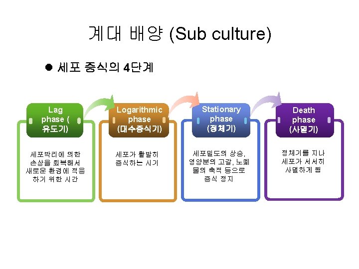
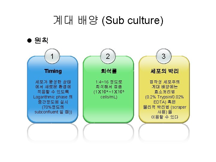
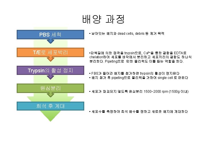
In vitro Hepatotoxicity test Cell Culture 2 MTT


 • A colorimetric assay • MTT [3 -(4, 5 -Dimethylthiazol-2")
 An increase in cell number results in an increase in")

 • 감염, 오염을 최소화하기 위해")


- Slides: 22
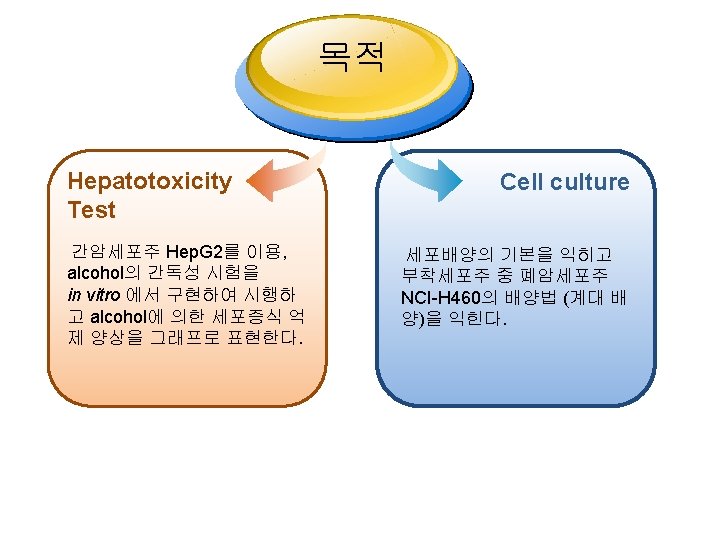
In vitro Hepatotoxicity test Cell Culture 독성학 실습 2 MTT 2011. 5. 13.
Hepatotoxicity test In vivo testing, assessed by histopathology is the traditional toxicology tool Several in vitro hepatotoxicity endpoints in human Hep. G 2 cells are measured using the Cell-based Assay Cell Loss/ Cell Cycle Arrest/ DNA Degradation/Apoptosis/ Nuclear Size/ Oxidative Stress/ Stress Kinase Activation/DNA Damage/ Mitochondrial Membrane Potential / Mitochondrial Mass/ Mitotic Arrest/ Cytoskeletal Integrity
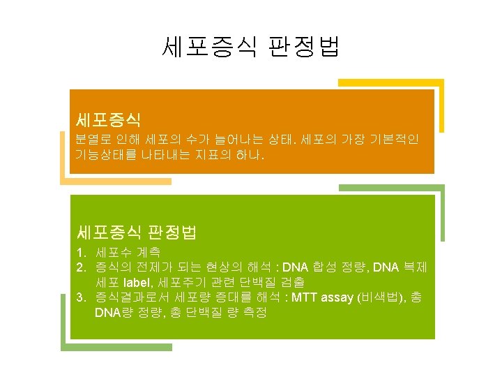
Methods for counting viable cells Trypan blue - A simple way to evaluate cell membrane integrity - Not sensitive, and cannot be adapted for high-throughput Screening Radioactive substances - tritium-labeled thymidine ([3 H]d. Thd) - accurate but time-consuming and involves handling of radioactive substances MTT assay WST-1
Cell proliferation assay (MTT) • A colorimetric assay • MTT [3 -(4, 5 -Dimethylthiazol-2 -yl)-2, 5 -diphenyltetrazolium bromide] a tetrazole, one of many categories of chemical structures is reduced to purple formazan in the mitochondria of living cells • This reduction takes place only when mitochondrial reductase enzymes are active (viable cells)
Cell proliferation assay (MTT) An increase in cell number results in an increase in the amount of MTT formazan formed an increase in absorbance To determine if particular drugs or conditions effect cell growth or cell death Solutions of MTT solubilized in tissue culture media or balanced salt solutions are yellowish in color purple MTT formazan crystals are insoluble in aqueous solutions The crystals can be dissolved in acidified isopropanol The resulting purple solution is spectrophotometrically measured
세포증식 그래프 • 그래프 그리기 – – O. D. value → % 로 표현 (Control을 100%로 놓는다) 평균 값으로 그래프를 그린다 (EXCEL 함수: “AVERAGE”) 표준편차도 오차막대로 나타낸다 (EXCEL 함수: “STDEV”) 결과를 해석한다 120. 00 Proliferaation (%) 100. 00 80. 00 60. 00 40. 00 20. 00 0 20 40 60 80 Alcohol (m. M) 100 120 140 m. M/% 1 2 3 Mean SD 0 103 100 97 100. 00 30 81 90 84 85. 00 4. 58 60 61 63 57 60. 33 3. 06 90 45 37 41 41. 00 4. 00 120 38 39 29 35. 33 5. 51
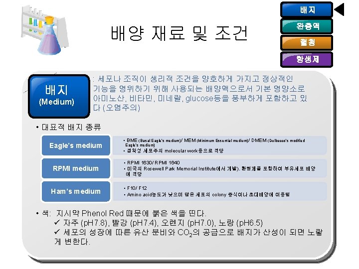
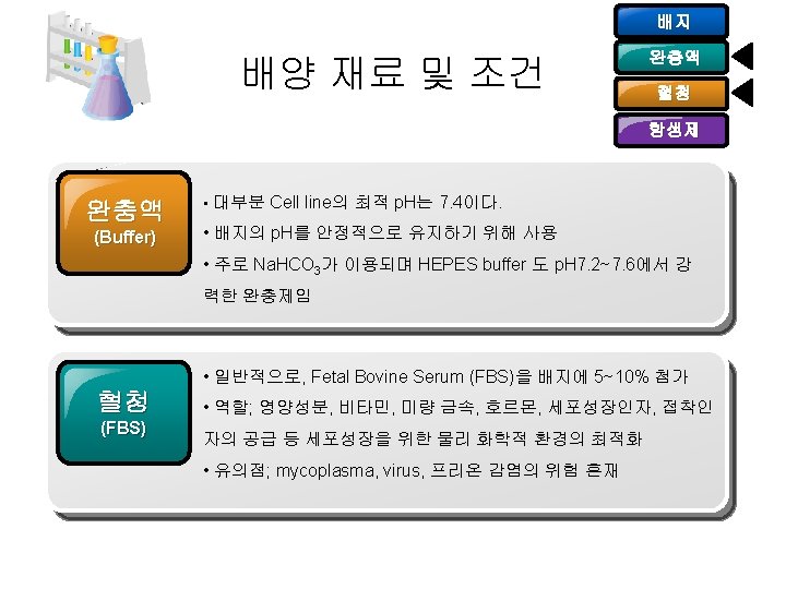
배지 배양 재료 및 조건 완충액 혈청 항생제 (Antibiotics) • 감염, 오염을 최소화하기 위해 사용하나 절대적이진 않다 • 단점 : 저항성 단백질의 발현을 조장할 수 있고 Mycoplasma의 감염 확인을 방해할 수 있다 • 세포배양 시 주로 사용되는 항생제 Application Amphotericin B Fungi, yeast Ampicillin Bacteria G+/- Ciprofloxacin Mycoplasma Polymixin B Bacteria G- Penicillin Bacteria G+ Streptomycin Bacteria G+/-
세포주 “Hep. G 2” l Hep. G 2 – 세계에서 12번째로 많이 사용 되는 세포주 – Origin : 15 year old Caucasian American male – Epithelial morphology (부착 세포주) – 다양한 혈장단백들을 분비 : e. g. , albumin, transferrin fibrinogen, alpha 2 -macroglobulin, alpha 1 -antitrypsin, transferrin and plasminogen. Hep. G 2 <Hep. G 2> <Floating cells> <NCI-H 460>
세포수 계산 l 세포수 계산법 cells/ml = average count per square × dilution factor × 10 4 Total cells = cells/ml × total original volume * The number 104 is the volume correction factor for the hemacytometer: each square is 1× 1× 0. 1 mm.
재료 준비 • 조별 받아갈 재료들 1. 2. 3. 4. 5. 6. 7. Alcohol (ethanol) 120 m. M 1 m. L Positive control - MTX (Methotrexate) 500 ppm PBS 1 m. L e-tube 조당 4개씩, e-tube rack Micropipet 1000/200 조당 하나씩 pipet tip 200/1000 Tip waste tube (50 m. L tube) * 96 well plate ; 실험 직전에 incubator에서 꺼내 받기