
In This Lesson Mitosis and the Cell Cycle


- Slides: 91
In This Lesson: Mitosis and the Cell Cycle From where do cells originate? Why? How?
Today’s Agenda • Chromosome basics. • The Cell Cycle. • Mitosis. – Where division = multiplication. mitois
By the end of this lesson… • You should be able to describe a normallyfunctioning somatic cell cycle and analyze what would happen if it no longer functioned properly. • You should be able to identify several chromosomal diseases using karyotypes. • You should be able to contrast mitosis and binary fission.
The Cell Theory • Think back to the cell theory… – All living things are made of cells. – Cells are the basic units of structure and function. – All cells come from preexisting cells. • That last one is the big one for us right now.
You again. • You started as a microscopic single cell – a zygote, the cell that resulted from the fusion of gametes – far smaller than the period at the end of this sentence.
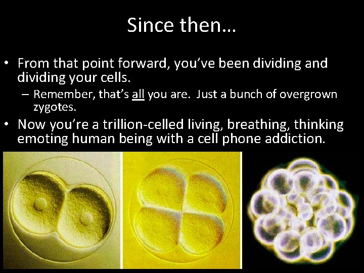
Since then… • From that point forward, you’ve been dividing and dividing your cells. – Remember, that’s all you are. Just a bunch of overgrown zygotes. • Now you’re a trillion-celled living, breathing, thinking emoting human being with a cell phone addiction.
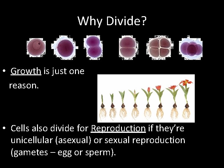
Why Divide? • Growth is just one reason. • Cells also divide for Reproduction if they’re unicellular (asexual) or sexual reproduction (gametes – egg or sperm).
• If they’re part of a multicellular organism, they may divide for: • Repair or renewal, such as when a cut heals or new skin cells to replace shedding ones. • Differentiation, since they need to grow up and get jobs. • Not to mention, cells also have to divide because they really can’t get bigger without ruining that whole surface area-to-volume ratio.
What cells divide often? • • Skin Stomach lining Red Blood cells Embryo Plant roots Hair Nails
What cells rarely/never divide? • Nervous System • Liver • Brain
Why do we age? • Eventually cells stop being replaced • “Apoptosis” – Cell death • “We die because our cells die. ” – William R. Clark
So what do we need to know? • To fully understand the cellular division process, which itself is mostly review of what you’ve already learned, we need to understand the following: – Chromosomes – The Cell Cycle
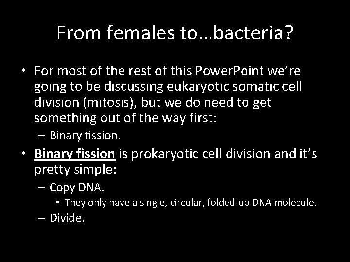
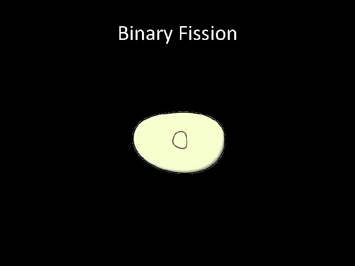
From females to…bacteria? • For most of the rest of this Power. Point we’re going to be discussing eukaryotic somatic cell division (mitosis), but we do need to get something out of the way first: – Binary fission. • Binary fission is prokaryotic cell division and it’s pretty simple: – Copy DNA. • They only have a single, circular, folded-up DNA molecule. – Divide.
Binary Fission
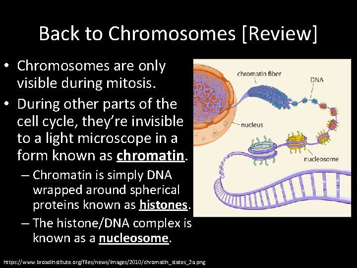
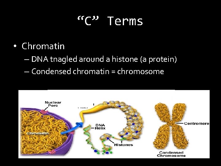
Back to Chromosomes [Review] • Chromosomes are only visible during mitosis. • During other parts of the cell cycle, they’re invisible to a light microscope in a form known as chromatin. – Chromatin is simply DNA wrapped around spherical proteins known as histones. – The histone/DNA complex is known as a nucleosome. https: //www. broadinstitute. org/files/news/images/2010/chromatin_states_2 a. png
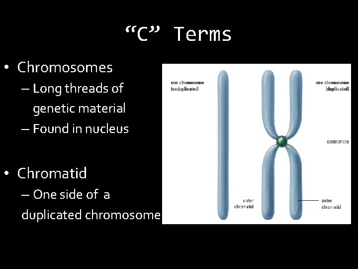
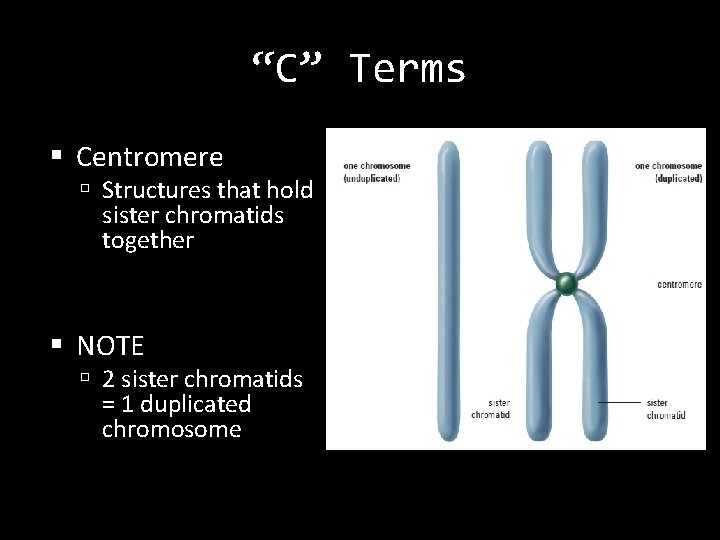
“C” Terms • Chromosomes – Long threads of genetic material – Found in nucleus • Chromatid – One side of a duplicated chromosome
“C” Terms Centromere Structures that hold sister chromatids together NOTE 2 sister chromatids = 1 duplicated chromosome
“C” Terms • Chromatin – DNA tnagled around a histone (a protein) – Condensed chromatin = chromosome
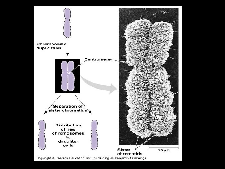
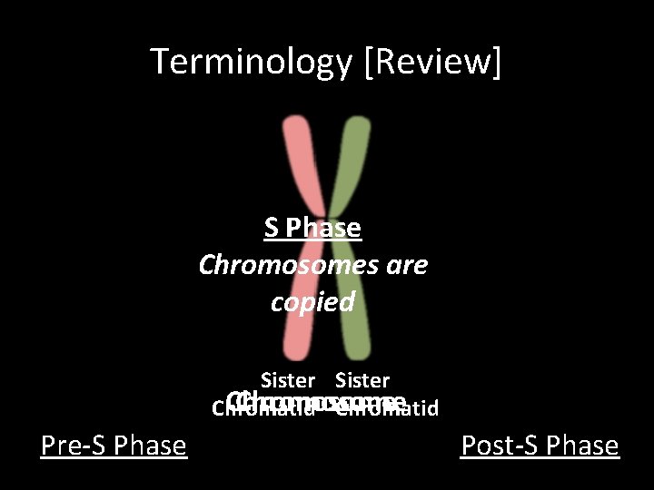
Terminology [Review] • Chromatid – Half of a duplicated (X-shaped) chromosome. • Sister Chromatids – The two identical chromatid copies that make up an X-shaped chromosome. • Centromere – The site at which the two sister chromatids join.
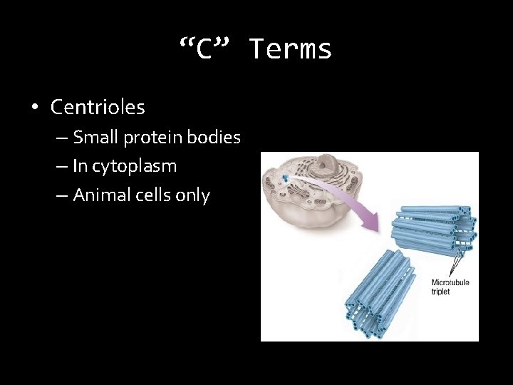
“C” Terms • Centrioles – Small protein bodies – In cytoplasm – Animal cells only
Cell Division in a Nutshell • Before: – Chromosome duplicates = 2 sister chromatids • During: – Sister chromatids separate • After: – 2 “daughter” cells – Genetically identical
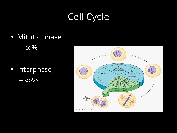
Cell Cycle • Mitotic phase – 10% • Interphase – 90%
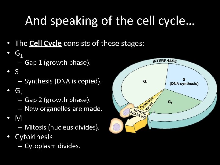
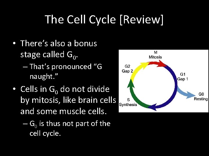
And speaking of the cell cycle… • The Cell Cycle consists of these stages: • G 1 – Gap 1 (growth phase). • S – Synthesis (DNA is copied). • G 2 – Gap 2 (growth phase). – New organelles are made. • M – Mitosis (nucleus divides). • Cytokinesis – Cytoplasm divides.
The Cell Cycle [Review] • There’s also a bonus stage called G 0. – That’s pronounced “G naught. ” • Cells in G 0 do not divide by mitosis, like brain cells and some muscle cells. – G 0 is thus not part of the cell cycle.
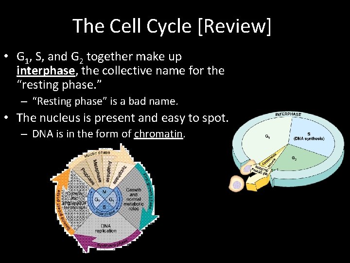
The Cell Cycle [Review] • G 1, S, and G 2 together make up interphase, the collective name for the “resting phase. ” – “Resting phase” is a bad name. • The nucleus is present and easy to spot. – DNA is in the form of chromatin.
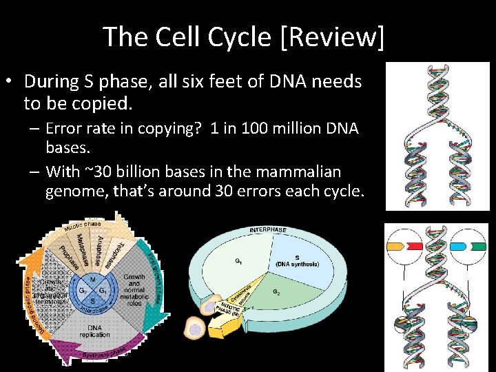
The Cell Cycle [Review] • During S phase, all six feet of DNA needs to be copied. – Error rate in copying? 1 in 100 million DNA bases. – With ~30 billion bases in the mammalian genome, that’s around 30 errors each cycle.
Terminology [Review] S Phase Chromosomes are copied Sister Chromosome Chromatid Pre-S Phase Post-S Phase
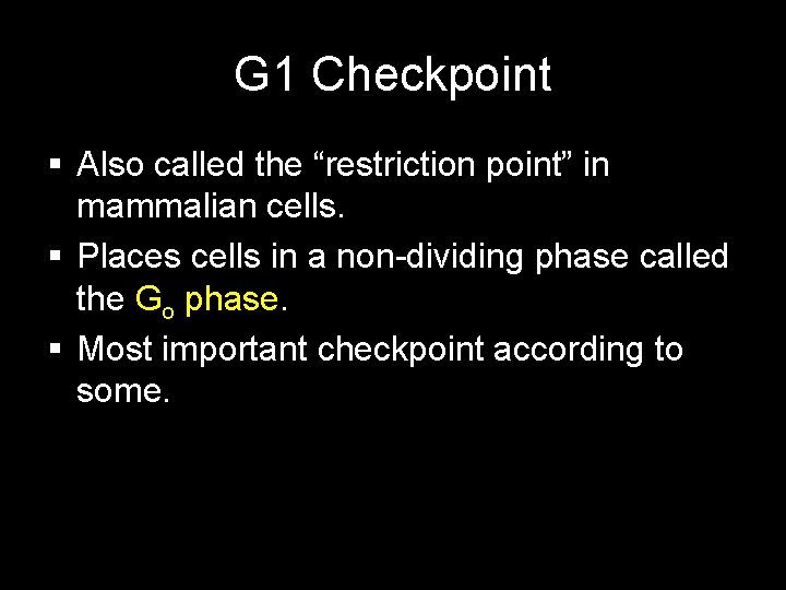
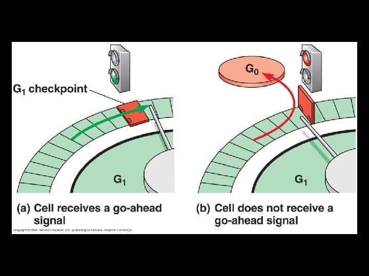
G 1 Checkpoint Also called the “restriction point” in mammalian cells. Places cells in a non-dividing phase called the Go phase. Most important checkpoint according to some.
GO
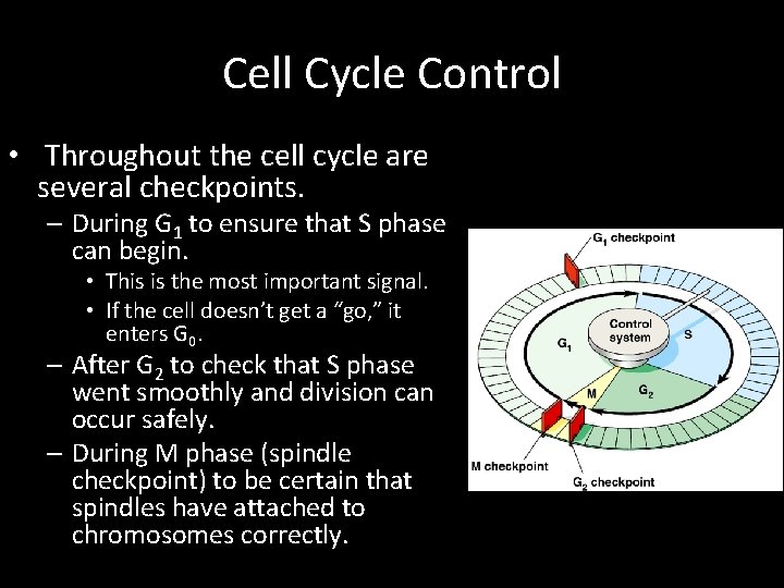
Cell Cycle Control • Throughout the cell cycle are several checkpoints. – During G 1 to ensure that S phase can begin. • This is the most important signal. • If the cell doesn’t get a “go, ” it enters G 0. – After G 2 to check that S phase went smoothly and division can occur safely. – During M phase (spindle checkpoint) to be certain that spindles have attached to chromosomes correctly.
Checkpoints http: //skreened. com/scienceforscientists/dna-checks-itself-before-it-wrecks-itself
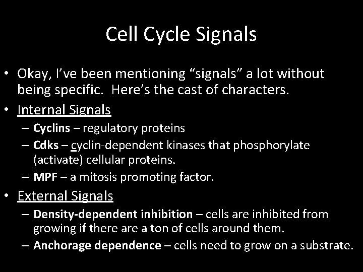
Cell Cycle Signals • Okay, I’ve been mentioning “signals” a lot without being specific. Here’s the cast of characters. • Internal Signals – Cyclins – regulatory proteins – Cdks – cyclin-dependent kinases that phosphorylate (activate) cellular proteins. – MPF – a mitosis promoting factor. • External Signals – Density-dependent inhibition – cells are inhibited from growing if there a ton of cells around them. – Anchorage dependence – cells need to grow on a substrate.
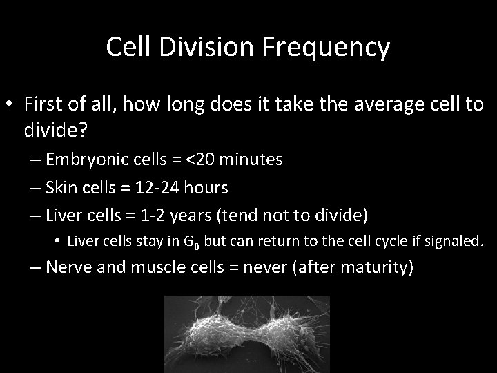
Cell Division Frequency • First of all, how long does it take the average cell to divide? – Embryonic cells = <20 minutes – Skin cells = 12 -24 hours – Liver cells = 1 -2 years (tend not to divide) • Liver cells stay in G 0 but can return to the cell cycle if signaled. – Nerve and muscle cells = never (after maturity)
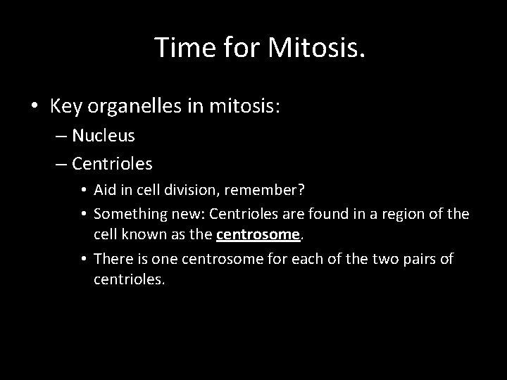
Time for Mitosis. • Key organelles in mitosis: – Nucleus – Centrioles • Aid in cell division, remember? • Something new: Centrioles are found in a region of the cell known as the centrosome. • There is one centrosome for each of the two pairs of centrioles.
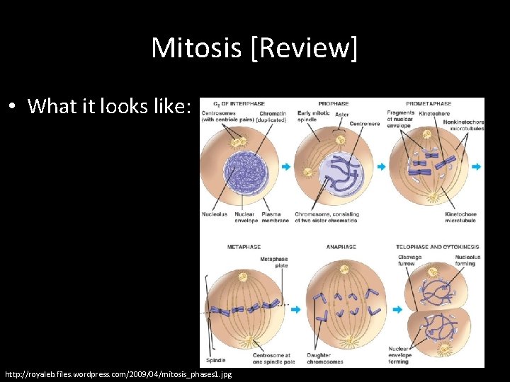
Mitosis [Review] • What it looks like: http: //royaleb. files. wordpress. com/2009/04/mitosis_phases 1. jpg
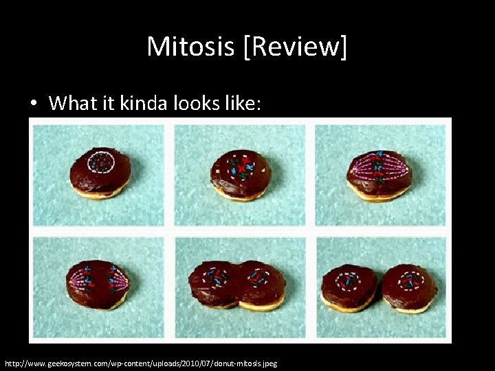
Mitosis [Review] • What it kinda looks like: http: //www. geekosystem. com/wp-content/uploads/2010/07/donut-mitosis. jpeg
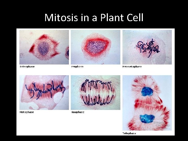
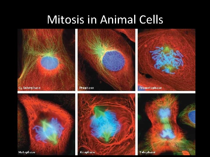
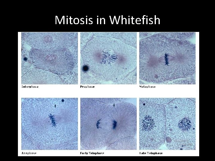
Mitosis [Review] • Mitosis (M phase) is the division of the nucleus. • There are four main phases of mitosis: – Prophase • Some people put an additional step here: Prometaphase. – Metaphase – Anaphase – Telophase
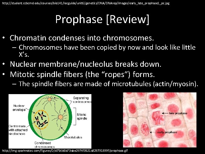
http: //student. ccbcmd. edu/courses/bio 141/lecguide/unit 6/genetics/DNArep/images/early_late_prophase 1_pc. jpg Prophase [Review] • Chromatin condenses into chromosomes. – Chromosomes have been copied by now and look like little X’s. • Nuclear membrane/nucleolus breaks down. • Mitotic spindle fibers (the “ropes”) forms. – The spindle fibers are made of microtubules (actin/myosin). http: //img. sparknotes. com/figures/D/d 756 b 5 b 73 abe 2974 f 3521 a 828791899 f/prophase. gif
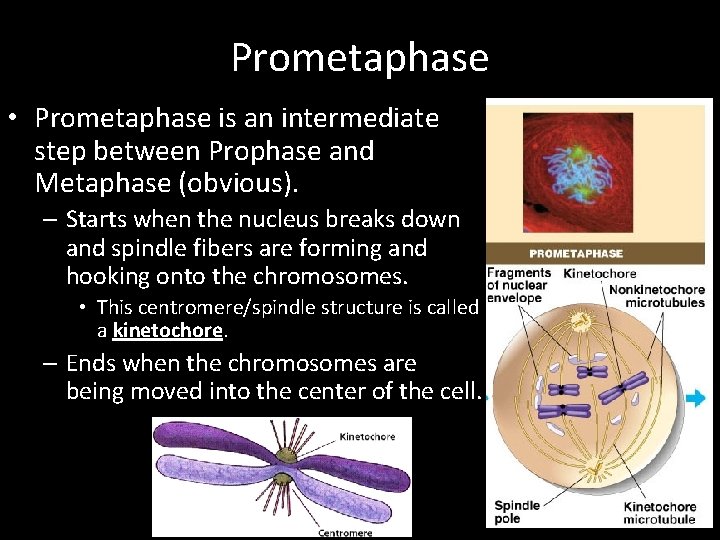
Prometaphase • Prometaphase is an intermediate step between Prophase and Metaphase (obvious). – Starts when the nucleus breaks down and spindle fibers are forming and hooking onto the chromosomes. • This centromere/spindle structure is called a kinetochore. – Ends when the chromosomes are being moved into the center of the cell.
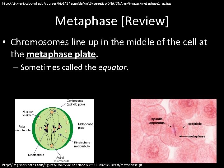
http: //student. ccbcmd. edu/courses/bio 141/lecguide/unit 6/genetics/DNArep/images/metaphase 1_ac. jpg Metaphase [Review] • Chromosomes line up in the middle of the cell at the metaphase plate. – Sometimes called the equator. http: //img. sparknotes. com/figures/D/d 756 b 5 b 73 abe 2974 f 3521 a 828791899 f/metaphase. gif
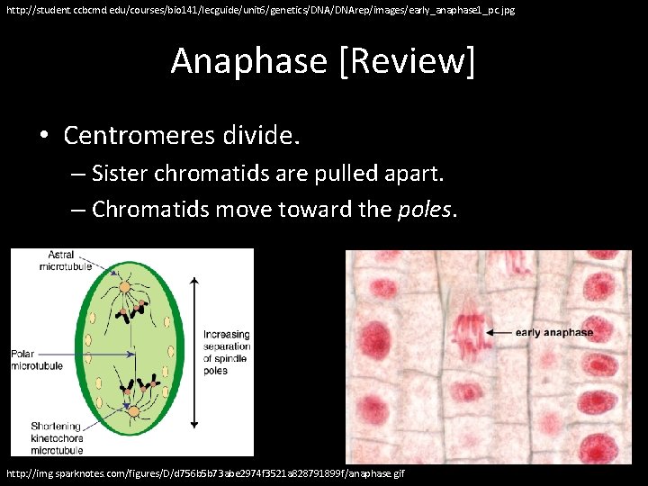
http: //student. ccbcmd. edu/courses/bio 141/lecguide/unit 6/genetics/DNArep/images/early_anaphase 1_pc. jpg Anaphase [Review] • Centromeres divide. – Sister chromatids are pulled apart. – Chromatids move toward the poles. http: //img. sparknotes. com/figures/D/d 756 b 5 b 73 abe 2974 f 3521 a 828791899 f/anaphase. gif
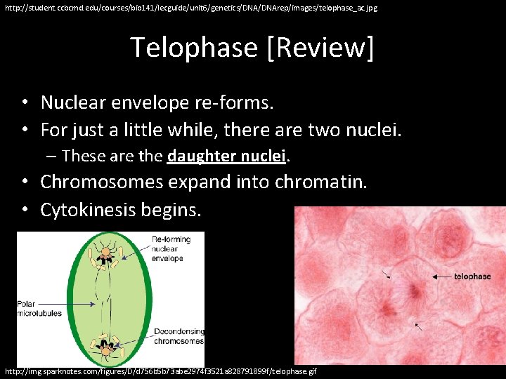
http: //student. ccbcmd. edu/courses/bio 141/lecguide/unit 6/genetics/DNArep/images/telophase_ac. jpg Telophase [Review] • Nuclear envelope re-forms. • For just a little while, there are two nuclei. – These are the daughter nuclei. • Chromosomes expand into chromatin. • Cytokinesis begins. http: //img. sparknotes. com/figures/D/d 756 b 5 b 73 abe 2974 f 3521 a 828791899 f/telophase. gif
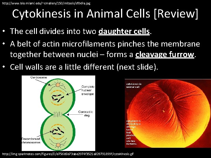
http: //www. bio. miami. edu/~cmallery/150/mitosis/sf 8 x 9 a. jpg Cytokinesis in Animal Cells [Review] • The cell divides into two daughter cells. • A belt of actin microfilaments pinches the membrane together between nuclei – forms a cleavage furrow. • Cell walls are a little different (next slide). http: //img. sparknotes. com/figures/D/d 756 b 5 b 73 abe 2974 f 3521 a 828791899 f/cytokinesis. gif
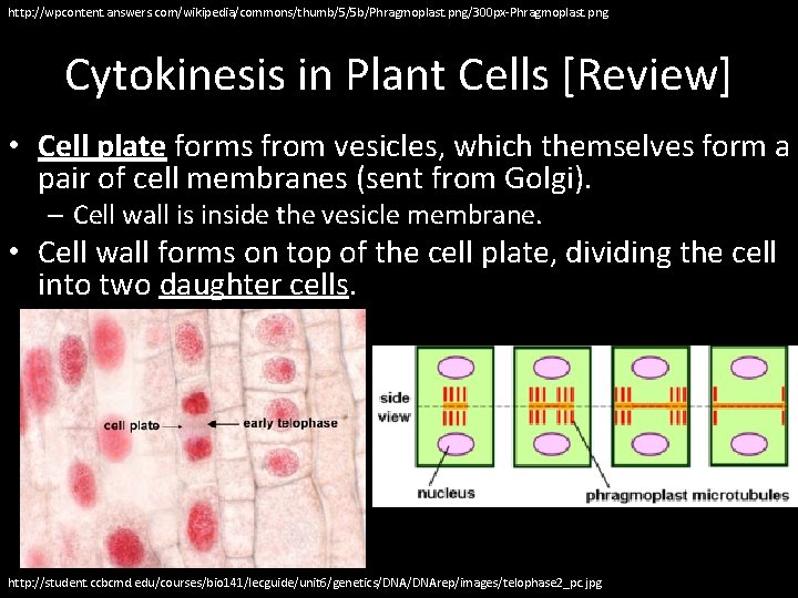
http: //wpcontent. answers. com/wikipedia/commons/thumb/5/5 b/Phragmoplast. png/300 px-Phragmoplast. png Cytokinesis in Plant Cells [Review] • Cell plate forms from vesicles, which themselves form a pair of cell membranes (sent from Golgi). – Cell wall is inside the vesicle membrane. • Cell wall forms on top of the cell plate, dividing the cell into two daughter cells. http: //student. ccbcmd. edu/courses/bio 141/lecguide/unit 6/genetics/DNArep/images/telophase 2_pc. jpg
Mitosis in a Plant Cell
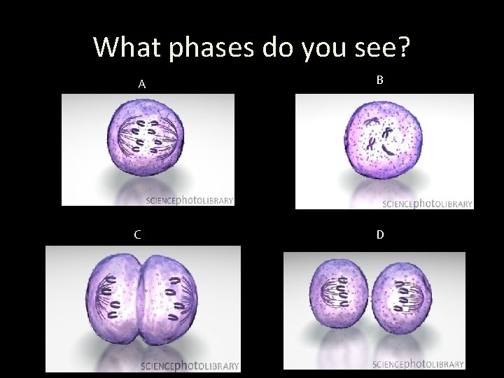
What phases do you see? A C B D
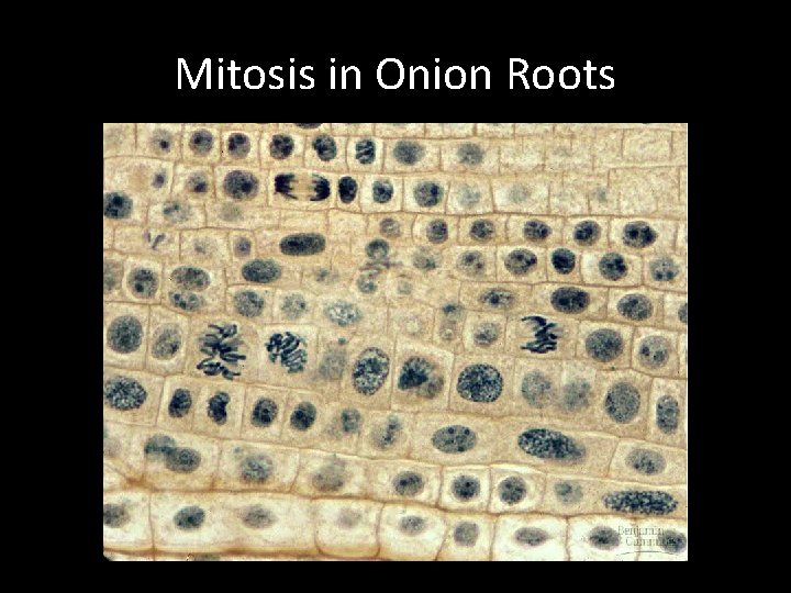
Mitosis in Onion Roots
Mitosis in Animal Cells
Mitosis in Whitefish
Mitosis Summary • This really cool company called Hybrid Medical Animations put together an awesome CGI look at mitosis in a cell. • http: //www. hybridmedicalanimation. com/wo rk/animation/the-stages-of-mitosis/
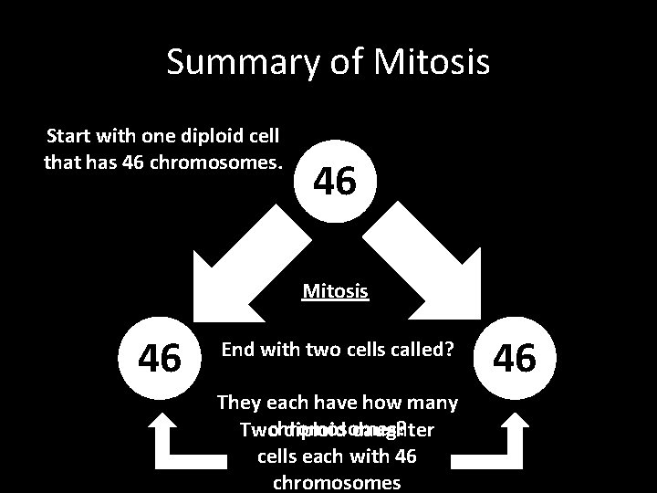
Summary of Mitosis Start with one diploid cell that has 46 chromosomes. 46 Mitosis 46 End with two cells called? They each have how many Twochromosomes? diploid daughter cells each with 46 chromosomes 46
Mitosis Fun Facts • 300, 000 cells die and are replaced every minute • 50, 000 cells are born in the time it takes me to read this. • Nerve cells do not divide but are replaced from glial cells through neurogenesis.
Aside: How Old Are You? • While it’s true that cells divide relatively quickly, they also don’t last forever. • Right now, the oldest cells in your body are 79 years old. – That’ll stay the same throughout your life – always 7 -9 years old, max, for your cells… • …which means that nothing living about you is actually as old as…you.
Concept Map
Cell Cycle Game • The Cell Cycle Game
Back to the Cell Cycle • You’ve now reviewed all of the cell cycle, from interphase through mitosis and cytokinesis. – That’s the “what, ” as in, “what is the cell cycle? ” • We now need to talk about the regulation of the cell cycle. – This is the “how, ” as in, “how does the cell manage this whole process and keep it from getting out of control?
Cell Cycle Regulation • Normal growth and maintenance of a multicellular creature thus requires careful coordination of cell division.
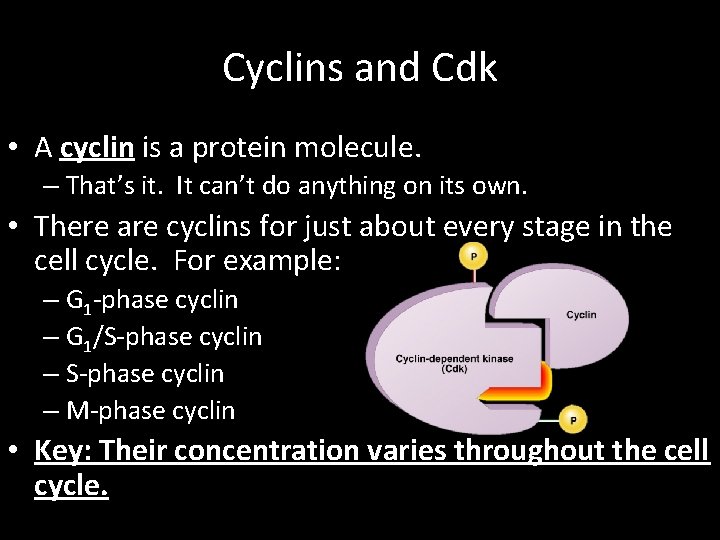
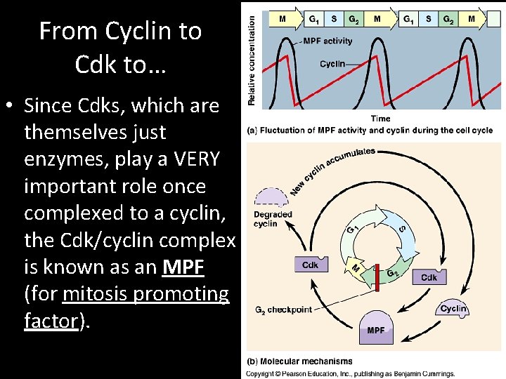
Cyclins and Cdk • A cyclin is a protein molecule. – That’s it. It can’t do anything on its own. • There are cyclins for just about every stage in the cell cycle. For example: – G 1 -phase cyclin – G 1/S-phase cyclin – M-phase cyclin • Key: Their concentration varies throughout the cell cycle.
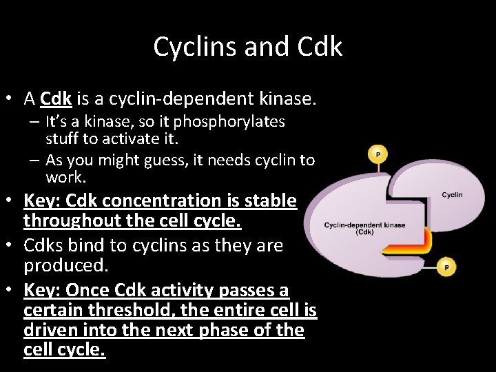
Cyclins and Cdk • A Cdk is a cyclin-dependent kinase. – It’s a kinase, so it phosphorylates stuff to activate it. – As you might guess, it needs cyclin to work. • Key: Cdk concentration is stable throughout the cell cycle. • Cdks bind to cyclins as they are produced. • Key: Once Cdk activity passes a certain threshold, the entire cell is driven into the next phase of the cell cycle.
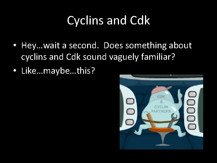
Cyclins and Cdk • Hey…wait a second. Does something about cyclins and Cdk sound vaguely familiar? • Like…maybe…this?
From Cyclin to Cdk to… • Since Cdks, which are themselves just enzymes, play a VERY important role once complexed to a cyclin, the Cdk/cyclin complex is known as an MPF (for mitosis promoting factor).
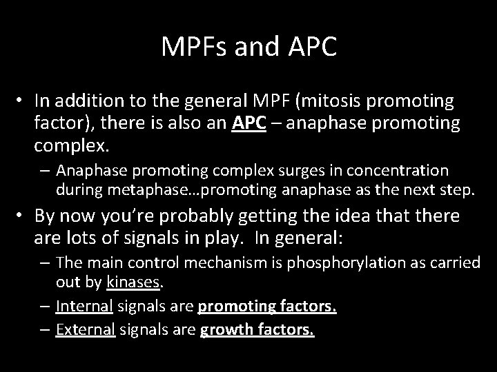
MPFs and APC • In addition to the general MPF (mitosis promoting factor), there is also an APC – anaphase promoting complex. – Anaphase promoting complex surges in concentration during metaphase…promoting anaphase as the next step. • By now you’re probably getting the idea that there are lots of signals in play. In general: – The main control mechanism is phosphorylation as carried out by kinases. – Internal signals are promoting factors. – External signals are growth factors.
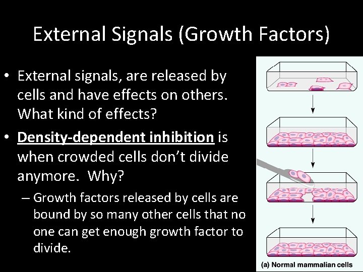
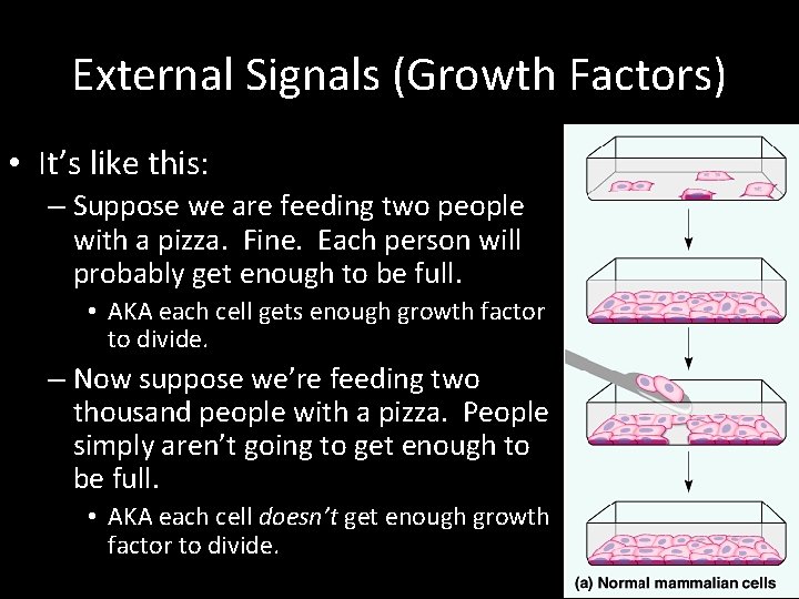
External Signals (Growth Factors) • External signals, are released by cells and have effects on others. What kind of effects? • Density-dependent inhibition is when crowded cells don’t divide anymore. Why? – Growth factors released by cells are bound by so many other cells that no one can get enough growth factor to divide.
External Signals (Growth Factors) • It’s like this: – Suppose we are feeding two people with a pizza. Fine. Each person will probably get enough to be full. • AKA each cell gets enough growth factor to divide. – Now suppose we’re feeding two thousand people with a pizza. People simply aren’t going to get enough to be full. • AKA each cell doesn’t get enough growth factor to divide.
Anchorage Dependence • Cells have touch receptors that need to be on a proper substrate to grow. – Substrate as in “growing surface, ” This is called anchorage dependence. • Cue the video: NOVA – Artificial Organ Growth
Case in Point: Growth Factor • Let’s take an in-depth look at a growth factor and related processes. • This is a fantastic review of a lot of stuff we’ve learned so far. – Anything important that we’ve reviewed will be underlined. – Anything new and important will be bold and underlined. • The growth factor? – Platelet-Derived Growth Factor (PDGF)
Case in Point: Growth Factor • The story starts with erythropoietin (EPO), which is a glycoprotein hormone made by the kidney. • When renal (kidney) oxygen levels drop too low, kidneys release EPO which stimulates bone marrow to make more red blood cells. – Thus the blood can now carry more oxygen, thus enabling greater ATP production through aerobic respiration. • This is a negative feedback loop.
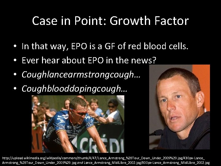
Case in Point: Growth Factor • • In that way, EPO is a GF of red blood cells. Ever hear about EPO in the news? Coughlancearmstrongcough… Coughblooddopingcough… http: //upload. wikimedia. org/wikipedia/commons/thumb/4/47/Lance_Armstrong_%28 Tour_Down_Under_2009%29. jpg/430 px-Lance_ Armstrong_%28 Tour_Down_Under_2009%29. jpg and Lance_Armstrong_Midi. Libre_2002. jpg/800 px-Lance_Armstrong_Midi. Libre_2002. jpg
Case in Point: Growth Factor • EPO has been used as a performance-enhancing drug (PED) (in this case known as “blood doping”) because it increases the oxygen-carrying capacity of the blood by making it extra rich with blood cells. • The downside? – All those extra blood cells makes your blood considerably thicker/more viscous. – Dehydration also makes your blood thicker/more viscous. – Thick/viscous blood = increased risk of stroke/heart attack/death. • Just say no to EPO.
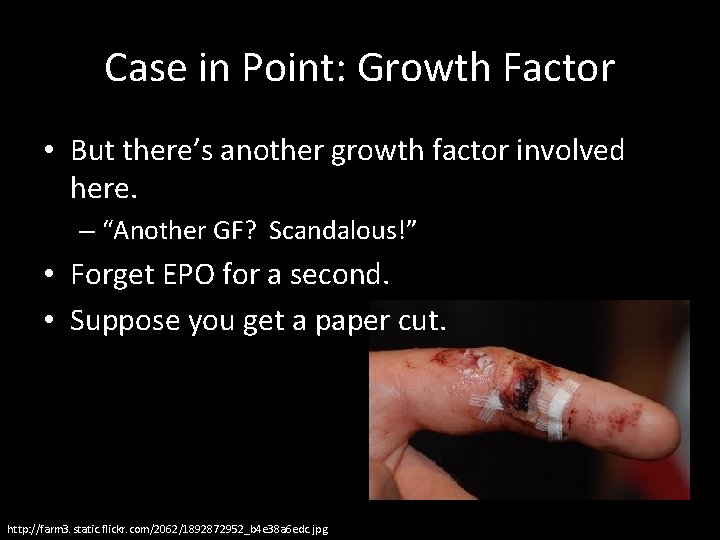
Case in Point: Growth Factor • But there’s another growth factor involved here. – “Another GF? Scandalous!” • Forget EPO for a second. • Suppose you get a paper cut. http: //farm 3. static. flickr. com/2062/1892872952_b 4 e 38 a 6 edc. jpg
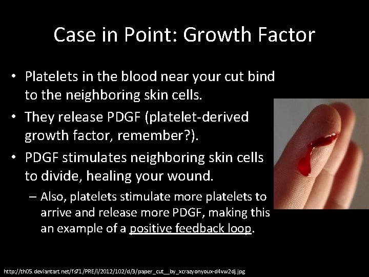
Case in Point: Growth Factor • Platelets in the blood near your cut bind to the neighboring skin cells. • They release PDGF (platelet-derived growth factor, remember? ). • PDGF stimulates neighboring skin cells to divide, healing your wound. – Also, platelets stimulate more platelets to arrive and release more PDGF, making this an example of a positive feedback loop. http: //th 05. deviantart. net/fs 71/PRE/i/2012/102/d/3/paper_cut__by_xcrazyonyoux-d 4 vw 2 dj. jpg
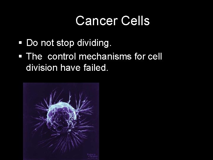
Cancer Cells Do not stop dividing. The control mechanisms for cell division have failed.
So the logical question… • We have all these checkpoints. • What happens if something goes wrong. • In a word? – Cancer. • Many cancers are caused by cells that escape from the normal cell cycle. – The brakes on cell division have been removed. • Normal cells divide up to 60 times or so. – Cancer cells? A lot more. • TED: George Zaidan – How Cancer Cells Behave Differently from Healthy Ones
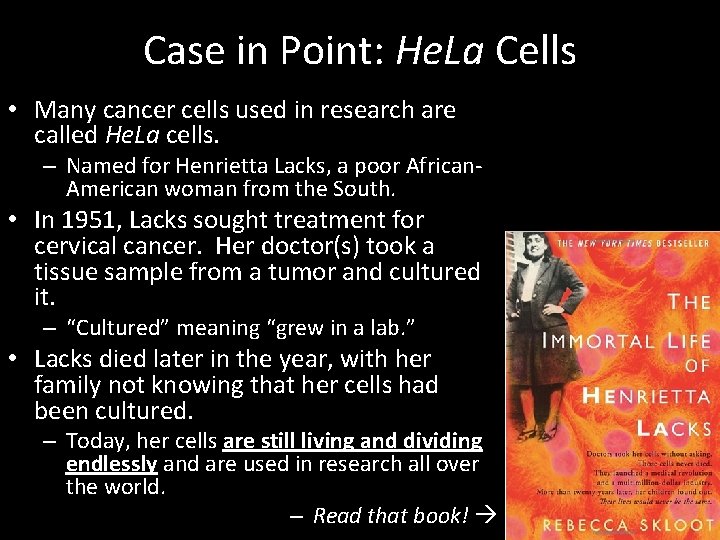
Case in Point: He. La Cells • Many cancer cells used in research are called He. La cells. – Named for Henrietta Lacks, a poor African. American woman from the South. • In 1951, Lacks sought treatment for cervical cancer. Her doctor(s) took a tissue sample from a tumor and cultured it. – “Cultured” meaning “grew in a lab. ” • Lacks died later in the year, with her family not knowing that her cells had been cultured. – Today, her cells are still living and dividing endlessly and are used in research all over the world. – Read that book!
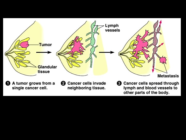
Cancer Terminology • Tumor – A mass of cancer cells. • Remember, cancer cells divide A LOT. – Benign tumors are sitting in their original spot. – Metastatic or malignant tumors are spreading throughout the body. • Carcinogen – Something that causes cancer. • Like smoking or chewing tobacco.
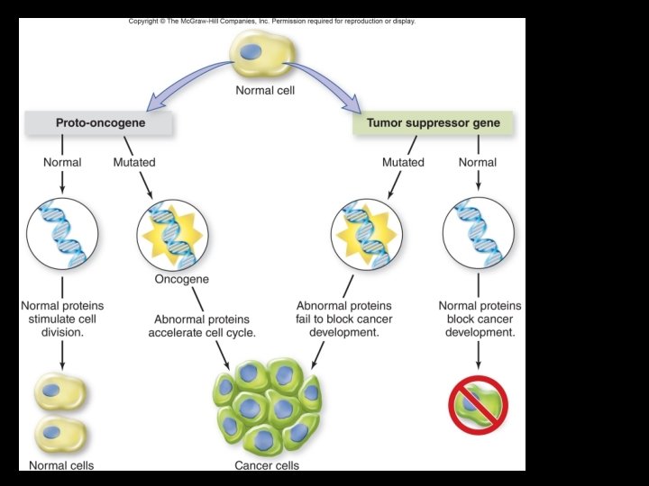
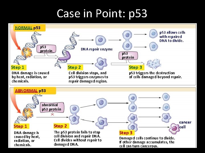
Cancerous Growth Factors • Proto-oncogenes are genes that cause cancer if switched “on” and mutated. – Ras is a group of genes like this and are involved in 30% of human cancers. • Tumor-suppressor genes cause cancer if switched “off. ” – p 53 is a tumor-suppressor gene and is involved in 50% of human cancers. • So it follows that mutagens are things that cause mutations and may also be carcinogens.
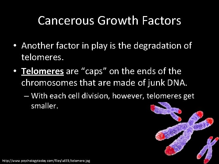
Cancerous Growth Factors • Another factor in play is the degradation of telomeres. • Telomeres are “caps” on the ends of the chromosomes that are made of junk DNA. – With each cell division, however, telomeres get smaller. http: //www. psychologytoday. com/files/u 693/telomere. jpg
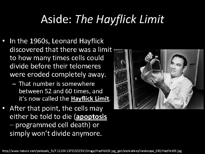
Aside: The Hayflick Limit • In the 1960 s, Leonard Hayflick discovered that there was a limit to how many times cells could divide before their telomeres were eroded completely away. – That number is somewhere between 52 and 60 times, and it’s now called the Hayflick Limit. • After that point, the cells may either be told to die (apoptosis – programmed cell death) or simply won’t divide anymore. http: //www. nature. com/polopoly_fs/7. 11184. 1372152253!/image/Hayflick 28. jpg_gen/derivatives/landscape_630/Hayflick 28. jpg
Cancer Growth Factors • Mutagens/Carcinogens – UV radiation • Tanning beds! – Chemicals – Radiation – Heat – Pollution – Cigarette smoke • Other Factors – Age – Genetics
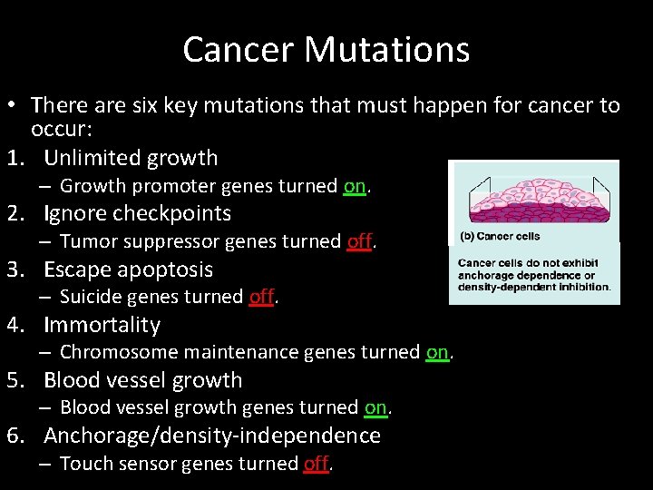
Cancer Mutations • There are six key mutations that must happen for cancer to occur: 1. Unlimited growth – Growth promoter genes turned on. 2. Ignore checkpoints – Tumor suppressor genes turned off. 3. Escape apoptosis – Suicide genes turned off. 4. Immortality – Chromosome maintenance genes turned on. 5. Blood vessel growth – Blood vessel growth genes turned on. 6. Anchorage/density-independence – Touch sensor genes turned off.
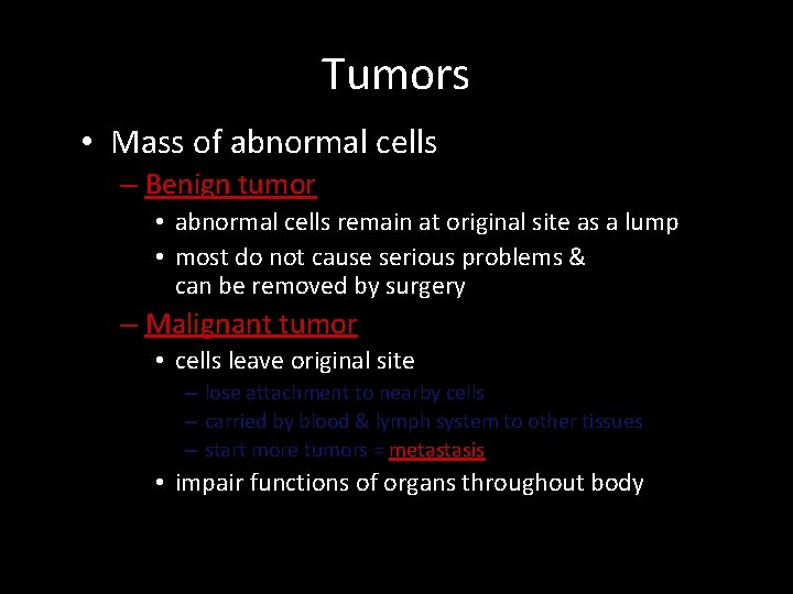
Tumors • Mass of abnormal cells – Benign tumor • abnormal cells remain at original site as a lump • most do not cause serious problems & can be removed by surgery – Malignant tumor • cells leave original site – lose attachment to nearby cells – carried by blood & lymph system to other tissues – start more tumors = metastasis • impair functions of organs throughout body
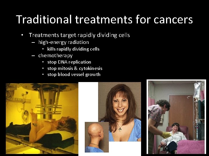
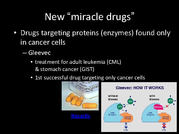
Traditional treatments for cancers • Treatments target rapidly dividing cells – high-energy radiation • kills rapidly dividing cells – chemotherapy • stop DNA replication • stop mitosis & cytokinesis • stop blood vessel growth
New “miracle drugs” • Drugs targeting proteins (enzymes) found only in cancer cells – Gleevec • treatment for adult leukemia (CML) & stomach cancer (GIST) • 1 st successful drug targeting only cancer cells without Gleevec Novartis with Gleevec
Now go to assignment
Cancer Treatments • High-Energy Radiation – Kill rapidly-dividing cells. • Chemotherapy – Stop DNA replication, mitosis/cytokinesis, and blood vessel growth. • Gleevec – A relatively new anti-cancer drug that targets enzymes found only in cancer cells.
How does cancer work? • Cancerous genes or cells are often found to be activating lots of cyclins, thus pushing the cell through the cell cycle too rapidly. • Tumor suppressor genes prevent errant DNA from being copied in mitosis. – p 53, for example, stops cell division if bad DNA is found, but if p 53 isn’t working…you get the idea. • Subsequent growth and spread of tumors can begin to block blood vessels, clog body systems, prevent other cells from functioning…the list goes on…
Case in Point: p 53 NORMAL p 53 allows cells with repaired DNA to divide. p 53 protein DNA repair enzyme p 53 protein Step 1 Step 2 Step 3 DNA damage is caused by heat, radiation, or chemicals. Cell division stops, and p 53 triggers enzymes to repair damaged region. p 53 triggers the destruction of cells damaged beyond repair. ABNORMAL p 53 abnormal p 53 protein Step 1 Step 2 DNA damage is caused by heat, radiation, or chemicals. The p 53 protein fails to stop cell division and repair DNA. Cell divides without repair to damaged DNA. Step 3 Damaged cells continue to divide. If other damage accumulates, the cell can turn cancerous. cancer cell