In the name of God Ovarian cysts and


 under ultrasound guidance")


 Lactate dehydrogenase (LDH) CA-125 Human chorionic gonadotropin (h. CG)")


- Slides: 61
In the name of God Ovarian cysts and neoplasms in infants, children, and adolescents Fariba Behnamfar Gyn. Oncology Fellowship Isfahan University of Medical Sciences
Case 1 A two months old Infant with palpable lower abdominal mass Sonography: 4 cm simple cyst in right ovary
Additional Imaging? Tumor markers? Surgery? Follow up? Emergency?
Introduction Ovarian masses may represent physiologic cysts, benign neoplasms, or malignant neoplasms. They may be associated with pain or present as an asymptomatic mass Although relatively rare, they are the most common genital neoplasms occurring in childhood
ovarian preservation as the standard Historically, all ovarian masses discovered in infants, children, and adolescents were removed surgically. However, the identification of tumor markers and advances in radiologic imaging allow a more conservative approach to the management of these neoplasms, with ovarian preservation as the standard except in cases of cancer.
CLASSIFICATION The World Health Organization classifies ovarian neoplasms based upon histologic cell type and benign versus malignant state The majority of ovarian tumors in girls and adolescents are of germ cell origin. By comparison, epithelial tumors account for the largest proportion of ovarian neoplasms in adults
Most childhood ovarian masses are benign. However, it is important for the clinician to establish an early diagnosis to reduce the risk of ovarian torsion with possible loss of adnexa and to improve the prognosis for those lesions that are malignant
OVARIAN CYSTS IN THE FETUS Follicular ovarian cysts in fetuses and neonates are common and increase in frequency with advancing gestational age and some maternal complications, such as diabetes mellitus, preeclampsia, rhesus isoimmunization Among live births, incidence of clinically significant ovarian cysts is 1 in 2500
Diagnosis is based upon sonographically determined presence of four criteria: female sex, nonmidline regular. D cyct i normal-appearing gastrointestinal tract a g n o Size and appearance are used to characterize cystic structure, normalappearing urinary tracts as probably physiologic or probably pathologic.
Follicular cysts Simple cysts less than 2 cm in diameter are considered physiologic Larger and complex cysts are more likely to be nonphysiologic Associated anomalies are rare since the cysts usually result from hormonal EFFECT
Follicular cysts are commonly detected incidentally on antenatal ultrasound examination The etiology is unclear, but they most likely arise from ovarian stimulation by maternal and fetal gonadotropin The majority of fetal ovarian cysts are unilateral, although both ovaries may be involved
Ovarian cyst in female fetus at 34 weeks' gestational age
Differential diagnosis The differential diagnosis of a fetal cystic intraabdominal mass includes › › genitourinary tract disorders gastrointestinal tract disorders miscellaneous disorders —[5]. The rate of malignancy is so low that it need not be considered in making therapeutic decisions.
Management and outcome › Spontaneous regression of both simple and complex cysts often occurs either antenatally or postpartum by six months of age › management is usually expectant › In one review of 66 published cases 90 percent regressed spontaneously by three months
Management and outcome Spontaneous regression of both simple and complex cysts often occurs either antenatally or postpartum by six months of age, therefore management is usually expectant In one review of 66 published cases of simple cysts, 90 percent resolved by three months The rate of malignancy is so low that it need not be considered in making therapeutic decisions
Ultrasound examination should be performed every three to four weeks antenatally After birth, neonatal management is as described below If in-utero torsion occurs, the ovary may undergo necrosis and develop into a calcified mass, a sessile mass, or disappear entirely
Complications that can occur include intracystic hemorrhage, rupture with possible intraabdominal hemorrhage, gastrointestinal or urinary tract obstruction, ovarian torsion and necrosis, incarceration in an inguinal hernia, difficulty with delivery due to fetal abdominal dystocia, respiratory distress at birth from a mass effect on the diaphragm
In a long-term follow-up study of 21 girls with prenatal ovarian cysts, sonographic follow-up was obtained in 14. There was inability to appreciate the ovary in 8 of 11 ovaries in which the cysts appeared complex on the first postnatal scan (two were treated with postnatal salpingooophorectomy; one was treated with postnatal aspiration; the remainder were observed)
These data suggest that prenatally detected ovarian cysts should be closely monitored, particularly if the cyst appears complex on postnatal sonography, due to the : increased risk of torsion and subsequent ovarian loss
Antenatal aspiration of large cysts (greater than 4 to 6 cm) under ultrasound guidance has been advocated to reduce the risk of complications : possible misdiagnosis and potential complications from the aspiration technique itself In particular, small anechoic cysts should be left alone a large cyst that undergoes torsion may lead to loss of the ovary and impair future fertility
Advantages of aspiration include elimination of the cyst with reduction of the risk of cystrelated complications and need for neonatal surgery. Disadvantages are risk of spillage. Complex cysts cannot be aspirated.
preferred route of delivery Most fetuses can be delivered vaginally with cesarean delivery reserved for the usual obstetric indications. Cesarean birth may be the preferred route of delivery of fetuses with very large cysts to prevent rupture and/or dystocia. Cyst aspiration antepartum is an alternative approach. There is no increased risk of recurrence in subsequent pregnancies
OVARIAN CYSTS IN NEONATES Clinical features and diagnosis A pelvic mass in a newborn is most likely a physiologic cyst on the fetal ovary resulting from maternal hormonal stimulation in-utero. The differential diagnosis is the same as that for fetuses Ultrasound examination may show a simple or complex sonographic pattern. A complex sonographic appearance makes a precise diagnosis more difficult.
Detection Neonatal cysts may have been initially detected on antenatal sonographic examination or may be identified as an asymptomatic abdominal mass because of displacement upward and out of the narrow neonatal pelvis. The ovary containing the cyst is generally freely mobile.
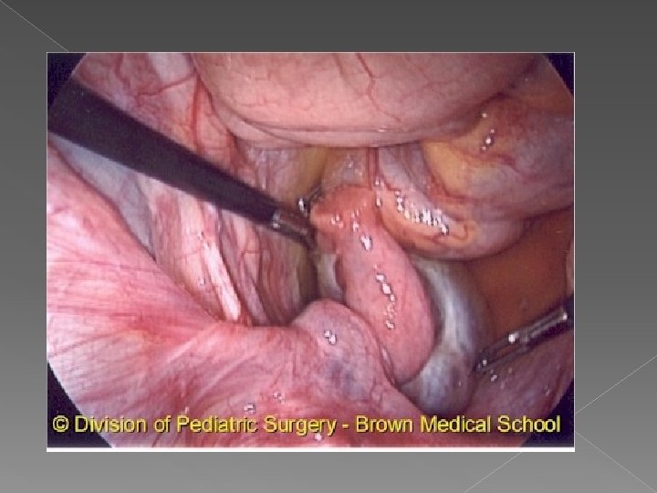
Ovarian torsion Torsion can occur with a cyst of any size, particularly when long pedicles are present Parents should be made aware of the signs and symptoms of torsion (lower abdominal pain of sudden onset, nausea, vomiting, low-grade fever) so they can seek emergent care
Ovarian tortion An attempt should always be made to salvage the torsed ovary by untwisting the vascular pedicle. A bivalve technique can be used to try to salvage a dark-appearing torsed ovary; this technique decreases the intraovarian pressure caused by venous occlusion and permits arterial flow into the ovary However, in rare instances, oophorectomy is necessary because of severe necrosis
Management Spontaneous regression usually occurs by four to six months of age management of neonatal cysts consists of: Serial ultrasound examinations at birth and every four to six weeks Aspiration of simple cysts ≥ 4 to 5 cm Surgical intervention for complex cysts, cysts that are increasing in size, symptomatic cysts, and cysts persisting for more than four to six months Laparoscopic surgery is feasible and safe in neonates with ovarian cysts 30 to 40 percent undergo torsion or another complication
OVARIAN CYSTS IN INFANTS AND PREPUBERTAL CHILDREN
Case 2 A 12 y old girl RLQ Abdominal pain Palpable Mass Sono: solid cystic mass
Additional Imaging Tumor Markers MRI Laparotomy versus laparoscopy Conservative Management Ovarian Preservation Frozen section Management of malignancy
Etiology Physiologic cysts are uncommon between the neonatal period and puberty because gonadotropin stimulation decreases most simple ovarian cysts in children are physiologic and result from enlargement of a cystic follicle Some ovarian cysts are hormonally active and result in precocious pseudopuberty (eg, Mc. Cune-Albright syndrome)
In girls with hormonally active cysts, the ovarian enlargement may be mistaken for an ovarian tumor, leading to unnecessary oophorectomy Girls presenting with premature vaginal bleeding and ovarian enlargement should be evaluated for features of Mc. Cune-Albright syndrome to avoid this potential mistake
Cafe-au-lait spots in Mc. Cune. Albright syndrome
Clinical manifestations An ovarian cyst in a young child is often discovered by a parent or clinician asymptomatic abdominal mass or because of increasing abdominal girth. in early life, the ovary is an abdominal organ and more susceptible to torsion Chronic abdominal aching pain, either periumbilical or localized to a lower quadrant, may be present. .
Acute severe pain may result from torsion, perforation, infarction, hemorrhage (into or from the ovarian mass) › intermittent pain: partial or intermittent torsion, which may resolve without therapy or act as a warning sign of impending torsion requiring emergency surgery › Torsion also causes nausea, vomiting, pallor, and leukocytosis followed by less severe localized pain
Evaluation Ultrasonography is the primary assessment tool. If torsion is suspected , Doppler ultrasound may be helpful. However, is not always conclusive. CT and MRI have also been used in an attempt to clarify equivocal findings
Children with recurrent, large, or multicystic ovarian masses and signs of early sexual development should be evaluated for precocious puberty In the absence of precocity, the possibility of a periovarian or mesothelial cyst should be considered
Management and outcome An ovarian mass that is purely cystic or has few internal echoes suggestive of hemorrhage and no complex features is almost certainly benign and can be managed by observation A follow-up ultrasound examination in four to eight weeks should be performed. If the cyst has not resolved and the ultrasonic characteristics are still reassuring, then continued observation is appropriate.
Management and outcome If acute rupture with hemorrhage occurs and bleeding is associated with hemodynamic instability, surgery should be done surgery can usually be performed laparoscopically , A hemoperitoneum is not a contraindication Laparotomy is indicated if the surgeon is not experienced in laparoscopy on children or if the patient is hypotensive In contrast, surgery is always indicated at the time of diagnosis of ovarian torsion for salvage
ovarian torsion Ovarian masses associated with torsion are usually benign. As an example, a series describing 102 girls aged 2 days to 20 years who underwent 106 consecutive separate ovarian operations found 42 percent (25/59) of those who presented with acute abdominal pain had ovarian torsion; the ovarian mass was malignant in only one of these girls In contrast, 26 percent of those presenting with asymptomatic abdominal masses had malignancies.
OVARIAN CYSTS IN ADOLESCENTS
an age group in which the development of both simple and complex cysts is quite common. Adolescent ovaries may contain multiple follicles in different stages of development. Most simple cysts result from failure of the maturing follicle to ovulate and involute. Cysts in the postmenarcheal adolescent may be asymptomatic , but can cause menstrual irregularities, pelvic pain, or heaviness.
Rupture leads to intraabdominal pain and bleeding, which can be minor or severe. Torsion also causes acute pain, as well as nausea, vomiting, pallor, and leukocytosis (with left shift), often followed by less severe localized pain. It is unclear why some nonruptured functional cysts cause symptoms and others do not
Differential diagnosis › The differential diagnosis of ovarian cysts in the adolescent patient is complex because of the functioning ovary, the onset of sexual activity, and the possibility of pregnancy.
Differential diagnosis Obstructive genital lesions Ovarian tumors Tubal conditions Uterine masses Gastrointestinal conditions
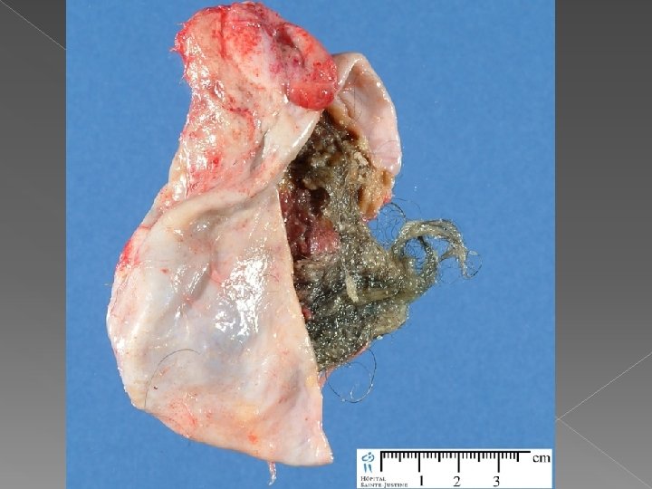
Evaluation should include a detailed menstrual and sexual history � The presence of calcification on ultrasound examination or an abdominal radiograph suggests a teratoma. � Color Doppler velocimetry of is used to detect low peripheral resistance, which can result from neovascularization related to malignancy � A pregnancy test and complete blood count are obtained, as indicated �
Management and outcome Follicular cysts Corpus luteum cysts
follicular cysts — Most resolve spontaneously in one to two months. Asymptomatic simple cysts <10 cm on ultrasound examination can be observed with or without administration of oral contraceptive pills. The patient should be evaluated monthly Ovarian cystectomy is preferred to cyst aspiration due to the high rate of recurrence after aspiration
follicular cysts If the cyst recurs or operative intervention is needed, the procedure should be conservative and preserve as much ovarian tissue as possible Patients incidentally found to have small follicular cysts at the time of surgery should not undergo cyst aspiration or cystectomy
Corpus luteum cysts common, can reach 5 to 12 cm In the absence of pain or intraperitoneal bleeding, observation for a time period between two weeks and three months The oral contraceptive pills will keep a new cyst from forming but do not help the current cyst regress. Corpus luteum cysts are at increased risk of torsion due to increased ovarian size and weight , management is removal of the cyst and cyst wall and conservation of ovary
Ovarian Cancer Ovarian neoplasms account for approximately 1 percent of all tumors in children and adolescents. approximately 35 percent of all ovarian neoplasms occurring during childhood and adolescence are malignant. Ovarian cancer is the most common gynecologic malignancy in women ≤ 25 years of age, and germ cell is the most common histology
Clinical manifestations Patients with an ovarian tumor may present with abdominal pain or complaints of increasing abdominal girth, nausea, and vomiting; or they may be asymptomatic abdominal palpation and rectal examination in the dorsal supine position are important Nonspecific symptoms may be more common with epithelial tumors
Imaging Sonography Doppler A solid ovarian mass in childhood is always considered malignant until proven otherwise by histological examination s
Tumor markers Alpha-fetoprotein (AFP) Lactate dehydrogenase (LDH) CA-125 Human chorionic gonadotropin (h. CG)
Treatment Surgical intervention is directed toward preservation of reproductive and sexual function. Unless a malignancy is diagnosed definitively on frozen section at the time of the procedure, conservative surgery should be undertaken with excision of the lesion and ovarian reconstruction
management If malignancy is suspected or confirmed, adequate staging includes abdominal and pelvic exploration, peritoneal washings, biopsies of suspicious areas, and periaortic and pelvic lymph node sampling