
In The Name Of God Heart Failure By

In The Name Of God Heart Failure By: M. Mohamadian Ph. D student of molecular medicine
HF & CHF Definition - Heart failure (HF) is a clinical syndrome that occurs because of an inherited or acquired abnormality of cardiac structure and/or function, that impairs ability of ventricle to fill with or eject blood, and as a result, cardiac output decreases and is unable to meet the metabolic demands of the body. - Because many patients present without signs or symptoms of volume overload, the term “Heart failure” is preferred over the older term “Conjestive heart failure”
Epidemiology -HF prevalence follows an exponential pattern, rising with age, and affects 6– 10% of people over age 65. -Incidence of HF is lower in women than in men, but women constitute at least one-half the cases of HF because of their longer life expectancy. - HF patients are now categorized into one of two groups: HF with a depressed EF (systolic failure) HF with a preserved EF (diastolic failure) And CO

Systolic vs Diastolic HF
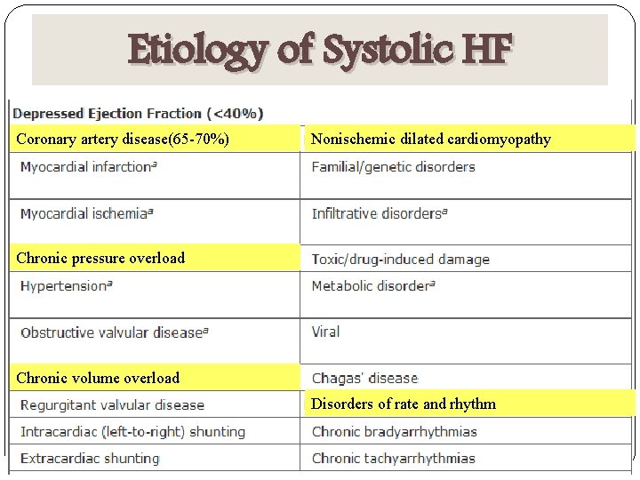
Etiology of Systolic HF Coronary artery disease(65 -70%) Nonischemic dilated cardiomyopathy Chronic pressure overload Chronic volume overload Disorders of rate and rhythm
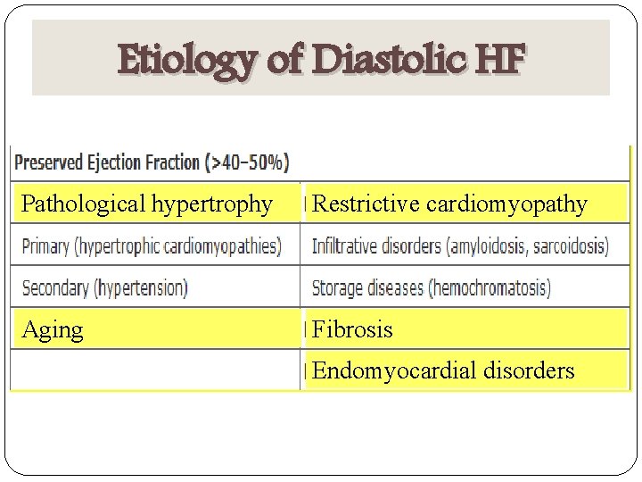
Etiology of Diastolic HF Pathological hypertrophy Restrictive cardiomyopathy Aging Fibrosis Endomyocardial disorders
Prognosis - Despite many recent advances in the evaluation and management of HF, the development of symptomatic HF still carries a poor prognosis. - Commonly, 30– 40% of patients die within 1 year of diagnosis and 60– 70% die within 5 years, mainly from worsening HF or as a sudden event (probably because of a ventricular arrhythmia). - Although it is difficult to predict prognosis in an individual, patients with symptoms at rest have a 30– 70% annual mortality rate, whereas patients with symptoms with moderate activity have an annual mortality rate of 5– 10%. Thus: Functional status is an important predictor of patient outcome.
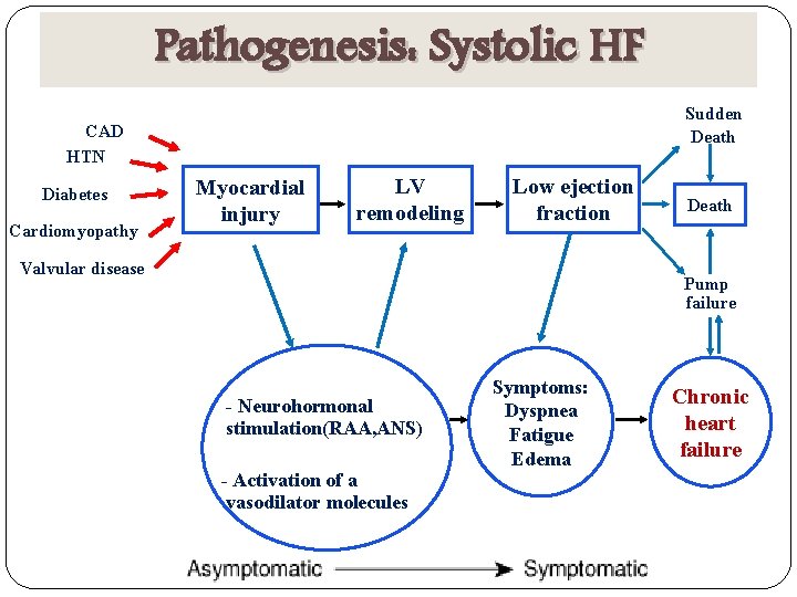
Pathogenesis: Systolic HF Sudden Death CAD HTN Diabetes Cardiomyopathy Myocardial injury LV remodeling Low ejection fraction Valvular disease Death Pump failure - Neurohormonal stimulation(RAA, ANS) - Activation of a vasodilator molecules Symptoms: Dyspnea Fatigue Edema Chronic heart failure
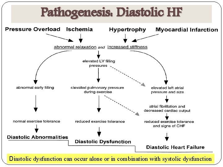
Pathogenesis: Diastolic HF Diastolic dysfunction can occur alone or in combination with systolic dysfunction
Clinical Manifestations Cardinal symptoms of HF are fatigue and shortness of breath - Fatigue: has been ascribed to the low cardiac output in HF. - Dyspnea: During exertion Less strenuous activity At rest Orthopnea: Nocturnal cough is a frequent manifestation. Paroxysmal Nocturnal Dyspnea (PND): acute episodes of severe shortness of breath and coughing that generally occur at night and awaken the patient from sleep, usually 1– 2 h after the patient retires. PND may be manifest by coughing or wheezing.
Clinical Manifestations Cheyne-Stokes Respiration (periodic respiration) (Apnea / Hypocapnea) - Is common in advanced HF and is usually associated with low CO. - Caused by a diminished sensitivity of the respiratory center to arterial PCO 2. Acute Pulmonary Edema Gastrointestinal symptoms. Anorexia, nausea, and early satiety associated with abdominal pain and fullness Cerebral symptoms, such as confusion, disorientation, and sleep and mood disturbances, particularly elderly patients with cerebral arteriosclerosis and reduced cerebral perfusion. Nocturia is common in HF and may contribute to insomnia.
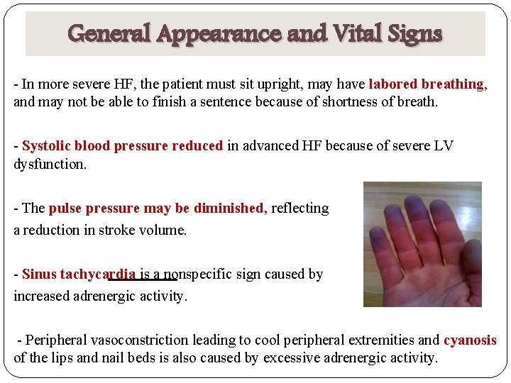
General Appearance and Vital Signs - In more severe HF, the patient must sit upright, may have labored breathing, and may not be able to finish a sentence because of shortness of breath. - Systolic blood pressure reduced in advanced HF because of severe LV dysfunction. - The pulse pressure may be diminished, reflecting a reduction in stroke volume. - Sinus tachycardia is a nonspecific sign caused by increased adrenergic activity. - Peripheral vasoconstriction leading to cool peripheral extremities and cyanosis of the lips and nail beds is also caused by excessive adrenergic activity.
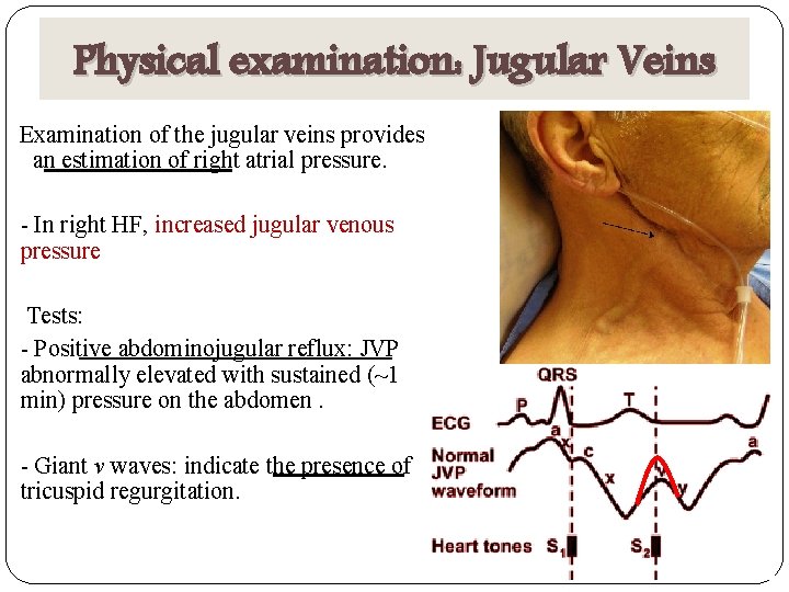
Physical examination: Jugular Veins Examination of the jugular veins provides an estimation of right atrial pressure. - In right HF, increased jugular venous pressure Tests: - Positive abdominojugular reflux: JVP abnormally elevated with sustained (~1 min) pressure on the abdomen. - Giant v waves: indicate the presence of tricuspid regurgitation.
Physical examination: Pulmonary Examination - Pulmonary crackles (rales or crepitations) result from the transudation of fluid from the intravascular space into the alveoli. ents without concomitant lung disease, rales are specificfor HF. - Pleural effusion result from the elevation of pleural capillary pressure and the resulting transudation of fluid into the pleural cavities. -Occur most commonly with biventricular failure, when unilateral they occur more frequently in the right pleural space.
Physical examination: Cardiac Examination - Cardiomegaly: the point of maximal impulse (PMI) is usually displaced below the fifth intercostal space and/or lateral to the midclavicular line. - An S 3 (or protodiastolic gallop) is most commonly present in patients with volume overload who have tachycardia and tachypnea. - A fourth heart sound (S 4) is not a specific indicator of HF but is usually present in patients with diastolic dysfunction. - The murmurs of mitral and tricuspid regurgitation are frequently present in patients with advanced HF.
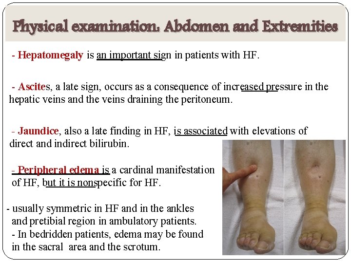
Physical examination: Abdomen and Extremities - Hepatomegaly is an important sign in patients with HF. - Ascites, a late sign, occurs as a consequence of increased pressure in the hepatic veins and the veins draining the peritoneum. - Jaundice, also a late finding in HF, is associated with elevations of direct and indirect bilirubin. - Peripheral edema is a cardinal manifestation of HF, but it is nonspecific for HF. - usually symmetric in HF and in the ankles and pretibial region in ambulatory patients. - In bedridden patients, edema may be found in the sacral area and the scrotum.
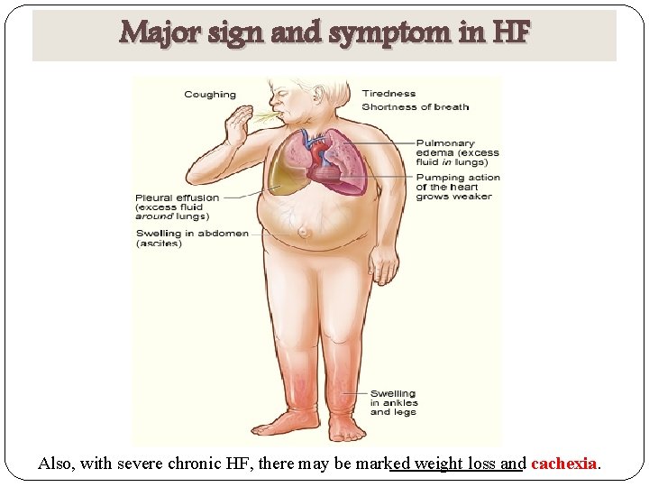
Major sign and symptom in HF Also, with severe chronic HF, there may be marked weight loss and cachexia.

Diagnosis - Electrocardiogram (ECG) -Assessment of cardiac rhythm, presence of LV hypertrophy or a prior MI (presence or absence of Q waves), as well as to determine QRS width. -A normal ECG virtually excludes LV systolic dysfunction. - Chest X-Ray sment of cardiac size and shape, as well as the state of the pulmonary vasculature, and noncardiac causes of the patient's symptoms. - 2 -D echocardiogram/Doppler Assessment of LV size and function and presence or absence of valvular and/or regional wall motion abnormalities (indicative of a prior MI). - Assessment of RV size and pulmonary pressures. - MRI: gold standard for assessing LV mass and volumes.

Diagnosis - Biomarkers - Circulating levels of natriuretic peptides : Both BNP and pro-BNP, are released from the failing heart, are relatively sensitive markers for the presence of HF with depressed EF. ut, natriuretic peptide levels increase with age, sex(woman) and renal impairmen, and decrease in obese patients. -Troponin T and I, C-reactive protein, TNF receptors, and uric acid, may be elevated in HF and provide important prognostic information. - Exercise Testing Not routinely advocated for patients with HF, but is useful for assessing the need for cardiac transplantation in patients with advanced HF.
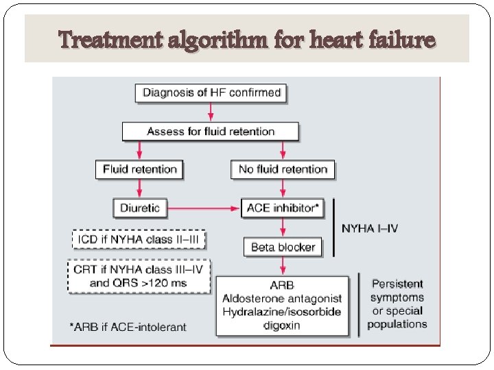
Treatment algorithm for heart failure

Staging & Treatment Stage A Risk factor reduction: treatment of High risk with no symptom HTN, diabetes, dyslipidemia If previous heart attack/ current diabetes mellitus or HTN, use ACE-I Stage B Structural heart disease, no Care measures in Stage A symptom Stage C Structural disease, previous or current symptoms Stage D Advanced symptoms, after assuring optimized medical care - ACE-I and beta blocker Care measures in Stage A and B - Diuretics - Digoxin - Dietary sodium restriction - Spironolactone Care measures in Stage A, B, C - Inotropes - Cardiac transplantation, VADs
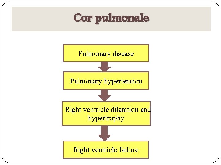
Cor pulmonale Pulmonary disease Pulmonary hypertension Right ventricle dilatation and hypertrophy Right ventricle failure
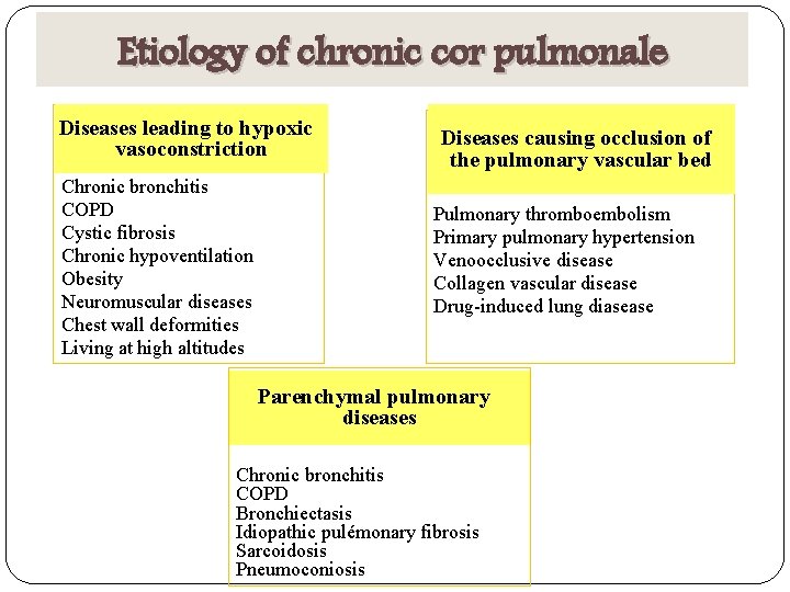
Etiology of chronic cor pulmonale Diseases leading to hypoxic vasoconstriction Chronic bronchitis COPD Cystic fibrosis Chronic hypoventilation Obesity Neuromuscular diseases Chest wall deformities Living at high altitudes Diseases causing occlusion of the pulmonary vascular bed Pulmonary thromboembolism Primary pulmonary hypertension Venoocclusive disease Collagen vascular disease Drug-induced lung diasease Parenchymal pulmonary diseases Chronic bronchitis COPD Bronchiectasis Idiopathic pulémonary fibrosis Sarcoidosis Pneumoconiosis
Symtoms and signs of cor pulmonale - Dyspnea - Effort-related syncope – because of the inability of the RV to deliver blood adequately to left side of the heart - Abdominal pain and ascite. - Lower extremity edema which can lead to peripheral vasodilatation Many of the signs that are encountered in cor pulmonale are also present in HF patients with a depressed EF, including tachypnea, elevated jugular venous pressures, hepatomegaly, Cyanosis and lower-extremity edema.
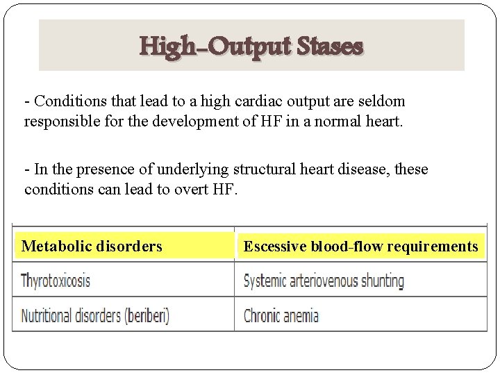
High-Output Stases - Conditions that lead to a high cardiac output are seldom responsible for the development of HF in a normal heart. - In the presence of underlying structural heart disease, these conditions can lead to overt HF. Metabolic disorders Escessive blood-flow requirements

Thank you for your time

- Slides: 28