
In The Name Of GOD Headache and visual




- Slides: 41
In The Name Of GOD
Headache and visual problem decreased libido Soheila sadeghi
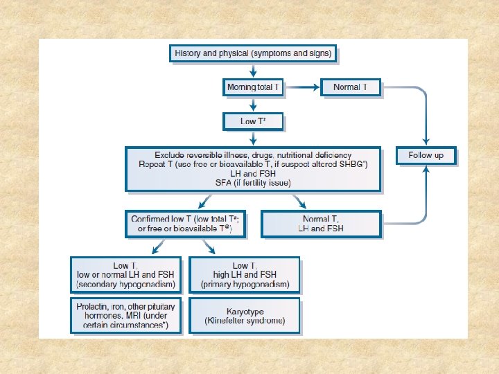
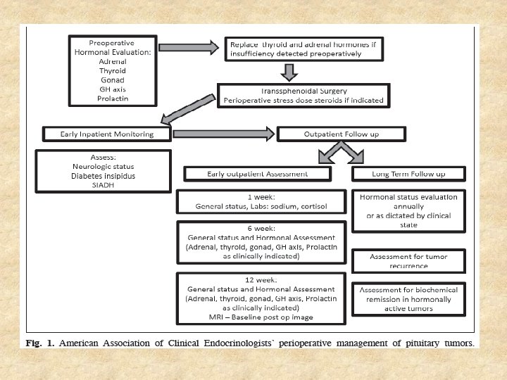
• Is there any difference between clinical feature of aneurysm and the other sellar mass? • Follow up after pituitary surgery? • Is hypogonadism reversible ? • Does the patient need testostrone replacement therapy?
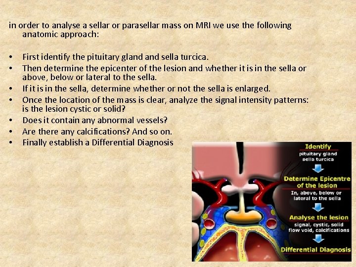
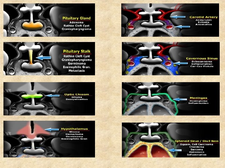
in order to analyse a sellar or parasellar mass on MRI we use the following anatomic approach: • • First identify the pituitary gland sella turcica. Then determine the epicenter of the lesion and whether it is in the sella or above, below or lateral to the sella. If it is in the sella, determine whether or not the sella is enlarged. Once the location of the mass is clear, analyze the signal intensity patterns: is the lesion cystic or solid? Does it contain any abnormal vessels? Are there any calcifications? And so on. Finally establish a Differential Diagnosis
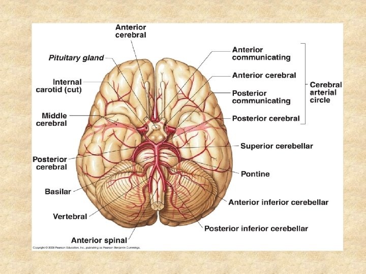
• • Aneurysms focal abnormal dilatation of a blood vessel typically occur in arteries Causes • Congenital • Atherosclerosis • Hypertension • vasculitis • hereditary connective tissue disorders • Marfan syndrome • Ehlers-Danlos syndrome • fibromuscular dysplasia • infection: mycotic aneurysm, syphilis (luetic aneurysm) • trauma • iatrogenic • myocardial infarction: may cause left a ventricular aneurysm • flow related (in cerebral AVM, contralateral ICA occlusion etc)
Complications • rupture • distal thromboembolism • pressure effects • A parasellar aneurysm may mimic a pituitary adenoma, and intraoperative rupture may be catastrophic, underlying the absolute need for preoperative diagnosis • Differentiating features of aneurysms from other pituitary masses may be subtle, including eye pain , very intense headaches, and relatively sudden onset of cranial nerve palsies • Although imaging techniques usually distinguish blood and hemorrhage from solid tumor or tissue, a highly vascular meningioma may be confused with an aneurysm.
• Intrasellar aneurysms that mimic pituitary tumors are rare with few previous reports • They may present with symptoms of mechanical compression or endocrinopathies • Though absence of endocrine symptoms may distinguish an aneurysm from a pituitary adenoma in most cases, mass effect from an enlarged aneurysm may prove the opposite in rare cases • Most symptoms of hypopituitarism secondary to intra or parasellar aneurysms have been reported from the effect of rupture or apoplexy
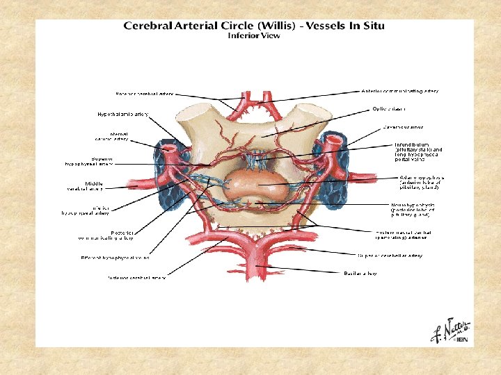
• Though the MRI is a very sensitive test. A wrong diagnosis may delay the management of a patient and lead to unwarranted complications such as intracerebral hemorrhage and even death • There are 2 primary anatomical subtypes of intrasellar aneurysms: q The first type is infradiaphragmantic (cavernous/clinoid segment of internal carotid artery) aneurysms with medial extension into the sella ( present with endocrinopathies) q The second type is supradiaphragmatic, which are suprasellar aneurysms originating from the ophthalmic segment of the ICA or the anterior communicating artery ( present with visual changes)
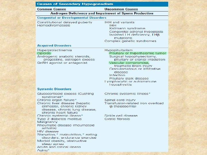
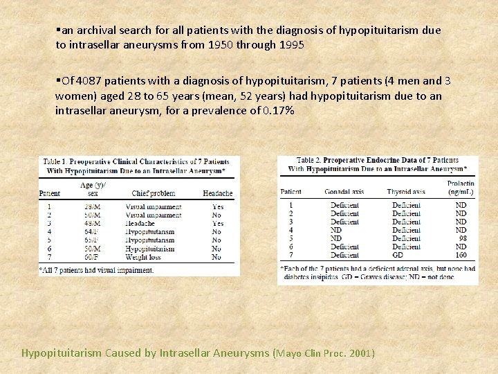
• An intrasellar aneurysm is a rare case of hypopituitarism. • It accounts for 0. 17% of all cases of hypopituitarism • The most common endocrinopathies are typically hyperprolactinemia and hypogonadism • Anterior communicating artery aneurysms usually indent the superior chiasmatic surface, while aneurysms of the ophthalmic segment of the internal carotid sometimes displace the optic nerve or chiasm superiorly, and occupy the sella turcica
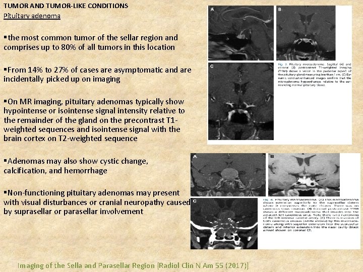
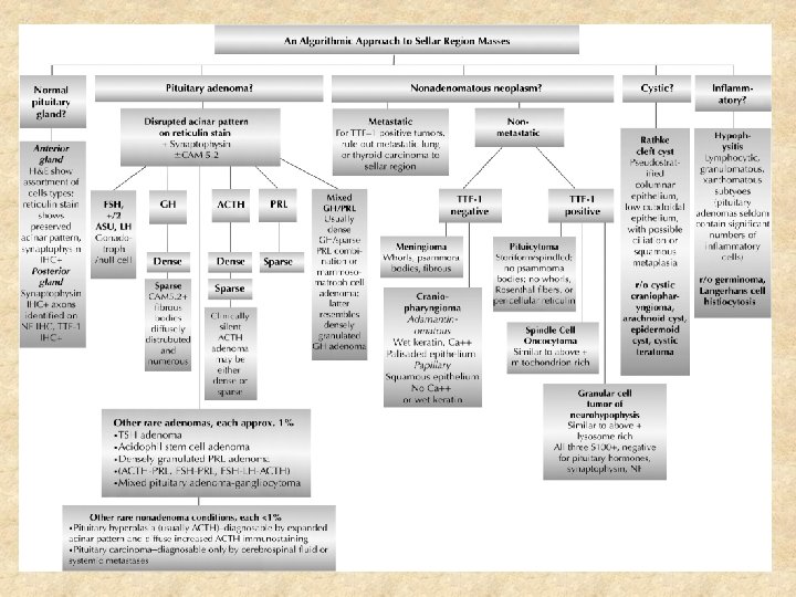
TUMOR AND TUMOR-LIKE CONDITIONS Pituitary adenoma §the most common tumor of the sellar region and comprises up to 80% of all tumors in this location §From 14% to 27% of cases are asymptomatic and are incidentally picked up on imaging §On MR imaging, pituitary adenomas typically show hypointense or isointense signal intensity relative to the remainder of the gland on the precontrast T 1 weighted sequences and isointense signal with the brain cortex on T 2 -weighted sequence §Adenomas may also show cystic change, calcification, and hemorrhage §Non-functioning pituitary adenomas may present with visual disturbances or cranial neuropathy caused by suprasellar or parasellar involvement Imaging of the Sella and Parasellar Region [Radiol Clin N Am 55 (2017)]
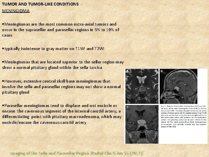
TUMOR AND TUMOR-LIKE CONDITIONS MENINGIOMA §Meningiomas are the most common extra-axial tumors and occur in the suprasellar and parasellar regions in 5% to 10% of cases §typically isointense to gray matter on T 1 WI and T 2 WI §Meningiomas that are located superior to the sellar region may show a normal pituitary gland within the sella turcica §However, extensive central skull base meningiomas that involve the sella and parasellar regions may not show a normal pituitary gland §Parasellar meningiomas tend to displace and not encircle or encase the cavernous segment of the internal carotid artery, a differentiating point with pituitary macroadenoma, which may encircle/encase the cavernous carotid artery Imaging of the Sella and Parasellar Region [Radiol Clin N Am 55 (2017)]
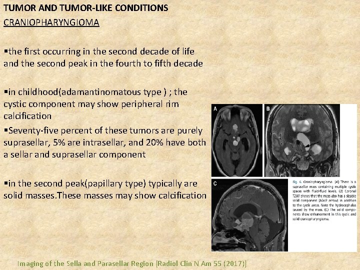
TUMOR AND TUMOR-LIKE CONDITIONS CRANIOPHARYNGIOMA §the first occurring in the second decade of life and the second peak in the fourth to fifth decade §in childhood(adamantinomatous type ) ; the cystic component may show peripheral rim calcification §Seventy-five percent of these tumors are purely suprasellar, 5% are intrasellar, and 20% have both a sellar and suprasellar component §in the second peak(papillary type) typically are solid masses. These masses may show calcification Imaging of the Sella and Parasellar Region [Radiol Clin N Am 55 (2017)]
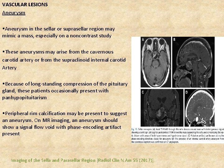
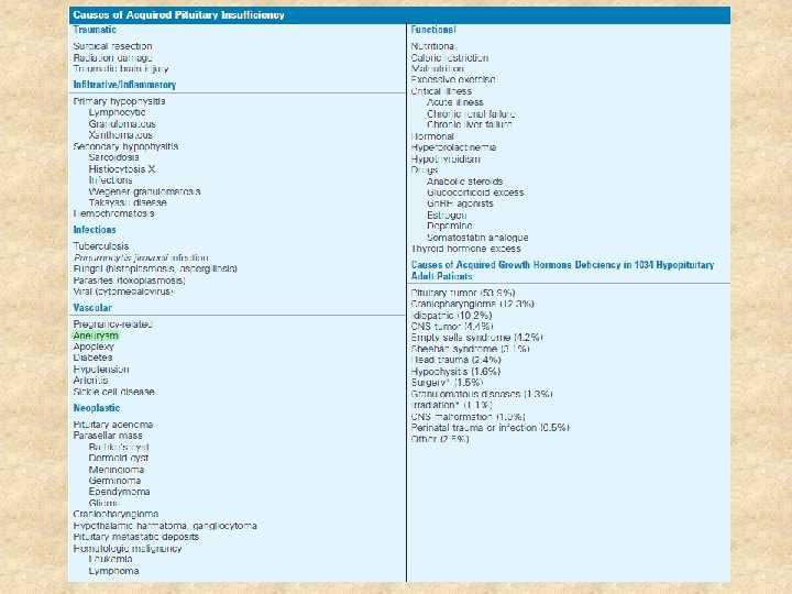
VASCULAR LESIONS Aneurysm §Aneurysm in the sellar or suprasellar region may mimic a mass, especially on a noncontrast study §These aneurysms may arise from the cavernous carotid artery or from the supraclinoid internal carotid Artery §Because of long-standing compression of the pituitary gland, these patients occasionally present with panhypopituitarism §Peripheral rim calcification may be present to suggest an aneurysm. On MR imaging, an aneurysm should show a signal flow void with phase-encoding artifact present Imaging of the Sella and Parasellar Region [Radiol Clin N Am 55 (2017)]
CONGENITAL/DEVELOPMENTAL LESIONS RATHKE CLEFT CYST §arise from the remnant of Rathke pouch during development of the pituitary gland §These cysts may lie within the sella, extend into the suprasellar cistern, or present purely as suprasellar cysts, but are always located inferior to the optic chiasm Imaging of the Sella and Parasellar Region [Radiol Clin N Am 55 (2017)]
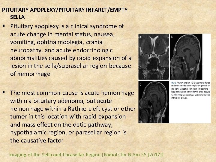
PITUITARY APOPLEXY/PITUITARY INFARCT/EMPTY SELLA § Pituitary apoplexy is a clinical syndrome of acute change in mental status, nausea, vomiting, ophthalmoplegia, cranial neuropathy, and acute endocrinologic abnormalities caused by rapid expansion of a lesion in the sella/suprasellar region because of hemorrhage § The most common cause is acute hemorrhage within a pituitary adenoma, but acute hemorrhage within a Rathke cleft cyst or other tumor in this location with rapid expansion and mass effect on the optic pathway, hypothalamic region, or parasellar region is the causative factor Imaging of the Sella and Parasellar Region [Radiol Clin N Am 55 (2017)]
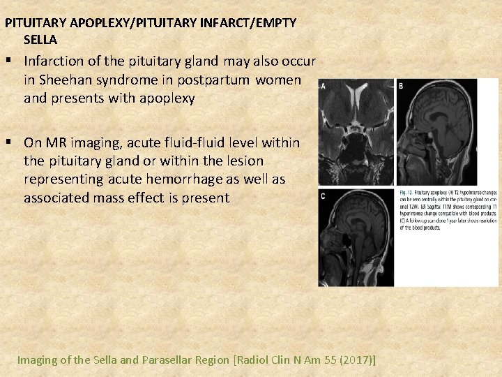
PITUITARY APOPLEXY/PITUITARY INFARCT/EMPTY SELLA § Infarction of the pituitary gland may also occur in Sheehan syndrome in postpartum women and presents with apoplexy § On MR imaging, acute fluid-fluid level within the pituitary gland or within the lesion representing acute hemorrhage as well as associated mass effect is present Imaging of the Sella and Parasellar Region [Radiol Clin N Am 55 (2017)]
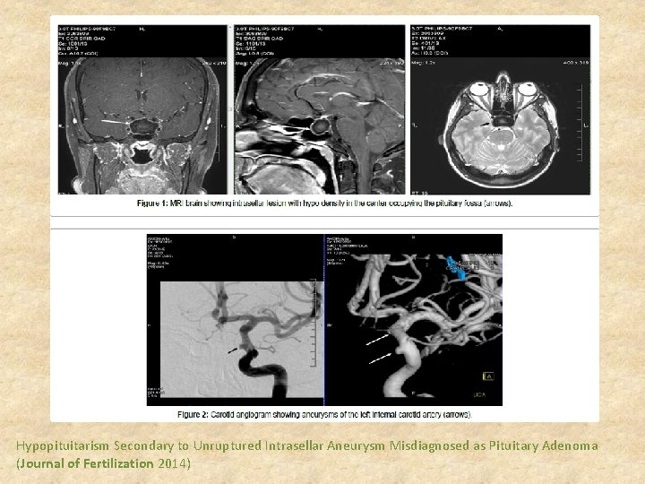
§ A 37 year-old female presented for management of secondary infertility with past medical history significant for long standing amenorrhea( At the age of 23, she started having new onset headaches and stopped having regular menstrual cycles)and a pituitary mass(<1 cm), not requiring treatment § She was taken to surgery for surgical excision of pituitary mass but on day of surgery was diagnosed to have an intrasellar aneurysm instead. § Fortunately the patient never experienced complications of aneurysm rupture and currently stable under management for hypopituitarism Hypopituitarism Secondary to Unruptured Intrasellar Aneurysm Misdiagnosed as Pituitary Adenoma (Journal of Fertilization 2014)
Hypopituitarism Secondary to Unruptured Intrasellar Aneurysm Misdiagnosed as Pituitary Adenoma (Journal of Fertilization 2014)
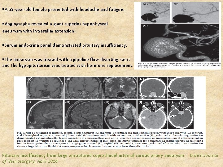
§A 59 -year-old female presented with headache and fatigue. §Angiography revealed a giant superior hypophyseal aneurysm with intrasellar extension. §Serum endocrine panel demonstrated pituitary insufficiency. §The aneurysm was treated with a pipeline flow-diverting stent and the hypopituitarism was treated with hormone replacement. Pituitary insufficiency from large unruptured supraclinoid internal carotid artery aneurysm British Journal of Neurosurgery, April 2014
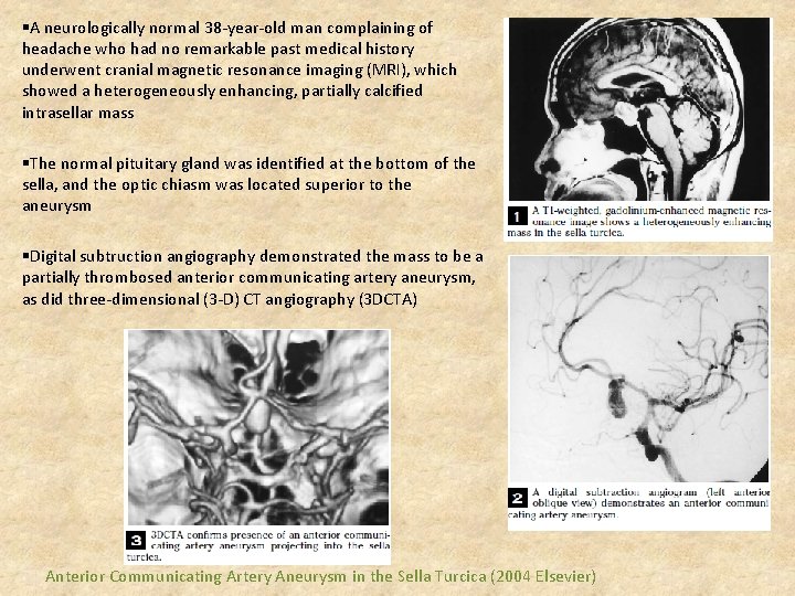
§A neurologically normal 38 -year-old man complaining of headache who had no remarkable past medical history underwent cranial magnetic resonance imaging (MRI), which showed a heterogeneously enhancing, partially calcified intrasellar mass §The normal pituitary gland was identified at the bottom of the sella, and the optic chiasm was located superior to the aneurysm §Digital subtruction angiography demonstrated the mass to be a partially thrombosed anterior communicating artery aneurysm, as did three-dimensional (3 -D) CT angiography (3 DCTA) Anterior Communicating Artery Aneurysm in the Sella Turcica (2004 Elsevier)
Coexistence of intracranial aneurysm with pituitary adenoma • The coexistence of PA and IA is a rare event, with an incidence of 0. 5% to 7. 4% • To date, many mechanisms have been proposed to explain the apparent increased incidence of IA in PA patients, including mechanical effects of PA on the vasculature, local circulatory changes (due to increased vascular demand), and hormonal influences • the growth of PA leads to microanatomic alterations in the cerebral circulation, which, in light of compression or traction caused by PA, leads to a modification in the blood flow and, consequently, greater hemodynamic stress, thus favoring the formation of the aneurysm
• an IA coexisting with a sellar lesion was more frequently observed in patients with aneurysms of the ICA and the anterior communicating artery because they supplied the pituitary region • a clear association between IA and PA has not been fully understood • The growth hormone and associated growth factors play a fundamental role in the formation of the aneurysm and the fact that in some series approximately 50% of the pituitary adenomas associated with cerebral aneurysms
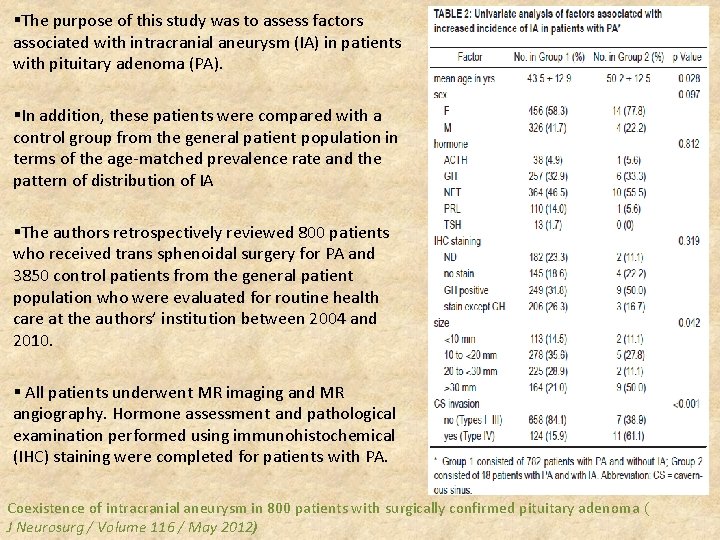
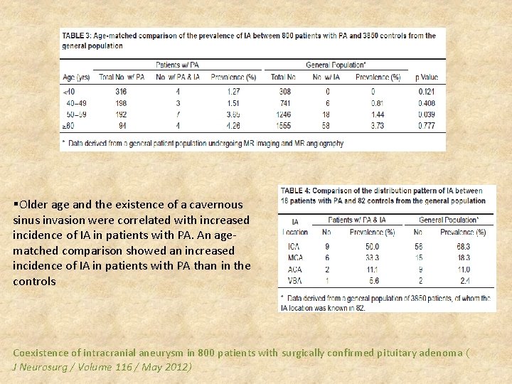
§The purpose of this study was to assess factors associated with intracranial aneurysm (IA) in patients with pituitary adenoma (PA). §In addition, these patients were compared with a control group from the general patient population in terms of the age-matched prevalence rate and the pattern of distribution of IA §The authors retrospectively reviewed 800 patients who received trans sphenoidal surgery for PA and 3850 control patients from the general patient population who were evaluated for routine health care at the authors’ institution between 2004 and 2010. § All patients underwent MR imaging and MR angiography. Hormone assessment and pathological examination performed using immunohistochemical (IHC) staining were completed for patients with PA. Coexistence of intracranial aneurysm in 800 patients with surgically confirmed pituitary adenoma ( J Neurosurg / Volume 116 / May 2012)
§Older age and the existence of a cavernous sinus invasion were correlated with increased incidence of IA in patients with PA. An agematched comparison showed an increased incidence of IA in patients with PA than in the controls Coexistence of intracranial aneurysm in 800 patients with surgically confirmed pituitary adenoma ( J Neurosurg / Volume 116 / May 2012)
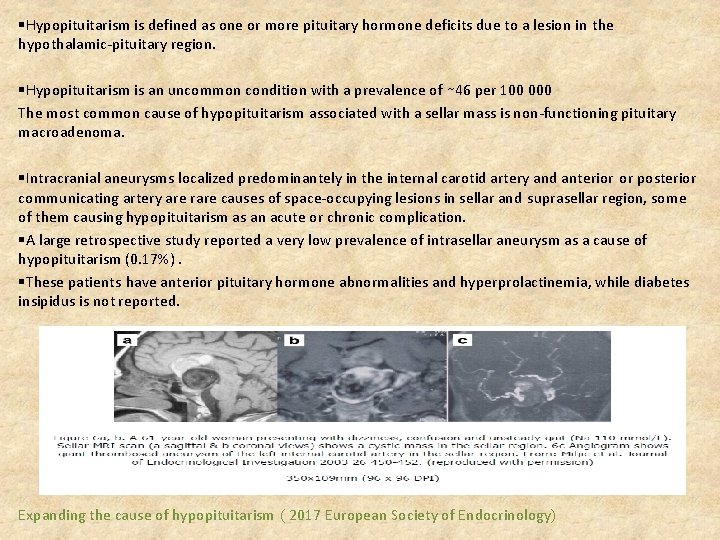
§Hypopituitarism is defined as one or more pituitary hormone deficits due to a lesion in the hypothalamic-pituitary region. §Hypopituitarism is an uncommon condition with a prevalence of ∼ 46 per 100 000 The most common cause of hypopituitarism associated with a sellar mass is non-functioning pituitary macroadenoma. §Intracranial aneurysms localized predominantely in the internal carotid artery and anterior or posterior communicating artery are rare causes of space-occupying lesions in sellar and suprasellar region, some of them causing hypopituitarism as an acute or chronic complication. §A large retrospective study reported a very low prevalence of intrasellar aneurysm as a cause of hypopituitarism (0. 17%). §These patients have anterior pituitary hormone abnormalities and hyperprolactinemia, while diabetes insipidus is not reported. Expanding the cause of hypopituitarism ( 2017 European Society of Endocrinology)
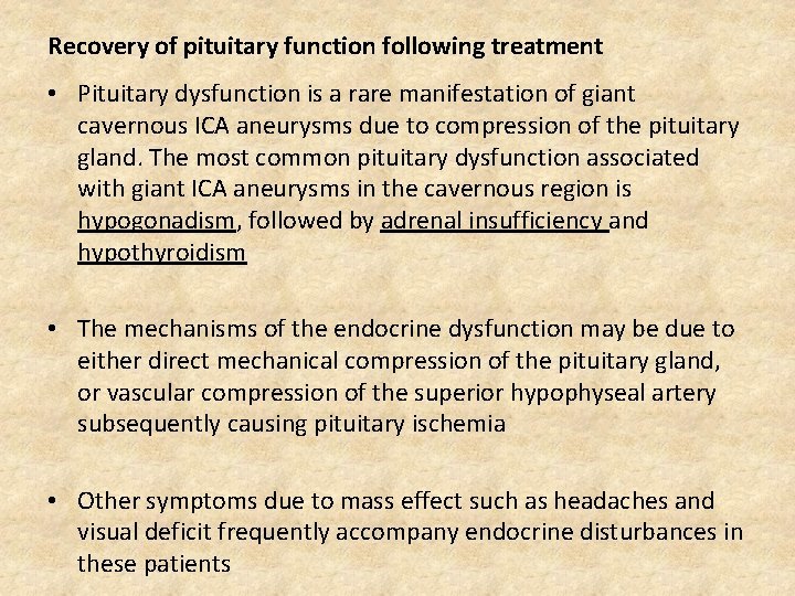
Recovery of pituitary function following treatment • Pituitary dysfunction is a rare manifestation of giant cavernous ICA aneurysms due to compression of the pituitary gland. The most common pituitary dysfunction associated with giant ICA aneurysms in the cavernous region is hypogonadism, followed by adrenal insufficiency and hypothyroidism • The mechanisms of the endocrine dysfunction may be due to either direct mechanical compression of the pituitary gland, or vascular compression of the superior hypophyseal artery subsequently causing pituitary ischemia • Other symptoms due to mass effect such as headaches and visual deficit frequently accompany endocrine disturbances in these patients
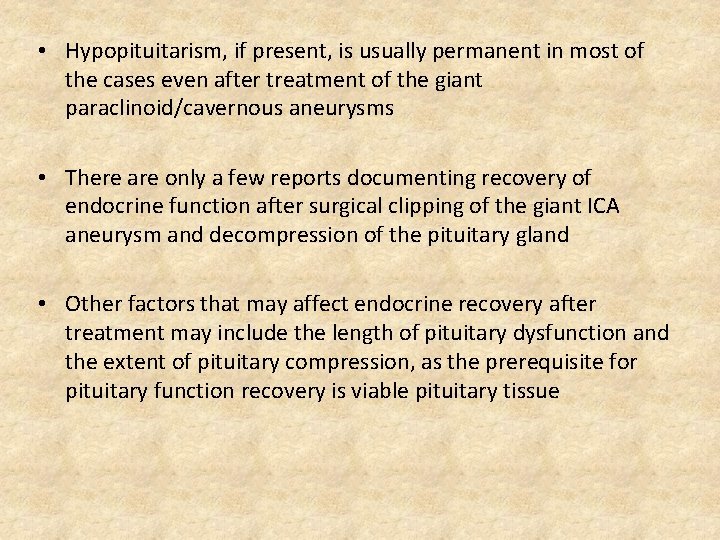
• Hypopituitarism, if present, is usually permanent in most of the cases even after treatment of the giant paraclinoid/cavernous aneurysms • There are only a few reports documenting recovery of endocrine function after surgical clipping of the giant ICA aneurysm and decompression of the pituitary gland • Other factors that may affect endocrine recovery after treatment may include the length of pituitary dysfunction and the extent of pituitary compression, as the prerequisite for pituitary function recovery is viable pituitary tissue
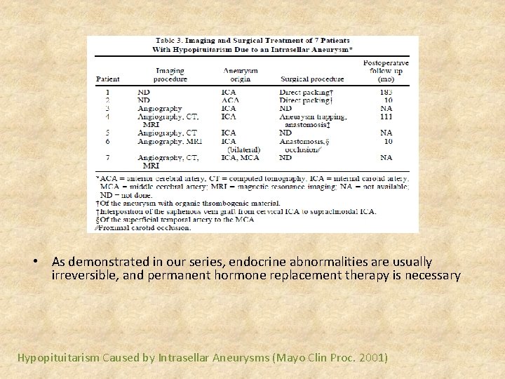
§an archival search for all patients with the diagnosis of hypopituitarism due to intrasellar aneurysms from 1950 through 1995 §Of 4087 patients with a diagnosis of hypopituitarism, 7 patients (4 men and 3 women) aged 28 to 65 years (mean, 52 years) had hypopituitarism due to an intrasellar aneurysm, for a prevalence of 0. 17% Hypopituitarism Caused by Intrasellar Aneurysms (Mayo Clin Proc. 2001)
• As demonstrated in our series, endocrine abnormalities are usually irreversible, and permanent hormone replacement therapy is necessary Hypopituitarism Caused by Intrasellar Aneurysms (Mayo Clin Proc. 2001)
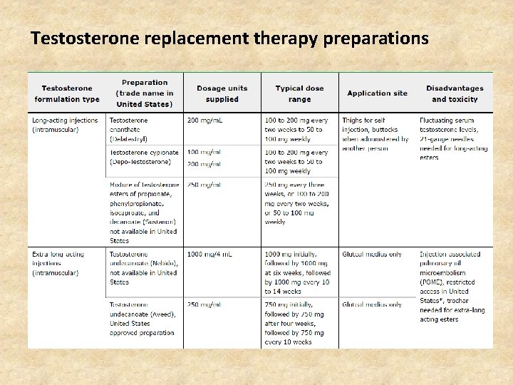
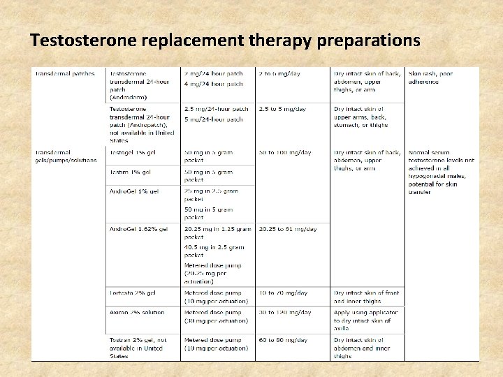
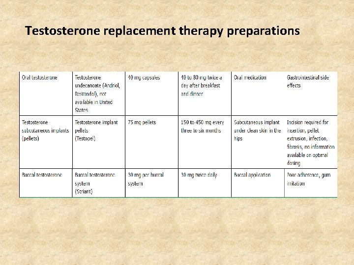
The following principles should guide testosterone therapy ●Testosterone should be administered only to a man who is hypogonadal, as evidenced by clinical symptoms and signs consistent with androgen deficiency and a distinctly subnormal serum testosterone concentration. ●Symptoms and signs suggestive of androgen deficiency include low libido, decreased morning erections, loss of body hair, low bone mineral density, gynecomastia, and small testes. Symptoms and signs such as fatigue, depression, anemia, reduced muscle strength, and increased fat mass are less specific ●Testosterone can be replaced satisfactorily whether the testosterone deficiency is due to primary or secondary hypogonadism
Testosterone replacement therapy preparations
Testosterone replacement therapy preparations
Testosterone replacement therapy preparations