Imunodeficiency states Primary Caused by defined genetic defects

Imunodeficiency states • Primary – Caused by defined genetic defects – Usually rare, but severe (exception: Ig. A deficiency) • Secondary – Consequence of some other disease, treatment, environmental factors… – Usually frequent, but usually clinically mild (exceptions: HIV disease, secondary aganulocytosis).
 • Early clinical manifestation (weeks-months) • Severe and complicated infections")
Severe combined immunodeficiency (SCID) • Early clinical manifestation (weeks-months) • Severe and complicated infections affecting respiratory and gastrointestinal tract and the skin • Failure to thrive • Frequent diarrhea • Usually lymphocytopenia • T-cell deficiency, B cell present in some patients • Decreased immunoglobulin levels

SCID, t-GVHR, generalised BCG-itis

SCID infections caused by atypical patogens • • Pneumocystis pneumonia Cytomegalovirus pneumonitis Disseminated BCG-itis Infections caused by atypical mycobacteria • Candidiasis of oropharynx, skin

Patient with SCID
. Susceptibility to infection")
Immunoglobulin Deficiencies Clinical manifestations begins at 6 -12 months (or late). Susceptibility to infection by encapsulated bacteria (Pneumococcus, Haemophilus). Respiratory tract predominantly affected; patients suffer from recurrent otitis media, bronchitis, sinusitis, pneumonia. Some patients also suffer from meningitis or chronic diarrhea.

X-linked agammaglobulinemia • Only boys affected • Clinical manifestation usually begins at 6 -12 months • Severe and complicated respiratory tract infections. • Very low levels of all immunoglobulin isotypes. • B-cell not detected
 • Both sexes affected • Clinical manifestation initiates at any")
Common variable immunodeficiency (CVID) • Both sexes affected • Clinical manifestation initiates at any age • Frequent and severe respiratory tract infections • Proneness to autoimmune diseases • Variable decrease of immunoglobulin isotypes, usually markedly decreased Ig. A and Ig. G leves • B-lymphocytes usually present

Selective Ig. A deficiency • • Frequency: 1: 400 Usually only mild manifestation Predominantly respiratory tract infections Patients are prone to autoimmune diseases • Beware of anti-Ig. A antibodies that can cause a severe anaphylactic reaction after artificial Ig. A administration (by blood, immunoglobulin derivates)!

T-cell Deficiences -Early onset of clinical manifestation. -Extreme susceptibility to viral, fungal, mycobacterial, and protozoal infections. - Respiratory system most frequently affected, but also other systems can be involved.

Di. George syndrome • Defect in embryonic development of the 3 rd and 4 th pharyngeal pouches. • Cardiovascular defects • Hypoparathyroidism ® hypocalcemia ® seizures • Thymic hypoplasia ® T cell deficiency • Typical facies: hypertelorism, micrognatia, low-set, posterior rotated ears.

Di. George syndrome

Di. George syndrome

Complement deficiencies • Deficiency of C 1 -C 4: autoimmune systemic disorders, susceptibility to bacterial infections • Deficiency C 5 -C 9: susceptibility to bacterial infections, mainly to meningococcal meningitis • Deficiency of C 1 INH: hereditary angioedema
 • Uncontrolled activation")
Hereditary angioedema • Deficiency of C 1 inhibitor (C 1 INH) • Uncontrolled activation of the complement system after trauma, infection, surgical operation. . • Vasoactive peptides (bradykinin, C 3 a, C 5 a) cause increased vascular permeability • Oedema of the skin, respiratory tract (dyspnoe), gastrointestinal tract (cramps, vomiting)
")
HEREDITARY ANGIOEDEMA (HAE)

Phagocytic dysfunction • Early onset of clinical manifestation • Susceptibility to bacterial and fungal infections • Abscess formation, mainly of the skin, periproctal area, liver, but any area may be affected.

Chronic granulomatous disease • Recurrent abscesses mainly of the liver, lungs, periproctal area, suppurative lymphadenitis, osteomyelitis • Infections are caused mainly by catalase-positive organisms: St. aureus, Candida sp. , Serratia marcescens • Usually early onset of symptoms • Production of reactive metabolites of oxygen is disturbed (defect of NADPH oxidase)

Wiskott-Aldrich syndrome • • • X-linked disease Thrombocytopemia ® bleeding tendency Severe eczema Immunodeficiency Severe allergic and autoimmune manifestations • B-cell lymphomas

Wiskott-Aldrich syndrome

Ataxia telangiectasia • Autosomal recessive • Progressive cerebellar ataxia • Telangiectasis especially on ear lobes and conjunctival sclera • Immunodeficiency • Frequent tumors • Cause: mutation in ATM gene

Ataxia telangiectasia

Treatment of primary immunodefciencies • SCID and other severe immunodeficiencies: bone marrow transplantation, gene therapy in some cases. • Antibody deficiencies: immunoglobulin replacement • Antibiotic prohylaxis

Clinical use of non-specific immunoglobulin derivates • Types of derivates: – „Normal immunoglobulin“ - for intramuscular use. Used very rarely at present bcause only low dose can be given. . – Intravenous immunoglobulins, subcutaneous immunoglobulins - can be used in high doses • Indications: – Replacement treatment in patients with antibody deficiencies – Prophylaxis of infections against which there is no specific immunoglobulin derivate (hepatitis A) – High doses of i. v. immunoglobulins are used in autoimmune diseases, systemic vasculitic diseases.

Causes of secondary immunodeficiency • • Metabolic - uremia, diabetes, malnutrition Iatrogenic – cytostatics, immunosuppressants Malignat tumors Viral infections - HIV, CMV, measles, infectious mononucleosis • Splenectomy • Stress • Injuries, operations, general anestesia

Imunodeficiency after splenectomy • Disturbed phagocytosis, decreased production of antibodies. • The most severe complication is hyperacute pneumococal sepsis. • Prevention: vaccination against Pneumococcus, Haemophilus infl. B, Meningococcus. PNC prophylaxis.

Secondary hypogammaglobulinemia • Decreased production of immunoglobulins – Chronic lymphatic leukemia – Lymphoma – Myeloma • Loss of immunoglobulins – Nephrotic syndrome – Exudative enteropathy

Ways of transmission 1. Sexual 2. Parenteral – intravenous drug addicts previously blood products 3. Vertical – mother to child – transplacental, during delivery, by brestfeeding

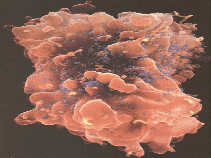
Infection of CD 4+ cell by HIV

HIV Cycle
 3 clinical categories A Asymptomatic disese B „small“ opportunistic")
Classification of HIV disease (CDC) 3 clinical categories A Asymptomatic disese B „small“ opportunistic infections C „big“ opportunistic infections and other states that define AIDS
 HIV infection • Asymptomatic HIV infection • Persistent")
Clinical category A • Accute (primary) HIV infection • Asymptomatic HIV infection • Persistent generalised lymphadenopathy (PGL)
 • Present in 50 -70% patients")
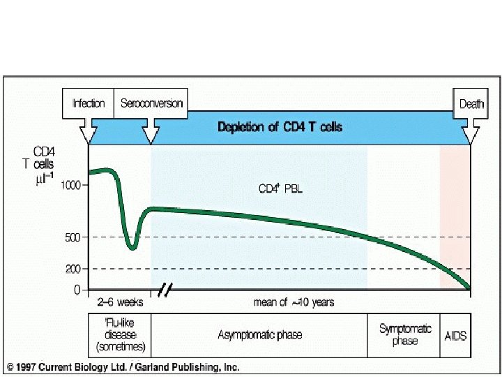
HIV PRIMOINFECTION • Acute retroviral syndrome, („mononucleosis-like syndrome“) • Present in 50 -70% patients • 2 -6 weeks after infection

Clinical presentaioon of HIV primoinfection • • Fever, lympadenopathy, pharyngitis Rash Myalgia, arthralgia, diarrhoea, cephalea nausea, vomiting thrush Neurologic symptoms Aphtous stomatitis

Perzistent generalized lympadenopathy • More than 3 months • 1/3 HIV-infected persons • Lymph nodes 0, 5 -2, 0 cm, painless

Clinical category B • • Fever >38, 5 C more than 1 month Diarrhoea more than 1 month Oropharyngea candidiasis Vulvovaginal candidiasis (chronic or difficult to treat) • Recurrent herpes zoster
 • Pneumocystis pneumonia • Brain abscess caused by toxoplasma •")
Clinical category C (AIDS) • Pneumocystis pneumonia • Brain abscess caused by toxoplasma • Esofageal, tracheal, bronchial or lung candidiasis • Chronic anal herpes, herpetic bronchitis, pneumonia • CMV retinitis, generalized CMV infection • Progressive multifocal leukoecephalopathy • Mycobacterial infections
 - Toxoplasma")
Opportunistic Infections in AIDS Patients - Pneumonia due to Pneumocystis jiroveci (carinii) - Toxoplasma brain abscess - Cytomegalovirus infection (retinitis, colitis) - Mycobacterial infections - Herpes virus and Varicella-Zoster infections
 - tumors • Kaposhi sarcoma • Brain lymphoma")
Clinical category C ( AIDS ) - tumors • Kaposhi sarcoma • Brain lymphoma

Kaposhi sarcoma

Kaposiho sarkom

Wasting syndrome
,")
Treatment of HIV-disease • Antiretroviral – Nucleoside inhibitors of reverse transcriptase: azidothymidin (syn. zidovudin), didanosin, zalcitabin, stavudin, lamivudin – Nonnucleoside inhibitors of reverse transcriptase: Nevirapin, delavirdin, efavirenz – HIV protease inhibitors: Saquinavir, ritonavir, indinavir • Prophylaxis of Pneumocystis carinii pneumonia (co-trimoxazol), antiviral and antimycotic antibiotics

Strategy of treatment • HAART - Highly Active Anti Retroviral Therapy • Mega-HAART

Diagnosis of HIV infection • Detection of anti-viral antibodies – ELISA – Western blott • Detection of antigen p 24
- Slides: 48