Important spotters 2 nd internalsBDS Important spotters II

-BDS • • Important spotters (II internals) – BDS 1.")
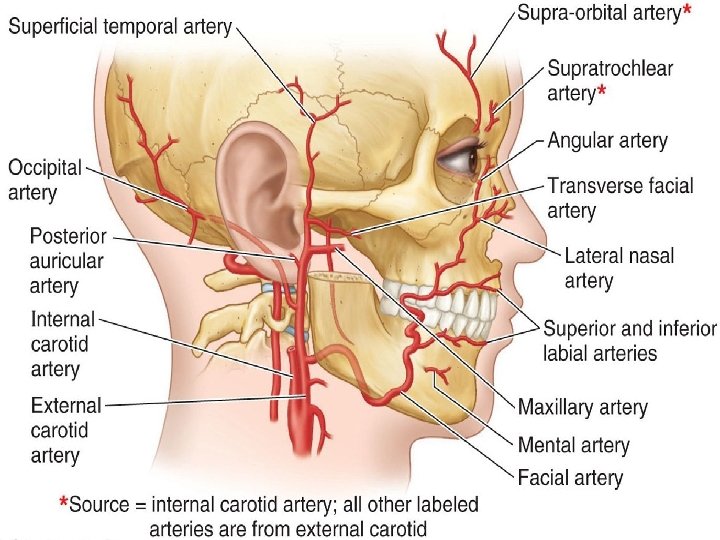
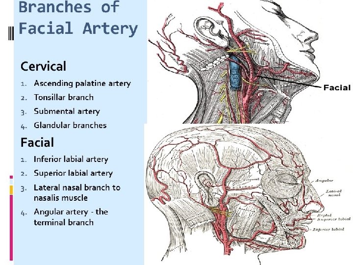
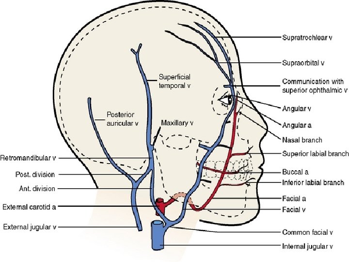
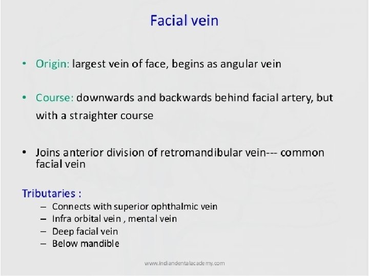
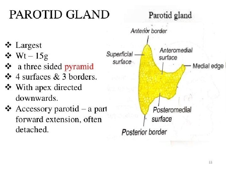
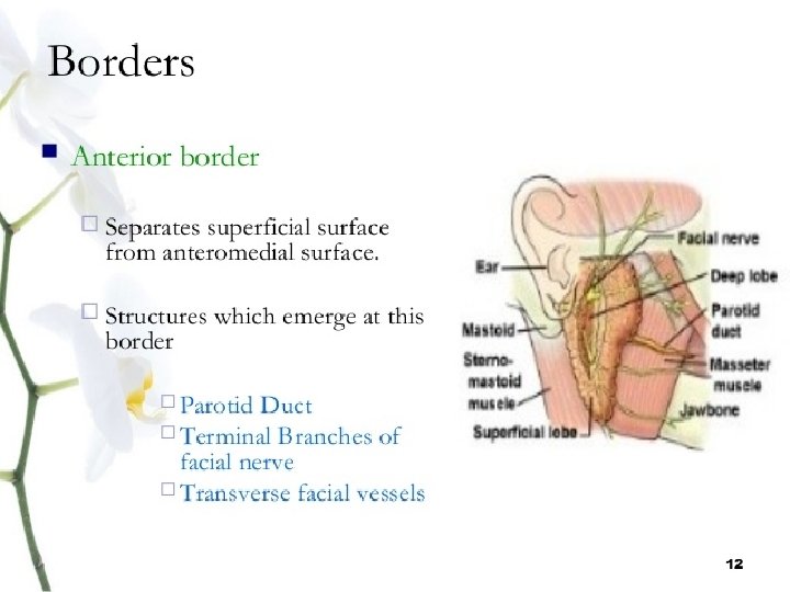
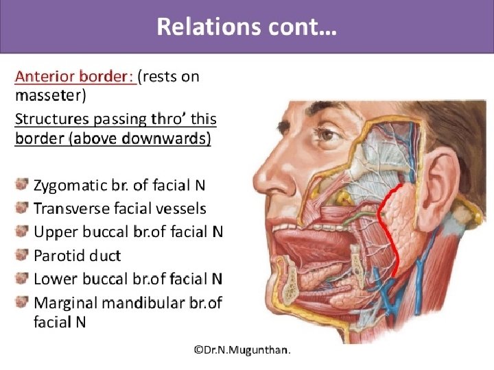
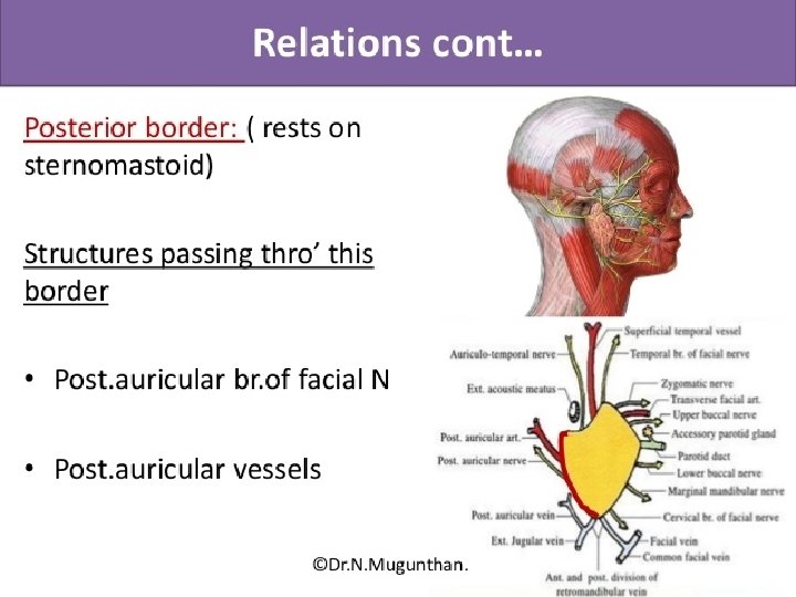
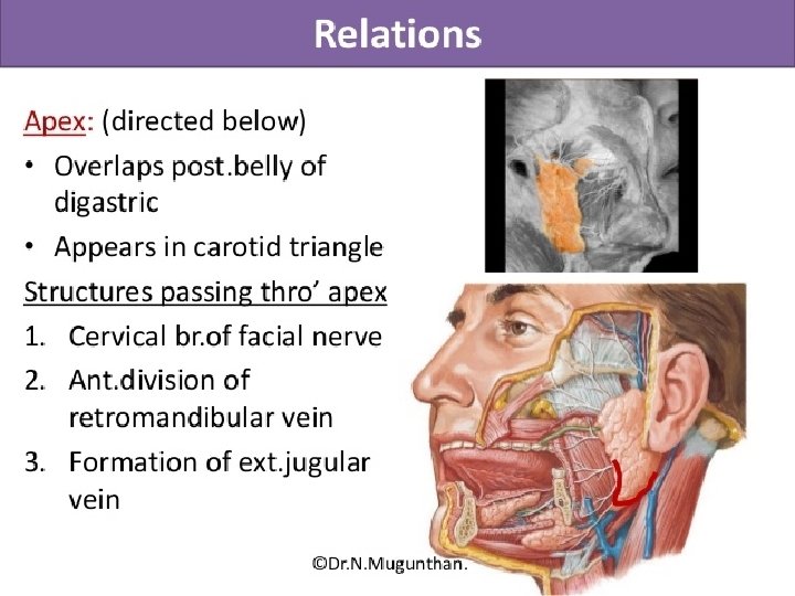
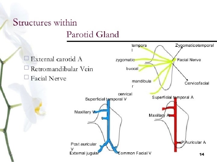
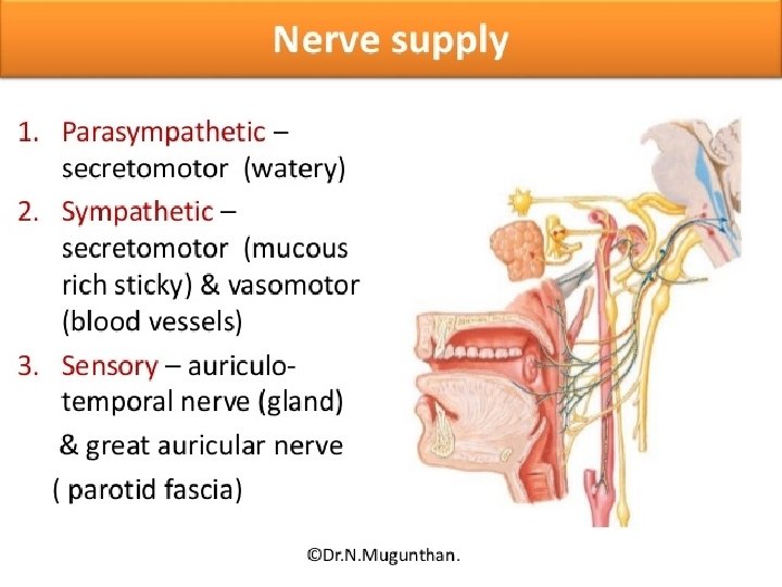
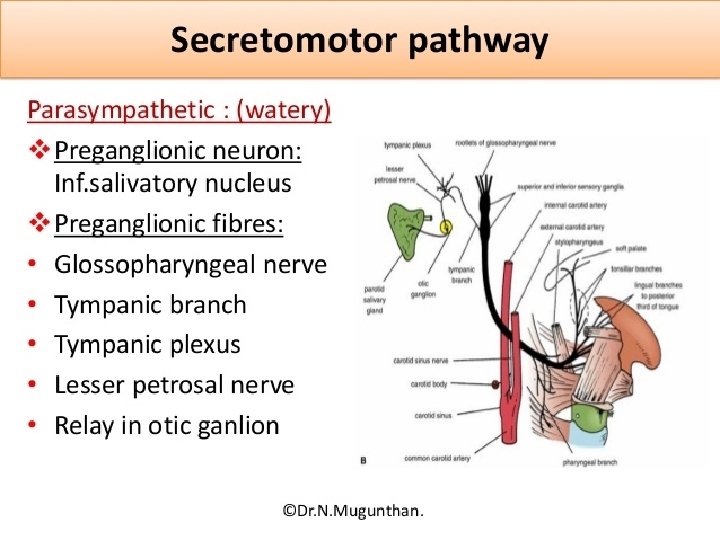
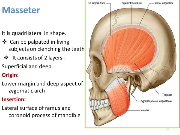
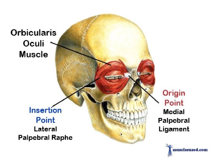
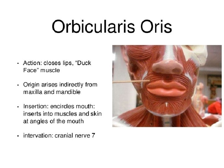
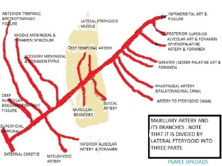
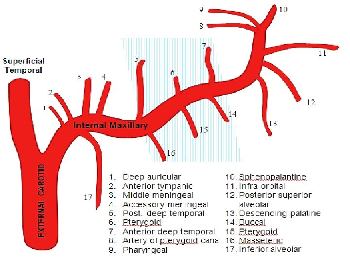
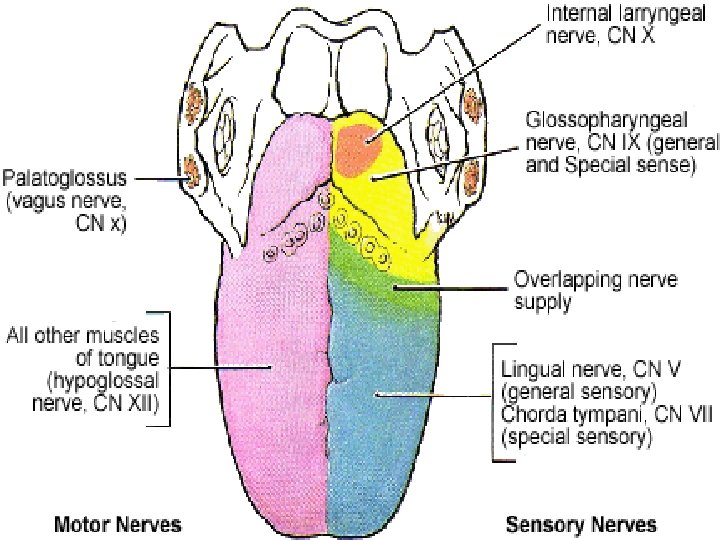
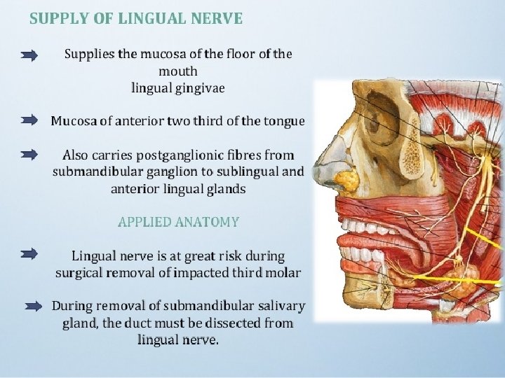
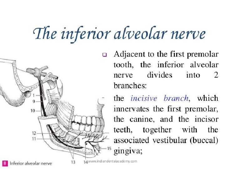
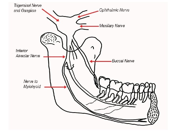
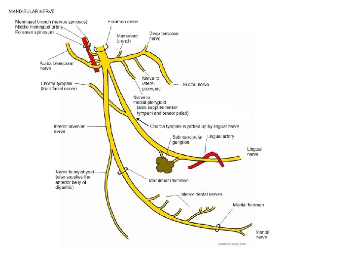
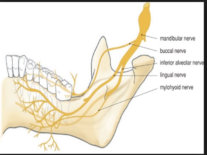
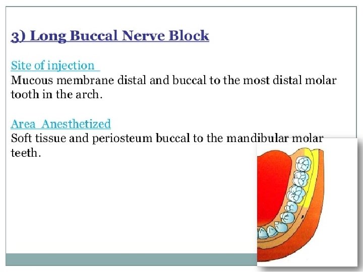
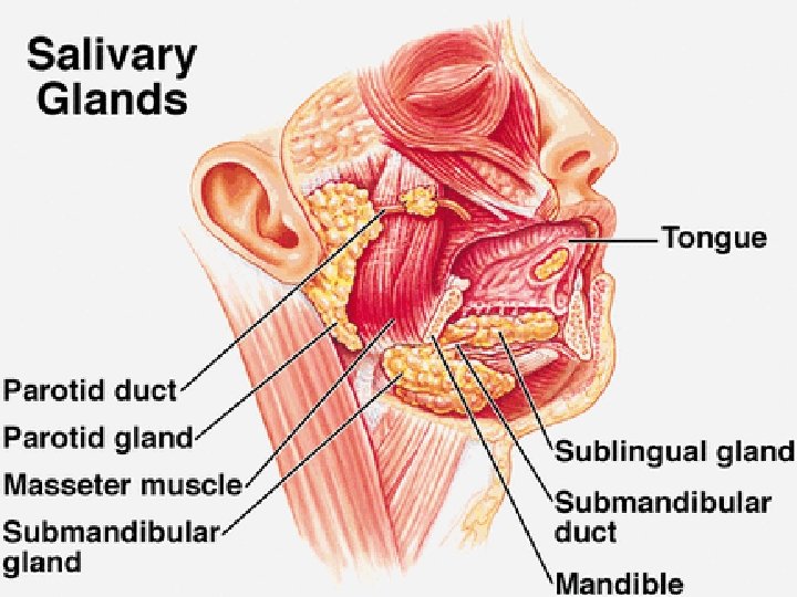
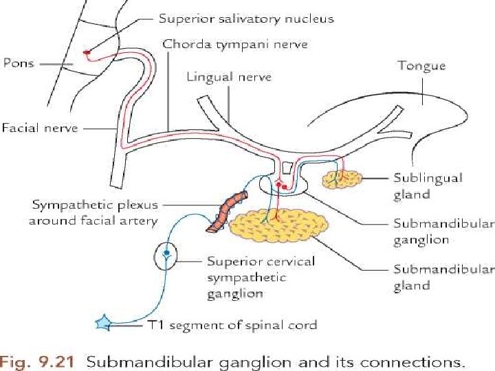
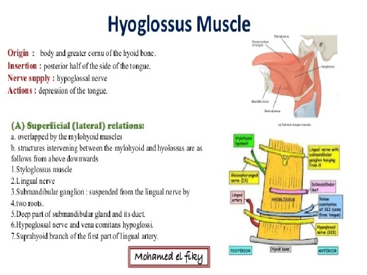
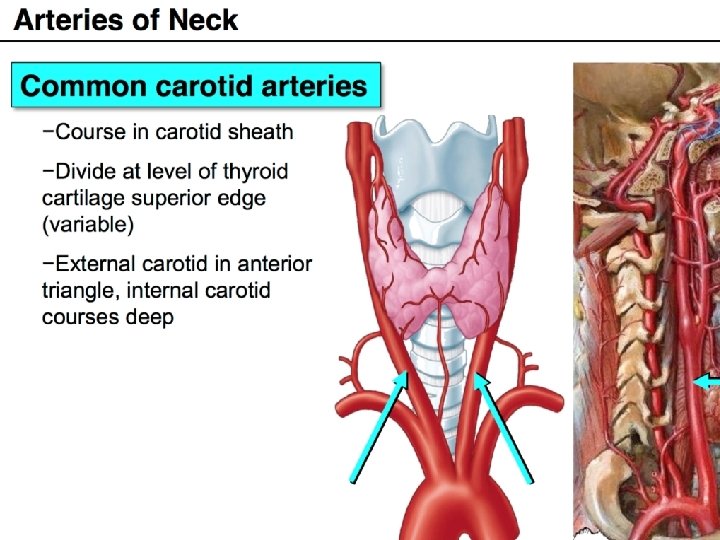
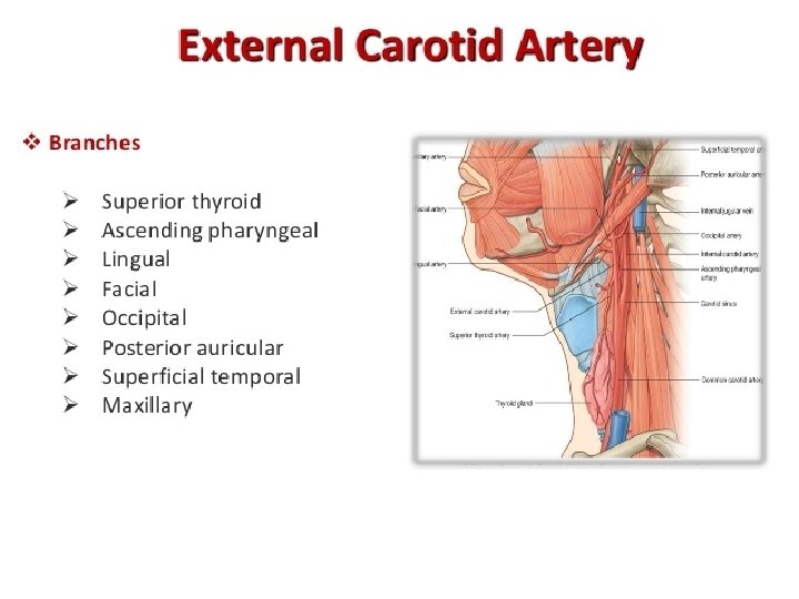
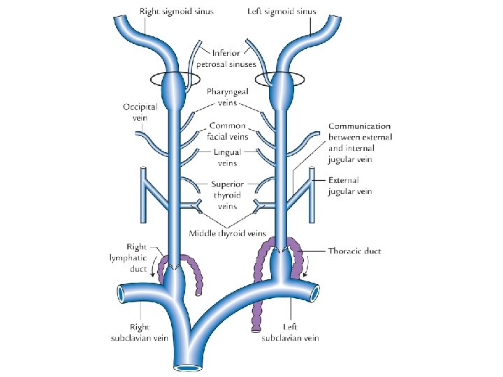
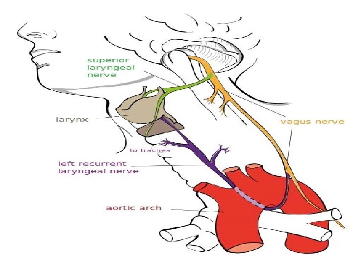
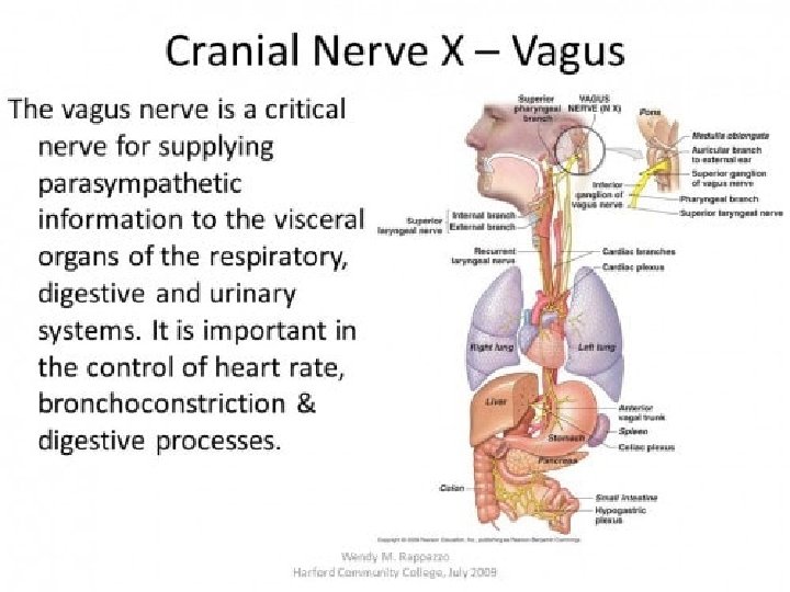
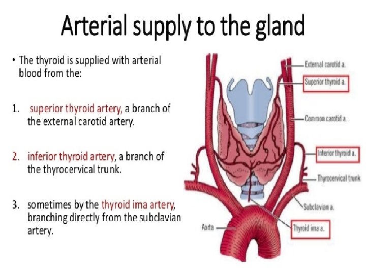
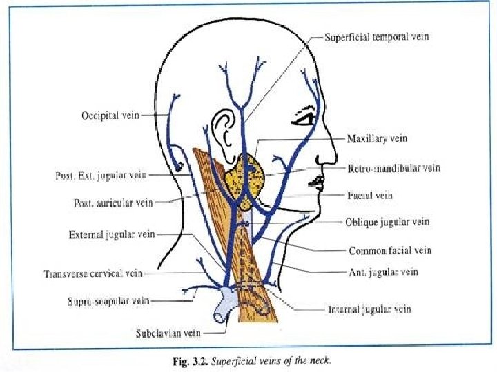
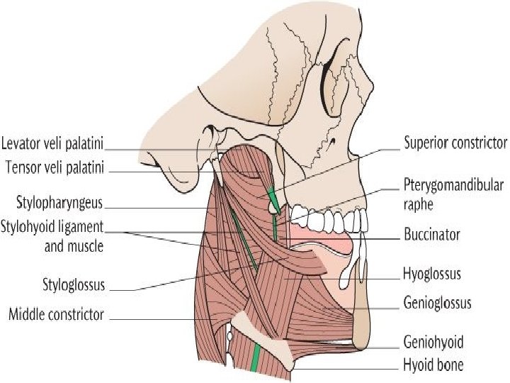
Important spotters (2 nd internals)-BDS • • Important spotters (II internals) – BDS 1. Facial artery – branches, origin , termination 2. Facial vein – formation, termination, tributaries 3. Parotid gland- parts, structures emerging from anterior border, apex or base, posterior border, secreto-motor pathway 4. Parotid duct – opening, structures pierced by it 5. Masseter muscle- origin, insertion, action, nerve supply 6. Temporalis- origin, insertion, action, nerve supply 7. Orbicularis oculi - origin, insertion, action, nerve supply 8. Orbicularis oris- origin, insertion, action, nerve supply 9. Maxillary artery – origin, branches from different parts 10. Lingual nerve – where does it innervates? 11. Inferior alveolar nerve – Terminal branches 12. Buccal branch of mandibular nerve - where does it innervates 13. Submandibular salivary gland – parts, secretomotor pathway, where does the duct opens? 14. Hyoglossus muscle - origin, insertion, action, nerve supply 15. Common carotid artery- branch of which artery? Terminal branches, level of bifurcation 16. External carotid artery - Branch of? ? , branches of it 17. Internal jugular vein – formation and termination, tributaries 18. Vagus nerve – branches in the neck 19. Superior thyroid artery – branch of ? ? , 20. Thyrohyoid membrane – structures piercing it?

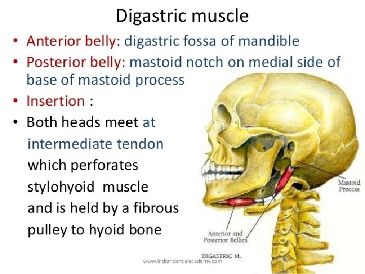
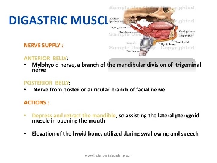
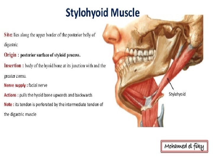
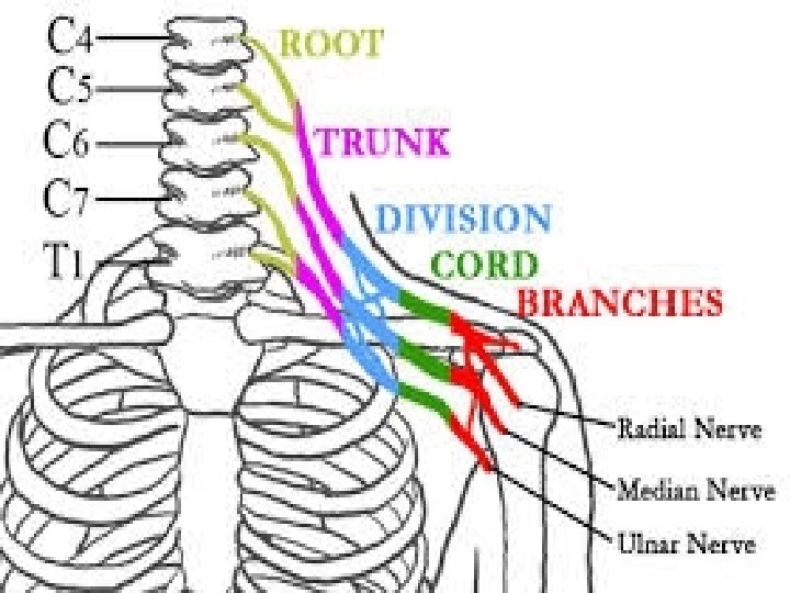
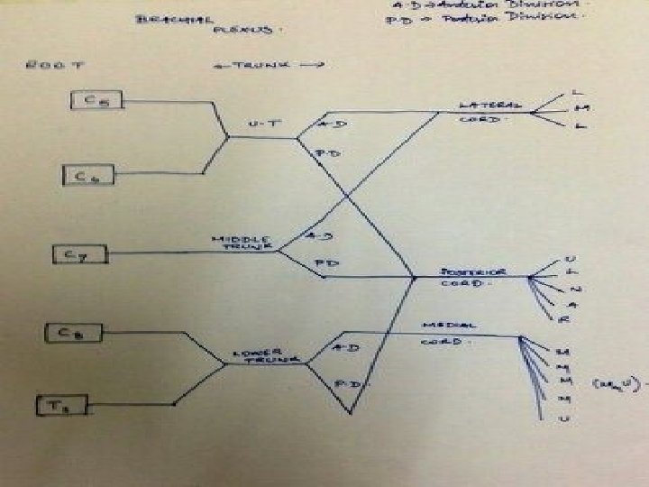
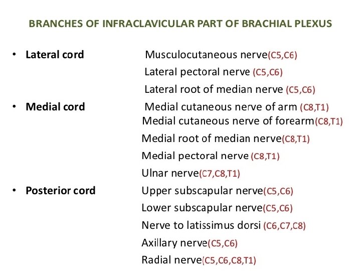
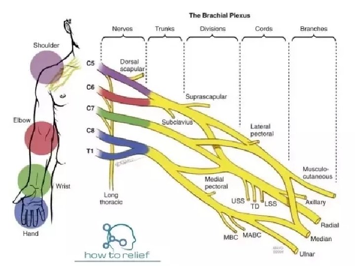
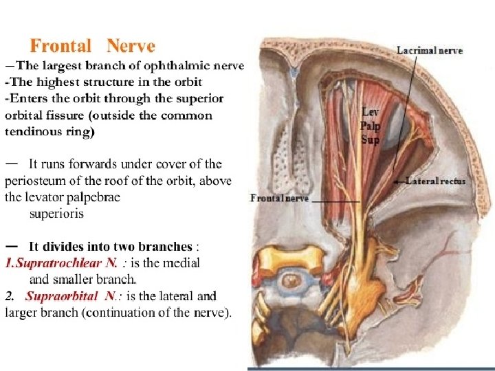
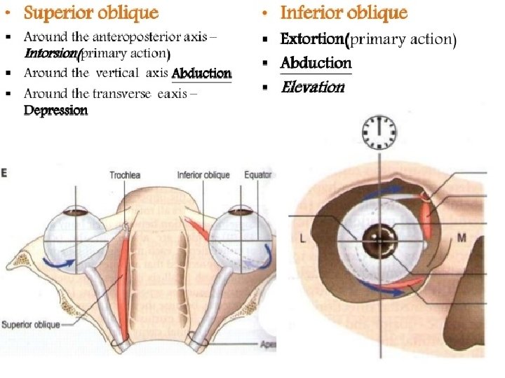
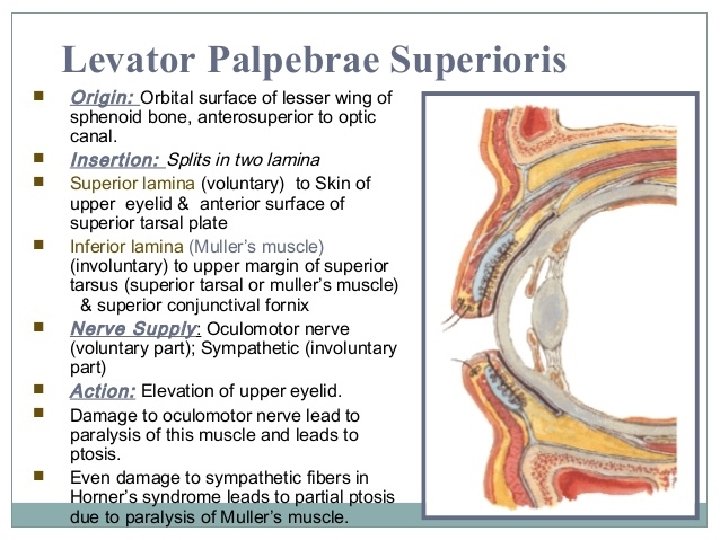
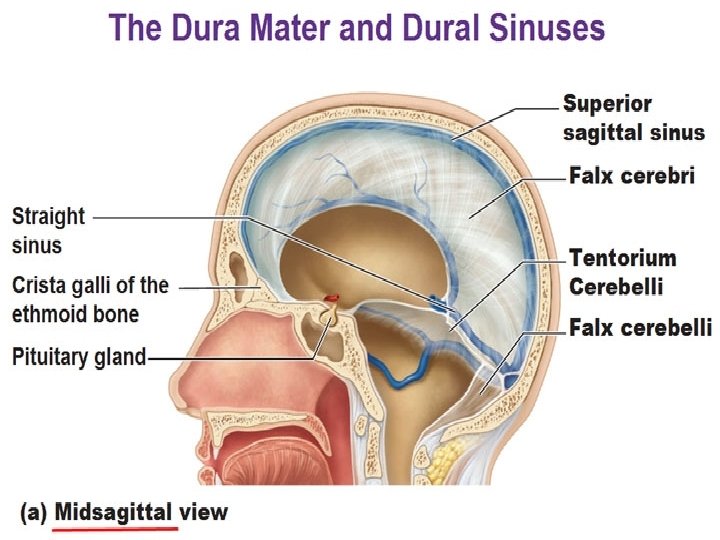
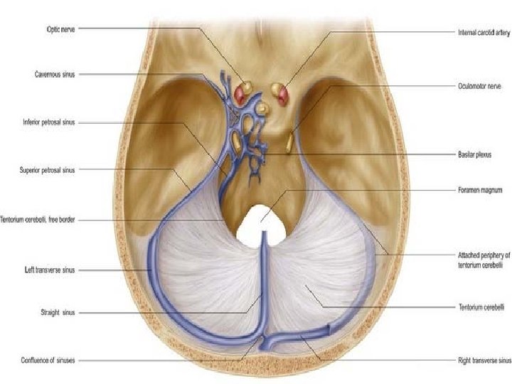
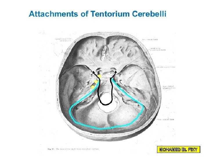
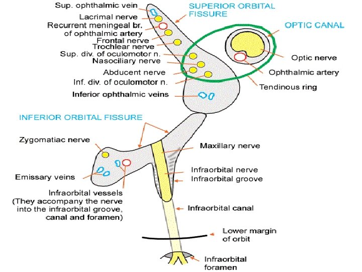
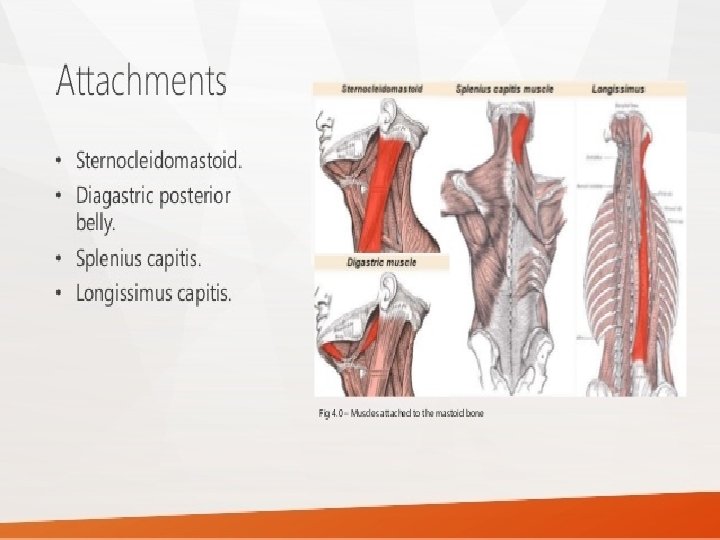
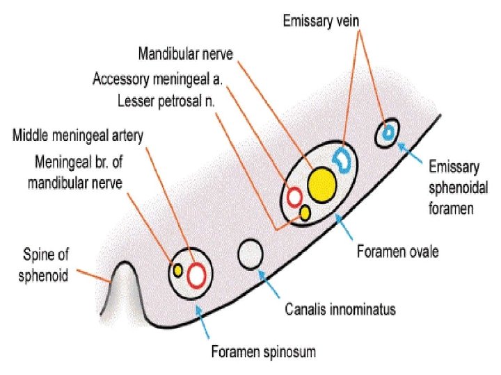
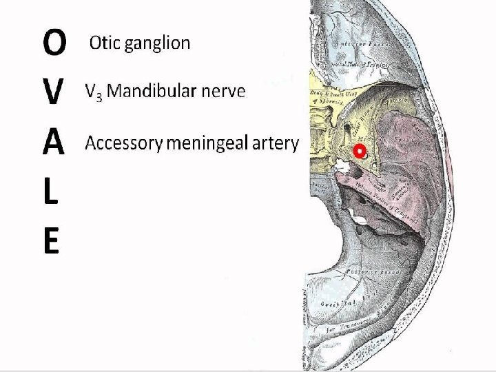
21. Anterior and posterior belly of digastric - origin, insertion, action, nerve supply 22. Stylohyoid muscle - origin, insertion, action, nerve supply 23. Thyroid gland – blood supply, parts, relation of medial surface of each lobe, function of gland 24. Inferior thyroid artery – branch of ? ? 25. Isthmus of thyroid gland - parts, relations 26. Spinal accessory nerve – muscles supplied by it? 27. External jugular vein- formation and termination, tributaries 28. Upper trunk of brachial plexus – branches? 29 Frontal nerve – terminal branches? ? , it is a branch of ? ? 30 Superior oblique muscle – nerve supply, action 31. Levator palpebrae superioris muscle – action, nerve supply 32. Falx cerebri – attachments, venous sinuses related to it 33. Tentorium cerebelli - attachments, venous sinuses related to it 34. Superior orbital fissure - contents 35. Inferior orbital fissure - contents 36. Spine of sphenoid – attachments , relations 37. Styloid process - attachments 38. Mastiod process – attachments, type of bone 39. Foramen ovale - contents 40. Foramen spinosum - contents

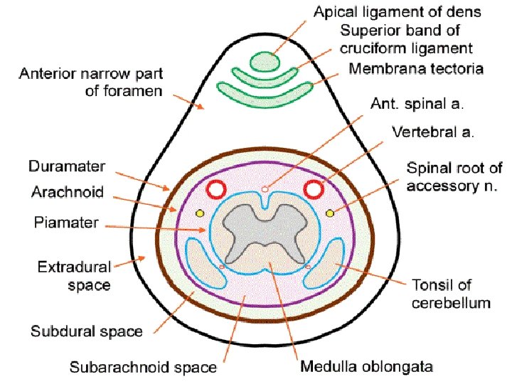
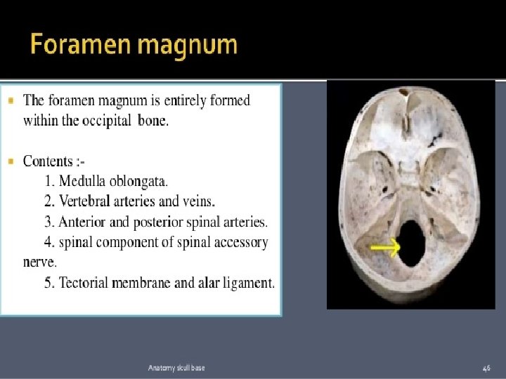
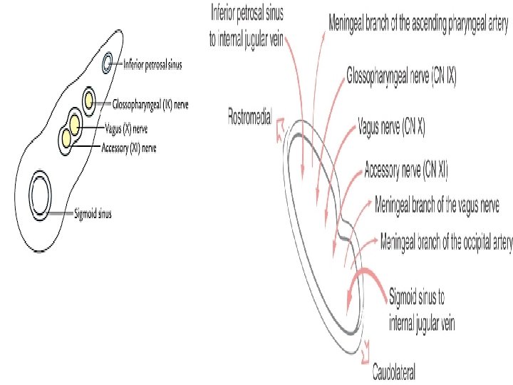
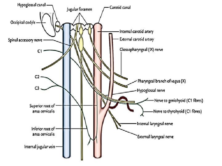
• • • • • • • • Foramen rotundum - contents Carotid canal - contents Internal acoustic meatus - contents Jugular foramen- contents Hypoglossal canal - contents Foramen magnum- contents Pterion – name the bones meeting, clinical importance Transeverse sulcus- related sinus Sigmiod sulcus – related sinus Anterior fontanellae - clinical importance Mandibular foramen- contents Pterygoid fovea- attached structure Coronoid process- attachments Condylar process- Joint formation, type of joint Symphysis menti – type of joint Mylohyoid line - attachment Angle of mandible – structure attached, normal angle External surface and internal surface of ramus of mandible- structures attached Mental foramen- contents Genial tubercles- structures attached Lingula of mandible - structures attached Atlas – identification points Axis - identification points 7 th cervical vertebra - identification points Typical cervical vertebra - identification points Posterior arch of atlas – structure related to the groove Odontoid process of axis – structures attached Foramen transversarium - contents Infra orbital foramen - contents Sternocleidomastoid muscle- origin. Insertion, nerve supply and action Hyoid bone- parts, attachments Ansa cervicalis- formation and name the muscles supplied by it

Spine of the sphenoid: It is a small pointed process projecting downward from the junction of posterior and squamosal borders of greater wing. The spine provides the attachments to three ligaments, two muscles, and related to important structures on its medial and lateral sides (Fig. 1. 12).

Clinical significance. The anterior fontanelle is useful clinically. Examination of an infant includes palpating theanterior fontanelle. A sunken fontanelle indicates dehydration, whereas a very tense or bulging anterior fontanelle indicates raised intracranial pressure.

- Slides: 63