Important Bacterial Diseases of Fish Dr Deepak Kumar

Important Bacterial Diseases of Fish Dr Deepak Kumar Assistant Professor Department of Veterinary Pathology Bihar Veterinary College, Patna Bihar Animal Sciences University, Patna - 14

Body Parts Of Fish

BACTERIAL DISEASES • Bacterial fish diseases and infection are very common in fish. The key to successful treatment of bacterial disease is early, accurate diagnosis and treatment. If treatment is delayed it can lead to substantial losses. • There are two types of pathogenic bacteria namely 1. ) Primary or obligate pathogens 2. )Opportunistic pathogens

BACTERIAL DISEASES Primary or obligate pathogens these are not part of the normal aquatic flora and are capable of causing disease in healthy individuals, eg. Aeromonas salmonicida. § Opportunistic pathogens These are normally free-living, either in the water or on the fish, but becomes pathogenic under certain circumstances. §

BACTERIAL DISEASES • Many of these are saprophytes, normally living on dead organic matter such as plant and animal remains or faeces, e. g. , Aeromonas hydrophila and Pseudomonas spp. • In general, most of the bacterial diseases that affect fishes are caused by opportunistic bacteria. Some of the common bacterial

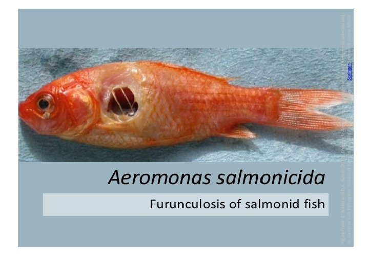
Furunculosis • The disesase is also known as – Salmon furunculosis, Ulcer disease, Goldfish ulcer disease. • In Asia goldfish mainly affected • Etiology – Aeromonas salmonida • 1 st reported by Emmerich & Weibel ( 1984) I Germany

Pathology • In chronic cases – Haemorrhage in vent, viscera, gills & muscles

Furunculosis

Post- Mortem Lesions • Haemorrhages are common in liver, spleen & kidney.

Infectious Dropsy/ Aeromoniasis • It is caused by – Aeromonas hydrophila which is associated with Aeromonas septcaemia or red spot. • It means generally swelling of soft tissues due to the accumulation of excess water. swollen and scales standing out from the body.

Infectious Dropsy/ Aeromoniasis Aeromonas hydrophila - is a heterotrophic, Gram-negative, rod-shaped bacterium mainly found in areas with a warm climate

Aeromonas hydrophila -

Sine • The infected fish suffers from inappetite • Abnormal swimming • Gill become pale I appearance.

Infectious Dropsy/ Aeromoniasis the swollen eyes and gill covers and the reddening of the belly and fin pits.

Lesions • Skin ulcer is common at any site of fish and is often surrounded by bright red area.

Infectious Dropsy/ Aeromoniasis

Bacterial gill disease • It is caused by – Flavobacterium branchiophilum

Bacterial gill disease • Flavobacterium is a of gramnegative, nonmotile and shaped bacteria genus motile, rod-

Flavobacterium branchiophilum • This bacterium can be found in fresh or brackish water. • It can survive in aerobic and anaerobic environments

Flavobacterium branchiophilum

Pathogenesis • The bacteria normally reside in gill mucous of fish. • After multiplication of bacteria, gill becomes proliferated, fusion of lamellae of gill occures. • The bronchial mucosa cells secrete more mucous.

Gills becomes proliferated.

Gill rot
")
Edwardsiellosis/fish Gangrene • It is also known as – Emphysematous putreactive disease ( EPD) • 1 st reported by Hoshima ( 1962) from Japan. • It is caused by – Edwardsiella tarda.

Edwardsiella tarda. • The bacterium is a facultatively anaerobic small, motile, gram negative, straight rod with flagella.

Pathogenesis • The bacteria produces hemorrhage is surrounded by large area of depigmentation, over skin. • After death of cell, purification ( pus formation in necrotic tissue by saprophytic bacteria), hydrogen ( H 2 S)sulphide gas is produced so emphysematous purification disease ( EPD).

Edwardsiellosis/fish Gangrene
 in channel catfish hemorrhage – pale skin and petechial.")
Edwardsiellosis (edwardsiella tarda) in channel catfish hemorrhage – pale skin and petechial.

Edwardsiellosis/fish Gangrene

Pseudomoniasis/ Pseudomonas Septicaemia. • Caused by – Pseudomonas fluorescens. • Pseudomonas fluorescens is a common Gramnegative, rod-shaped bacterium. • Ascites is most common.

Pseudomoniasis/ Pseudomonas Septicaemia. • After death of cell, inflammation starts, after puterification hyderogen sulphide gas is produced as termed as emphysematous putrifactive disease. • Carps are severely affected.

Pseudomoniasis/ Pseudomonas Septicaemia.

Enteric Red mouth/Haemorrhagic Septicemia • Caused by – Yersinia ruckeri • Yesinia ruckeri is a species of Gram-negative bacteria, known for causing enteric red mouth disease in some species of fish. • Strain 2396 -61 (= ATCC 29473) is its type strain. A draft genome for Yersinia ruckeri

Yersinia ruckeri

Classical lesions 1. Exophthalmus 2. Partly opened lower jaw 3. Haemorrhages present in the roof of mouth 4. Mouth becomes darken due to improper melanin control • 5. Anemia due to necrosis haematopoetic tissue. • •
,")
Enteric Red mouth/Haemorrhagic Septicemia A: darkening of the skin, enlarged abdominal valley (black arrow), and hemorrhages in the dorsal fin (white arrow). B: hemorrhages in and around the mouth (arrows). C: enlarged and black spleen (white arrow), and reddened intestine (black arrow).

Enteric Red mouth/Haemorrhagic Septicemia Histological sections of spleen and kidney organs of rainbow trout infected with Y. ruckeri. A: multifocal necrosis can be seen in the spleen. B: degeneration of interstitial tissue and a marked increase in melanomacrophages can be seen in the kidney. Sections were stained with
")
Common Freshwater prawn diseases in India I. III. IV. V. Larval midcycle disease (MCD) Bacterial necrosis – mortality up to 100% Black spot disease caused – by Beneka sp. Branchiostegite melanization – unknown eti Idiopathic muscle necrosis by environmental dismanagement. VI. Isopod parasitic disease by probopyrus origin.

Common Freshwater prawn diseases in India • VII. Epibiont fouling disease caused by various bacteria, algae, fungi, protoza etc. • VIII. White muscle syndrome/white tail disease- by Nodavirus.

BACTERIAL DISEASES diseases are: Bacterial disease Columnaris/ Saddleback Disease Causative agent Flavobacterium columnare Symptoms Edwardsiellosis/ Fish Gangreen Edwardsiella tarda Brown to yellowish brown lesion (sores) on their gills, skin, and/or fins. Characteristic lesion produced by columnaris is a pale white band (often persists as whitish plaques) encircling the body, often referred to as saddle back. Red cutaneous lesions located dorso-ventrally on the body Abscesses resulting in loss of pigmentation with a large amount of necrotized tissue.

BACTERIAL DISEASES Mycobacteriosis Acid-fast bacteria of the genus Mycobacterium fortuitum and Mycobacterium marinum Anorexia, emaciation and loss of equilibrium, inflammation of the skin, exophthalmia, ascites, and open lesions, and ulceration characterize tuberculosis. Internally, grey-white granulomas develop in the liver, kidney, spleen, heart and muscles. Skin discolourations on one side of head or body Motile aeromonad septicemia (MAS) Aeromonas hydrophila Manifested by several clinical signs like ulceration, exophthalmia, abdominal distention etc. Tail rot and fin rot Various bacteria such as Aeromonas, Pseudomonas and Mycobacterium Fin rot and tail rot leads to the destruction of the fins especially the caudal fin. This is usually caused due to poor environmental conditions, poor nutrition and stress.

Vibriosis Vibrio anguillarum Vibriosis of shell fish Vibrio spp. Necrotizing Hepatopancreatitis Bacterial infection in marine fish Proteobacteria (alpha) group Reddening of mouth in catfish Red spots on the ventral and lateral areas of the fishes. Swollen dark skin lesions releasing blood exudate. In acute epizootics, the infected fish die without showing any clinical signs High mortalities in post-larvae, young juveniles Moribund shrimp appear hypoxic and often come to the pond surface or edge. Presence of luminescence in tanks Reduced feed intake, empty gut, anorexia, poor length: weight ratios, pallid hepatopancreas, reduced lipid droplets, melanization of tubules Bacteriial ulcer

Furunculosis Aeromonas salmonicidae Enteric red mouthdisease Yersinina ruckeri Flavobacteriosis Flavobacterium spp. Hemorrhages at the base of fins and erosion of the pectoral fins Bloody or hemorrhagic vents and petechial hemorrhages Furuncles or blisters all over the body of the fish Septicemia with exophthalmus, ascites, hemorrhage and ulceration of the jaw, gills and operculum, swelling of the kidneys This disease is a cause of concern to primarily hobbyist and producers of ornamental fish (Mollie granuloma, Mollie madness, Mollie popeye). Infected fish are usually emaciated and pale. Multifocal white nodules are observed in the visceral organs, the retina and choroid and the brain.

Streptococcosis Streptococcus iniae Acute fulminating septicemia, haemorrhage of the fins, skin, and serosal surfaces, granulomas or granulomatous inflammation are evident in the liver, kidney, and brain (meningoenceph alitis). Rainbow trout fry anemia Cytophaga psychrophila Fish develop abdominal distention, exophthalmus, increased pigmentation, lethargy, loss of balance, pale gills, and occasional cutaneous ulcers and necrosis of tail fins. Splenomegaly and hepatomegaly are common with multifocal necrosis of the liver spleen and kidney.

Bacterial kidney disease Renibacterium salmoninarum Exophthalmus, skin darkening, and hemorrhage at the base of the fins. Cutaneous vesicles and ulcers may develop in mature trout "spawning rash". The large swollen kidney and spleen have numerous white nodules visible in the parenchyma Numerous granulomas (containing gram positive bacteria) are observed in the kidney and may be also present in the spleen, heart and liver

Epitheliocystis Chlamydia sp. Fish infected with mycobacteriosis Fish infected with Aeromonas sp Clinically infected fish may be asymptomatic or show respiratory distress or excessive mucus secretions. Multiple white cysts are observed on the gill lamella and skin.

Thanks • Open for Discussions ……. .
- Slides: 48