immunostaining Lab 2 What is IHC Immunohistochemistry IHC

immunostaining Lab 2
 is an important application of monoclonal as well")
What is IHC? • Immunohistochemistry (IHC) is an important application of monoclonal as well as polyclonal antibodies to determine the tissue distribution of an antigen of interest in health and disease. IHC is widely used for diagnosis of cancers; specific tumor antigens are expressed de novo or upregulated in certain cancers. This article deals with the various applications of IHC in diagnosis of diseases, with IHC playing an important role in diagnostic and research laboratories.

The Principle of IHC • IHC involves specific antigen–antibody reactions, it has apparent advantage over traditionally used special enzyme staining techniques that identify only a limited number of proteins, enzymes, and tissue structures. • Fluorescein isothiocyanate (FITC)-labeled antibodies with a fluorescent dye to localize pneumococcal antigens in infected tissues. With the expansion and development of IHC technique, enzyme labels have been introduced, such as peroxidase and alkaline phosphatase. Colloidal gold label has also been discovered and used to identify immunohistochemical reactions at both light and electron microscopy levels.

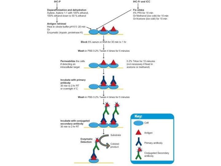
Procedure • Sample preparation Preparation of the sample is critical to maintain cell morphology, tissue architecture and the antigenicity of target epitopes. This requires proper tissue collection, fixation and sectioning. A solution of paraformaldehyde is often used to fix tissue, but other methods may be used.

Preparing tissue slices • The tissue may then be sliced or used whole, dependent upon the purpose of the experiment or the tissue itself. Before sectioning, the tissue sample may be embedded in a medium, like paraffin wax or cryomedia. Sections can be sliced on a variety of instruments, most commonly a microtome, cryostat, or vibratome. Specimens are typically sliced at a range of 3 µm-5 μm. The slices are then mounted on slides, dehydrated using alcohol washes of increasing concentrations (e. g. , 50%, 75%, 90%, 95%, 100%), and cleared using a detergent like xylene before being imaged under a microscope.

Sample labeling • The antibodies used for specific detection can be polyclonal or monoclonal. Polyclonal antibodies are made by injecting animals with the protein of interest, or a peptide fragment and, after a secondary immune response is stimulated, isolating antibodies from whole serum. • Thus, polyclonal antibodies are a heterogeneous mix of antibodies that recognize several epitopes. • Monoclonal antibodies are made by injecting the animal and then taking a specific sample of immune tissue, isolating a parent cell, and using the resulting immortalized line to create antibodies. This causes the antibodies to show specificity for a single epitope.

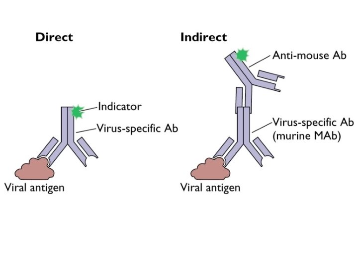
• For immunohistochemical detection strategies, antibodies are classified as primary or secondary reagents. Primary antibodies are raised against an antigen of interest and are typically unconjugated (unlabeled), while secondary antibodies are raised against immunoglobulins of the primary antibody species. The secondary antibody is usually conjugated to a linker molecule, such as biotin, that then recruits reporter molecules, or the secondary antibody itself is directly bound to the reporter molecule.

IHC reporters • Reporter molecules vary based on the nature of the detection method, the most popular being chromogenic and fluorescence detection mediated by an enzyme or a fluorophore, respectively. With chromogenic reporters, an enzyme label reacts with a substrate to yield an intensely colored product that can be analyzed with an ordinary light microscope. While the list of enzyme substrates is extensive, alkaline phosphatase (AP) and horseradish peroxidase (HRP) are the two enzymes used most extensively as labels for protein detection.

Counterstains • After immunohistochemical staining of the target antigen, a second stain is often applied to provide contrast that helps the primary stain stand out. Many of these stains show specificity for specific classes of biomolecules, while others will stain the whole cell. • Both chromogenic and fluorescent dyes are available for IHC to provide a vast array of reagents to fit every experimental design, and include: hematoxylin, Hoechst stain and DAPI are commonly used.

Diagnostic IHC markers • Brd. U: used to identify replicating cells. Used to identify tumors as well as in neuroscience research. • Cytokeratins: used for identification of carcinomas but may also be expressed in some sarcomas. • CD 15 and CD 30 : used for Hodgkin's disease • Alpha fetoprotein: for yolk sac tumors and hepatocellular carcinoma • CD 117 (KIT): for gastrointestinal stromal tumors (GIST) and mast cell tumors • CD 10 (CALLA): for renal cell carcinoma and acute lymphoblastic leukemia • Prostate specific antigen (PSA): for prostate cancer • estrogens and progesterone receptor (ER & PR) staining are used both diagnostically (breast and gyn tumors) as well as prognostic in breast cancer and predictive of response to therapy (estrogen receptor) • Identification of B-cell lymphomas using CD 20 • Identification of T-cell lymphomas using CD 3
- Slides: 12