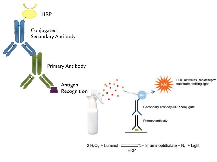
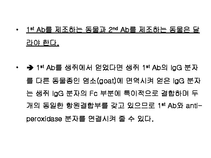
Immunohistochemistry Secondary antibody 2 nd Ab link antibody
")
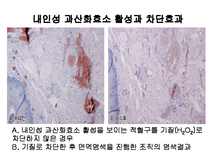
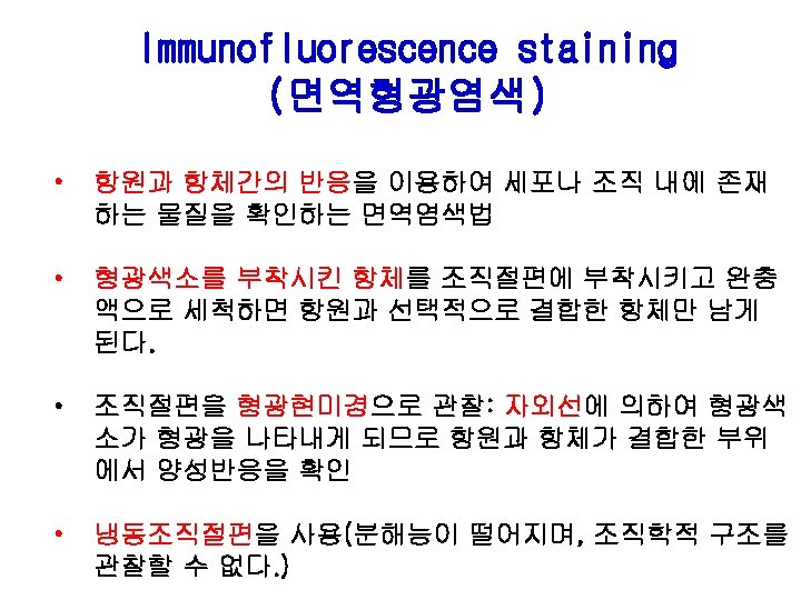
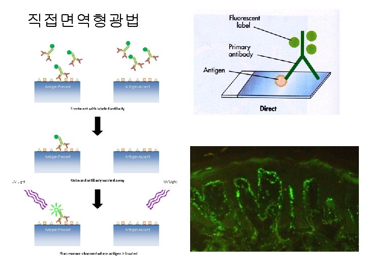
Immunohistochemistry (면역조직화학)
 = link antibody Primary antibody (1 st Ab) Tissue")
Secondary antibody (2 nd Ab) = link antibody Primary antibody (1 st Ab) Tissue Antigen
 • 면역세포화학염색법 (immunocytochemistry) • 면역형광염색법 (immunofluorescence, IF) • 유세포검사 (flow")
면역진단법 • 면역조직화학염색법 (immunohistochemistry) • 면역세포화학염색법 (immunocytochemistry) • 면역형광염색법 (immunofluorescence, IF) • 유세포검사 (flow cytometry) – FACS
")
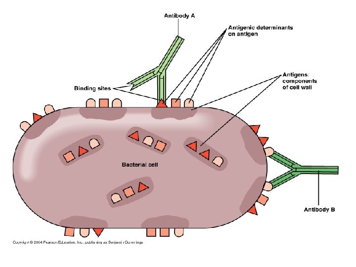
Polyclonal antibody ( 다클론항체)
")
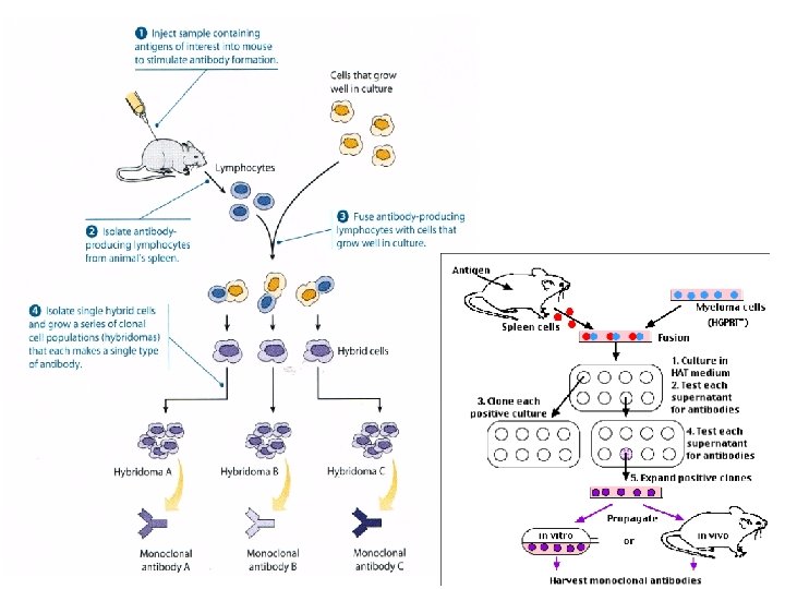
Monoclonal antibody (단클론항체)
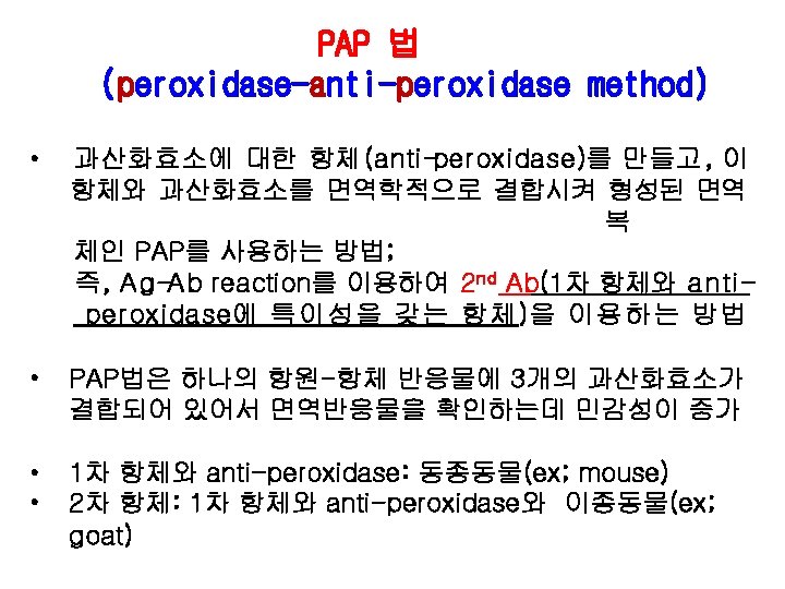

anti-peroxidase secondary antibody primary antibody Ag tissue peroxidase anti-peroxidase method

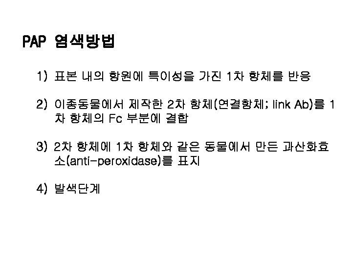
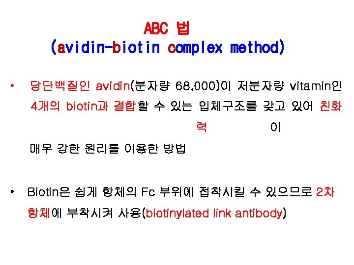
PAP 법 APAAP 법
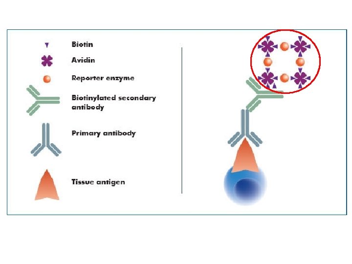
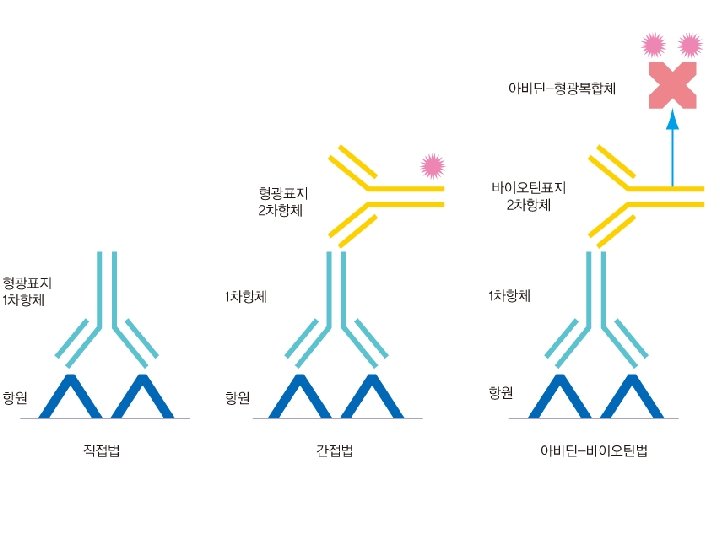
 1차 항체를 조직표면에 존재하는 항원에 결합 2) biotin이 부착된 2차 항체(biotinylated")
ABC 염색방법 1) 1차 항체를 조직표면에 존재하는 항원에 결합 2) biotin이 부착된 2차 항체(biotinylated antibody) 를 1차 항체의 Fc portion에 부착 3) 효소(peroxidase)가 결합된 avidin/biotin(enzyme conjugated avidin/biotin complex)을 2차 항체의 biotin과 결합 4) 기질(substrate)로 발색
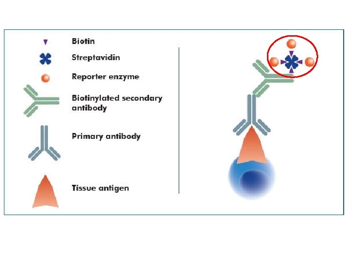
 • Streptomyces avidinii 라는 세균을 배양하면 배양액 으로부터 streptavidin(분자량 60")
LSAB법 (Labeled streptavidin-biotin method) • Streptomyces avidinii 라는 세균을 배양하면 배양액 으로부터 streptavidin(분자량 60 k. Da)을 추출 • streptavidin은 avidin과 유사한 성질을 갖고 있으며 , 비 특 이 적인 결합의 단점이 없어서 avidin의 결점을 보완 • streptavidin에 enzyme(streptavidin-HRP)을 직접 부착 시켜서 사용
")
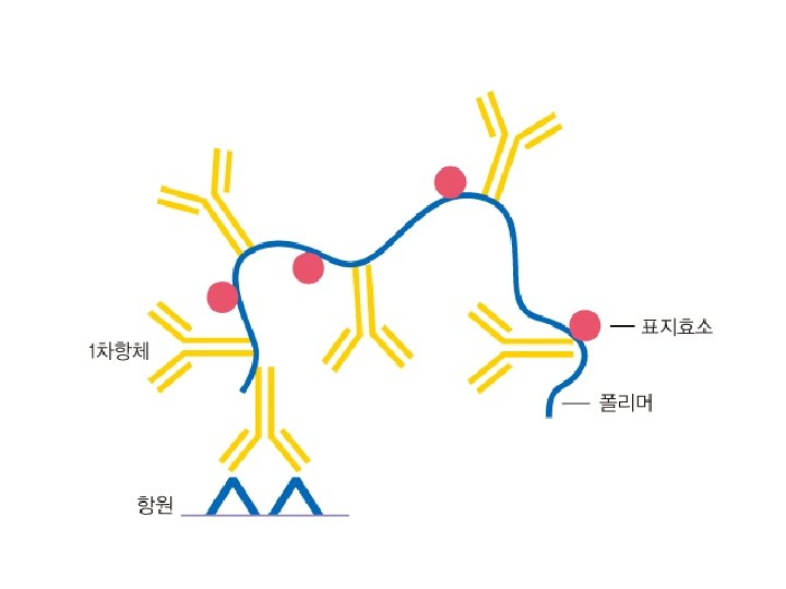
EPOS법 (Polymer based method)
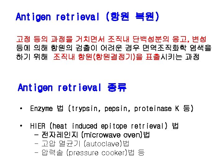
 전자레인지(Microwave oven) Pressure cooker")
항원 복원 장비 고압멸균기(Autoclave) 전자레인지(Microwave oven) Pressure cooker
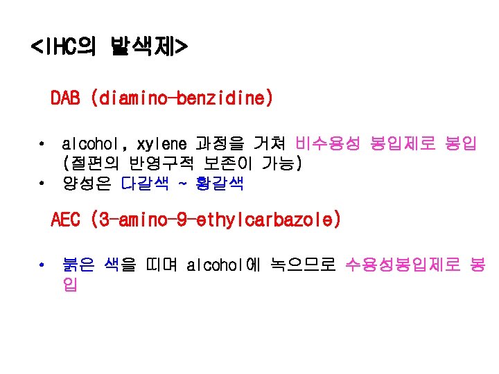
 : Peroxidase DAB Permanent AEC Aqueous")
IHC의 발색제 (developer) : Peroxidase DAB Permanent AEC Aqueous
 BCIP/INT Aqueous (갈색 :")
IHC의 발색제 : Alkaline Phosphatase BCIP/NBT Permanent (흑청색 : 비수용성) BCIP/INT Aqueous (갈색 : 수용성) New Fuchsin Permanent (적색 : 비수용성) Fast Red Aqueous (적색 : 수용성)
 10분 acetone으로 고정 2) 10분 air dry 3) TBS로 10분 수세")
동결절편 염색과정 1) 10분 acetone으로 고정 2) 10분 air dry 3) TBS로 10분 수세 4) Blocking reagent에 10분 처리 5) Primary antibody를 1~2시간 반응 6) TBS에 10 min 수세 7) Secondary antibody에 30분 반응 8) TBS에 10분 수세 9) Streptavidin solution에 30분 반응 10)Tris buffer에 10분 수세
 Deparaffinize and Rehydrate to tap water washing 2) D/W에 3~5 min")
파라핀절편 염색과정 1) Deparaffinize and Rehydrate to tap water washing 2) D/W에 3~5 min 수세 3) 가온된 retrieval solution에 silde를 담근 후 microwave oven 에 5 min간 3번 처리 4) 안정을 위하여 20 mim 동안 microwave oven에 그대로 둔다. ( 매우 중요) 5) Slide를 꺼낸 후(절대 마르지 않게 한다) Tris buffer에 5 min 수세 6) 0. 3% H 2 O 2 -methanol solution에 10 min 반응 (endogenous peroxidase 의 activity를 억제) 7) Tap water에 5 min 간 rinse한 후 D. W. 에 수세 8) Tris buffer에 10 min 수세
 Blocking reagent에 20 min 처리 (blocking reagent를 사용하지 않는 경우는 바로 primary antibody를")
9) Blocking reagent에 20 min 처리 (blocking reagent를 사용하지 않는 경우는 바로 primary antibody를 반응) 10) Primary antibody를 1~2 hour incubate 11) Tris buffer에 10 min 수세 12) Secondary antibody에 30 min 반응 13) Tris buffer에 10 min 수세 14) Streptavidin solution에 50 min 반응 15) Tris buffer에 10 min 수세 16) AEC (or DAB)에서 약 4~5분 발색 17) 흐르는 물에 수세 18) Mayer hematoxylin으로 counterstain 19) 흐르는 물에 수세 20) D/W에 담근 후 수용성 봉입제 (비수용성 봉입제)로 봉입

Paraffin section Xylene, alcohol Antigen retrieval 0. 3% H 2 O 2 -methanol - 15 min 1 -5% normal serum(secondary Ab animal) -10 min Primary antibody- 30 min PBS wash – 5 min x 3 Biotinylated secondary antibody - 30 min PBS wash- 5 min x 3 Streptavidin-peroxidase complex - 10 -60 min PBS wash - 5 min x 3 Chromogen(DAB or AEC) d. H 20 wash Nuclear stain Mount – 3 -10 min
 using")
Microphotograph of a histological section of human skin prepared for direct immunofluorescence (DIF) using an anti-Ig. A antibody. Ig. A deposits are found in the walls of small superficial capillaries (yellow arrows).
")
Ig. G linear basement membrane zone (20 x)

A. H 460/Endo ARTS DAPI B. SNUC-4/Endo ARTS Mitotraker Merged
- Slides: 74