Immunoglobulin Collage of Medical Henan University Contents Introduction

Immunoglobulin Collage of Medical, Henan University

Contents Introduction SectionⅠ Molecular Structure of Ig SectionⅡ Characteristics and Functions of the 5 Classes of Ig SectionⅢ Fc Receptors for Ab Molecules SectionⅣ Biological Activity of Ab SectionⅤ Immunogenicity of Ig SectionⅥ Artificial Ab
: Glycoprotein molecules that are produced by plasma cells and can combine")
Concepts Antibody (Ab): Glycoprotein molecules that are produced by plasma cells and can combine with the corresponding Ag specifically are called Ab. Ab is produced by B cells in the response to a stimulation of Ag. Ab possesses a high degree of specificity and affinity
: It refers to all globulins that possess the activity of Ab")
• Immunoglobulin(Ig): It refers to all globulins that possess the activity of Ab or show a similar structure to Ab • Therefore, All Abs are Igs, but not all Igs possess the functions of Abs
")
Other Concepts γ- Globulin Antiserum Humoral Immunity s. Ig and m. Ig(BCR)

SectionⅠ Molecular Structure of Ig
 l Ig is composed of four polypeptide chains")
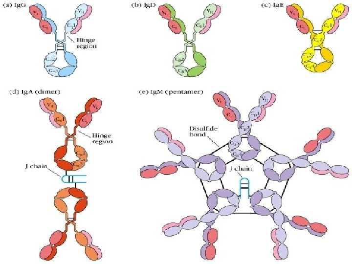
Ⅰ. Basic structure (four polypeptide chains) l Ig is composed of four polypeptide chains joined by S-S bonds. inter-chain disulfide bonds (S-S) intra-chain disulfide bonds (S-S) l It shows “T” or “Y” shape.
: 450 ~ 550 aa, 50")
1. H and L chain: . Heavy chains (H): 450 ~ 550 aa, 50 ~ 75 KD . Light chains (L): 214 aa, 25 KD

Two terminal ends for each peptide chain “N” “C” terminal end “C” L chains attach to H chains from “N” end
 According to the differences of H chains")
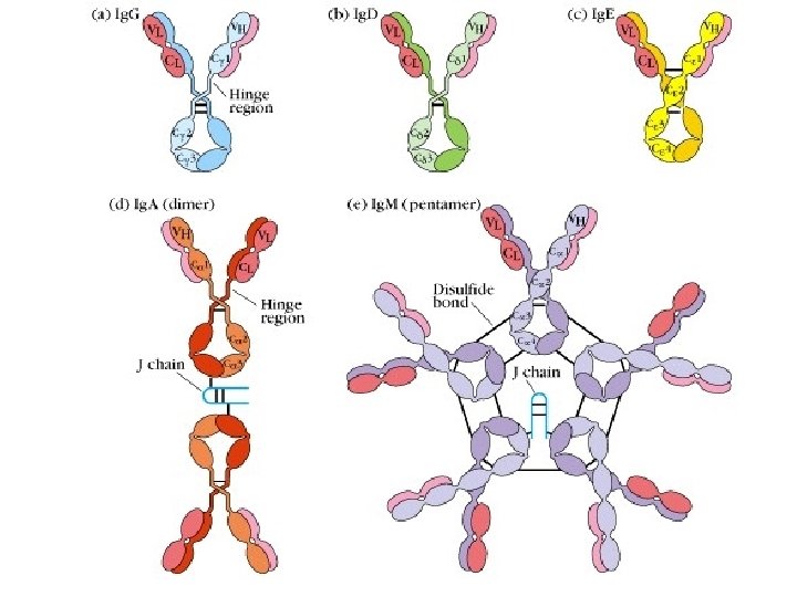
2. classes and types of Ig (1) According to the differences of H chains (amino acid composition, sequence) Igs can be divided into 5 classes subclasses • Five classes of H Chain: • Five classes of Igs: Ig. G Ig. A Ig. M Ig. D Ig. E Ig. G 1~ Ig. G 4 Ig. A 1, Ig. A 2
 According to the differences of L chains • Two types of L chain:")
(2) According to the differences of L chains • Two types of L chain: , : human) 1~ 4 subtypes 20: 1 (in mice); 2: 1 (in
 Constant region (C) (2) Variable region")
3. Two regions of each peptide chain (1) Constant region (C) (2) Variable region (V) (3) Hinge region
 Constant region ( C ) •")
3. Two regions of each peptide chain (1) Constant region ( C ) • CH: 3/4 or 4/5 ( , ) of H chain from the c end • CL: 1/2 of L chain from the c end (2) Variable region ( V ) • CH: 1/4 or 1/5 ( , ) of H chain from the N end • CL: 1/2 of L chain from the N end
 Variable region ( V ): Ø Hypervariable region(HVR) There are three highly diversity")
(2) Variable region ( V ): Ø Hypervariable region(HVR) There are three highly diversity stretches within the V egion, they are called HVR. Ø Framework region(FR): FR 1 -FR 4

Ag-binding sites
")
Complementarity determining regions(CDR)
 Variable region (V) Complementarity determining regions(CDR) L: CDR 1, CDR 2, CDR 3")
(2) Variable region (V) Complementarity determining regions(CDR) L: CDR 1, CDR 2, CDR 3 H: CDR 1, CDR 2, CDR 3

Idiotype of Ig Igs produced by different B cells possess unique structure respectively in hypervariable region (HVR), the unique structure of Ig is called idiotype or idiotypic determinant

In fact: HVR CDR Idiotype are in the same sites of Ig
 Hinge region: • Flexible and suitable for CDR of Ig binding to antigenic")
(3) Hinge region: • Flexible and suitable for CDR of Ig binding to antigenic determinants. • Sensitive to proteolytic enzyme • Ig. M, Ig. E
 l Secretory piece(SP)")
Other structures of Ig • Joining chain(J) l Secretory piece(SP)
 : l Produced by plasma cells l Functions: linker, to compose")
Joining chain(J ) : l Produced by plasma cells l Functions: linker, to compose dimer、 pentamer or polymer(Ig. A, Ig. M)
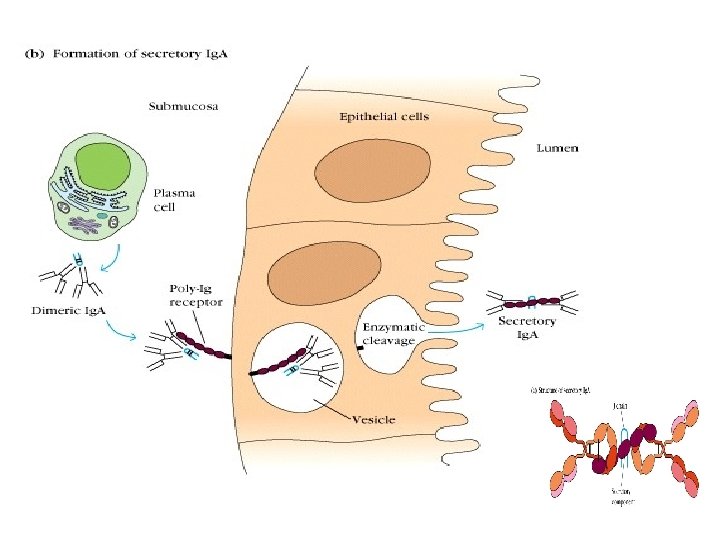
: . Produced by mucosa epithelial cells. Secretory Ig. A (s. Ig.")
Secretory piece( SP): . Produced by mucosa epithelial cells. Secretory Ig. A (s. Ig. A). Functions: protect s. Ig. A, resist proteolysis in extra secretory liquid. Ig. A

Ⅱ. Domains of Ig

1. Domain : Polypeptide chains of Ig are folded into a globular structure by intra chain s-s bond within each 110 aa region which is called a domain
 : VL, CL • H chain(4~5): VH,")
2. Domains of Ig • L chain(2) : VL, CL • H chain(4~5): VH, CH 1, CH 2, CH 3 CH 4(in Ig. M, Ig. E) hinge region
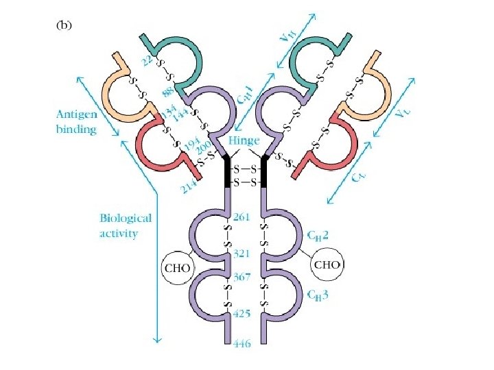

3. Function of each domain • VH, VL: antigen-binding site • CH 1, CL: allogeneic marker • CH 2/CH 3: complement-fixing site, permeate placenta(Ig. G) • CH 3/CH 4: cell-binding site l Hinge region : flexible and suitable for CDR of Ig binding to antigenic determinants

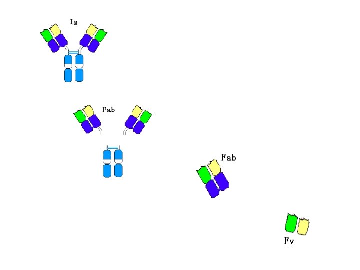
Ⅲ. Hydrolytic fragments of Ig Ig can be digested by papain and pepsin • Position • Fragments • Function

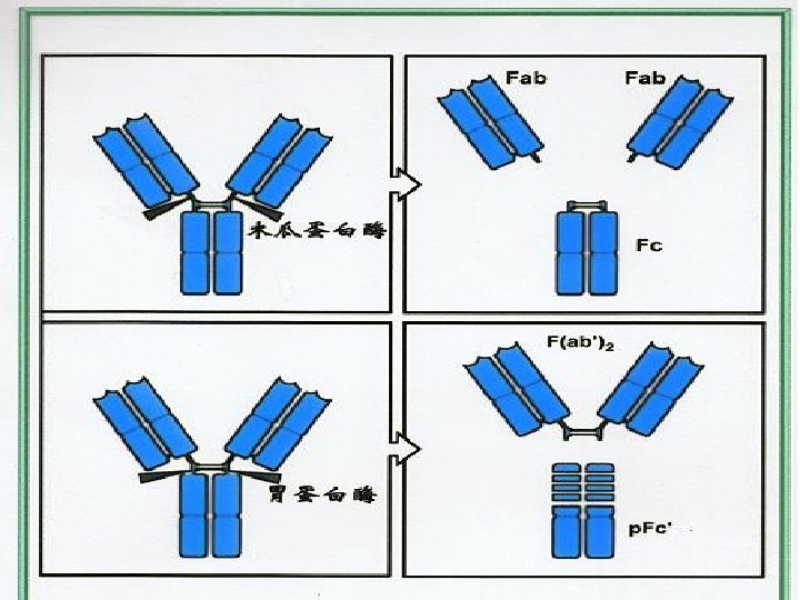
1. Digested by papain • Position: near the S-S bonds of H inter-chains fromthe N end l Fragments: l Function: • 2 Fab: fragment antigen-binding Fc: fragment crystallizable Fab: recognize and bind Ag Fc: (1) fix complement (2) crossing the placenta (3) bind to Fc. R in different cells

Ag: Ab ratio

2. Digested by pepsin • Position: near the S-S bond of H inter-chains from the C end • Fragments and function : F(ab′)2: bind antigen(2 valence) p. Fc′: no function

Significance l Elucidating the relationships between the structure and function of Igs l Decrease the immunogenicity of Ig for clinical treatment

SectionⅡ Characteristics and Functions of the 5 Classes of Igs
")
Ⅰ. Ig. G 1. Highest concentration in serum(75% of total Ig)

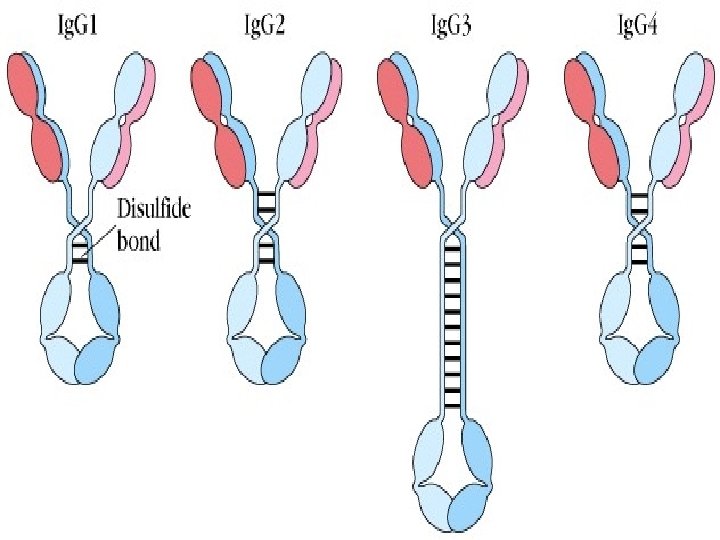
2. Four subclasses: Ig. G 1, Ig. G 2, Ig. G 3, Ig. G 4

3. Unique Ig that can pass through placenta 4. Half-life is longer( 16 -24 days ) 5. Starts to be produced at 2 -3 month after birth and reach the level of adult at 5 years old

6. Functions of Ig. G: • • • Against bacteria and virus, neutralize toxin Combine with the Fc receptor(FcγR) Activate complement Combine with SPA Some belong to the auto-antibodies • Take part in type Ⅱ and Ⅲ hypersensitivity
, 10 valences")
Ⅱ. Ig. M 1. Highest MW:pentamer(90 KD), 10 valences
 3. The first Ig to be synthesized • Appear")
2. Half-life is shorter(4~5 days) 3. The first Ig to be synthesized • Appear in the early stage after infection • Be produced during fetus • The first m. Ig of the B cells, act as the antigen receptors(BCR)

4. Functions: • Ig. M is more effective in binding Ag and activating C, and play an important role in anti-infection • Natural Ab for blood-type antigen • Auto-antibody: rheumatoid factor(RF) • Take part in type Ⅱ and Ⅲ hypersensitivity
: dimer,trimer")
Ⅲ. Ig. A 1. Two types Serum type :monomer Secretary type(s. Ig. A): dimer,trimer or polymer 2. Two subclasses:Ig. A 1,Ig. A 2

3. To be produced at 4 months after birth 4. Exist in almost all body fluid

6. Local mucosal immunity • • Immune barrier Neutralize virus/toxin Rich in colostrum Activate C by alternative pathway • Take part in type Ⅲ hypersensitivity

Ⅳ. Ig. D 1. The concentration in serum is low and sensitive to proteinase 2. Act as the antigen receptor on B cells (m. Ig. D): Regulate the differentiation of B cells

Ⅴ. Ig. E 1. Concerntration of Ig. E in serum is the lowest in normal individual, but is very high in some patients. 2. Related to typeⅠpersensitivity FcεRⅠ: mast cell, basophil

SectionⅢ Fc Receptors for Ab Molecules
---phagocyte Fc RⅡ(CD 32)---immune complex Fc RⅢ(CD 16)--NK, macrophage,")
Ig. G---Fc R: Fc RⅠ(CD 64)---phagocyte Fc RⅡ(CD 32)---immune complex Fc RⅢ(CD 16)--NK, macrophage, T cell Ig. E---Fc R: Fc RⅠ--- mast cell, basophil Fc RⅡ--- macrophage, B cell Ig. A---FcαR(CD 89)---phagocyte, neutrophil

SectionⅣ Biological Activity of Ab

1. Recognize and bind to antigen specifically 2. Fix complement 3. Bind to Fc receptor on some cells 4. Transfer selectively : . Planceta transfer (Ig. G). Mucosa transfer (s. Ig. A)

Affinity and Avidity

Neutralization

Ig. M, Ig. G 1~3: classical pathway Ig. A, Ig. G 4, Ig. E: alternative pathway

MAC
 Opsonization(Ig. G, Ig. M): Enhance the phagocytosis of MΦ")
(1) Opsonization(Ig. G, Ig. M): Enhance the phagocytosis of MΦ
 ADCC( antibody dependent cell mediated cytotoxicity)")
(2) ADCC( antibody dependent cell mediated cytotoxicity)
 Hypersensitivity typeⅠ - mast cell, basophil(Fc RⅠ) allergen Ig. E Fce. RI degranulation")
(3) Hypersensitivity typeⅠ - mast cell, basophil(Fc RⅠ) allergen Ig. E Fce. RI degranulation inflammation

SectionⅤ Immunogenicity of Ig

Isotype: CH, CL

Allotype:CH, CL

Idiotype: VH, VL Anti-idiotype antibody

SectionⅥ Artificial Ab Ø Ø Ø Polyclonal Ab Monoclonal Ab Gene engineering Ab

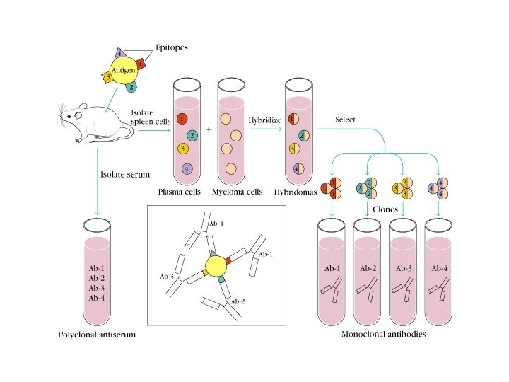
1. Polyclonal Ab l A mixture Ab with different specificities and affinities l Generate in a natural response or artificial immunization l Cross reaction

Cross-reactivity: if two antibody antigens share recognizes an chemically similar, epitope. an epitope unrelated, an but
 Ab produced by single clone (or one hybridomas clone")
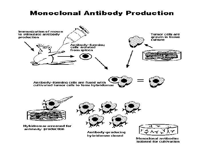
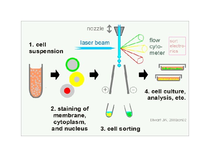
2. Monoclonal Ab (m. Ab) Ab produced by single clone (or one hybridomas clone ) and having a single specificity
 hybride")
m. Ab / Mc. Ab u Prepared by hybridomas technique: Immunized spleen cells(B) hybride with myeloma cells----hybridomas

Artificial antibodies POLYCLONAL. MONOCLONAL. Derived from different B Lymphocytes cell lines Derived from a single B cell clone Batch to Batch variation affecting Ab reactivity & titre m. Ab offer Reproducible, Predictable & Potentially inexhaustible supply of Ab with exquisite specificity NOT Powerful tools for clinical diagnostic tests Enable the development of secure immunoassay systems.
")
Cell fusion technique: Secrete Ab Short life No Ab Long life Splenocyte (B cell) Myeloma cell Secrete Ab Long life Hybridoma cell Köhler and Milstein, 1975 1984, Nobel Prize
 Hybridoma cell Myeloma cell S/ S Short")
Screening of hybridoma cell: Splenocyte (B cell) Hybridoma cell Myeloma cell S/ S Short life Yes M/ M splenocyte Short life Myeloma cell

Screening with HAT selective medium: H, 次黄嘌呤; A, 氨基喋呤; T, 胸腺�� (Hypoxanthine guznine phosphoribosyl transferase) H 次嘌呤� 嘌呤磷酸核糖� 移酶 ( HGPRT ) Extrinsic pathway(alternative) 谷氨� 胺 or � 磷酸尿苷酸 T 二� 叶酸� 原酶 A DNA Intrinsic pathway(major) Extrinsic pathway(minor) 胸腺�� 激酶 ( TK ) (Thymidine kinase) B cell: HGPRT+, TK+ live Myeloma cell: HGPRT-, TK- die

PRODUCTION OF MONOCLONAL ANTIBODY HYBRIDOMA TECHNOLOGY

Applications of Monoclonal Antibodies • Diagnostic Applications Biosensors & Microarrays • Therapeutic Applications Transplant rejection Muronomab-CD 3 Cardiovascular disease Abciximab Cancer Rituximab Infectious Diseases Palivizumab Inflammatory disease Infliximab • Clinical Applications Purification of drugs, Imaging the target • Future Applications Fight against Bioterrorism


EVOLUTION OF MONOCLONAL ANTIBODY 1. TRANSGENIC DNA SPLICING / GENE KNOCK OUT 2. LIBRARIES a. BACTERIOPHAGE b. m. RNA c. Cell Surface

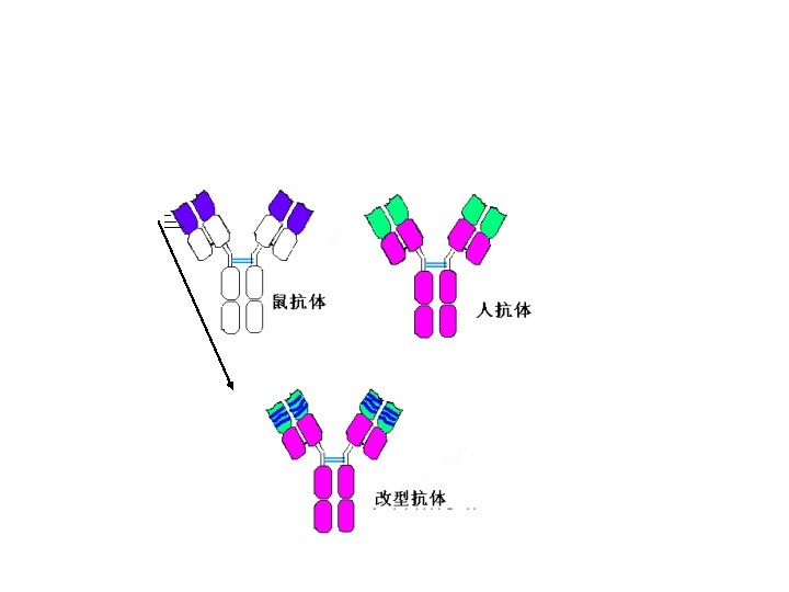
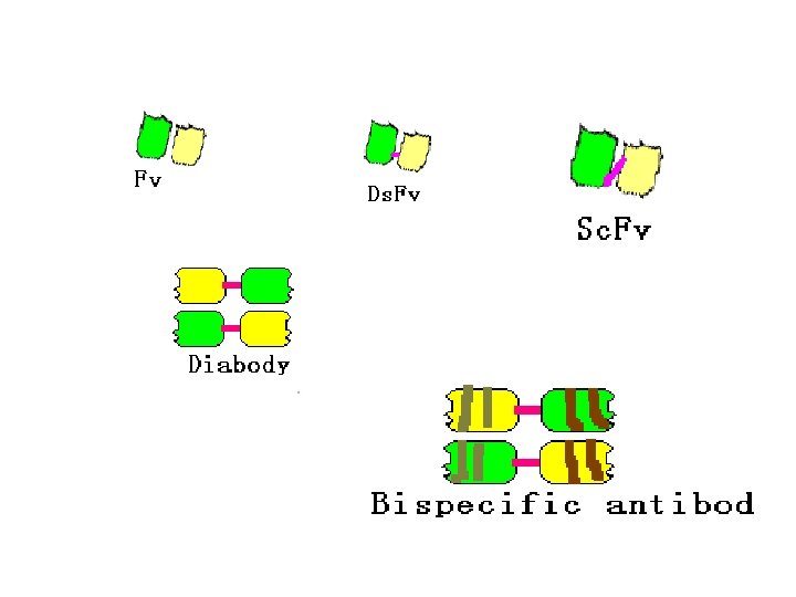
3. Gene engineering Ab • Abs prepared by the method of gene recombination • Chimeric Ab: human Fc bind with mice Fab • Recombinant single chain Ab: VH-linker-VL
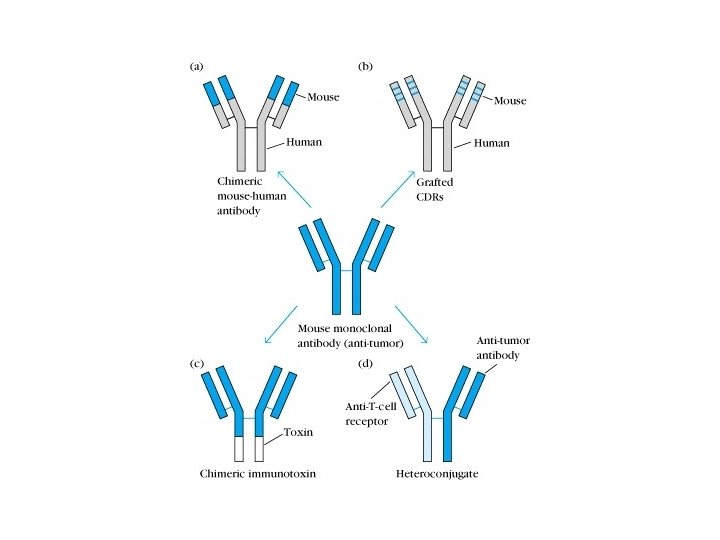

Human-mouse chimeric Ab

Niels K Jerne Denmark Basel Institute for Immunology Basel, Switzerland J. Kohler Germany Basel Institute for Immunology Basel, Switzerland S. Milstein Argentina MRC Laboratory of Molecular Biology Cambridge,
- Slides: 89