Immunoblistering Skin Diseases Dr Ahmed Abdulhussein ALHuchami 1

Immunoblistering Skin Diseases Dr. Ahmed Abdulhussein AL-Huchami 1
")
Bullous Skin Disorders (BSD)

INTRODUCTION �BSD are skin conditions characterised by blister formation. �A blister is an accumulation of fluid between cells of the epidermis or upper dermis. �Causes of blister could be genetic, physical, inflammatory, immunologic and as a reaction to drugs. �BSDs are mostly autoimmune.
![Immunoblistering Skin Diseases Group of disorders sharing two common features: 1. Immunological [Autoimmune] Pathogenesis.](http://slidetodoc.com/presentation_image_h/009f9176c98834a00be8c7e6b20ff556/image-5.jpg "Immunoblistering Skin Diseases Group of disorders sharing two common features: 1. Immunological [Autoimmune] Pathogenesis.")
Immunoblistering Skin Diseases Group of disorders sharing two common features: 1. Immunological [Autoimmune] Pathogenesis. 2. The primary presenting lesion is a bulla or blister. Immunoblistering skin diseases include: 1. Pemphigus group (P. vulgaris, P. vegetans, P. foliaceus, P. erythematosus and Paraneoplastic Pemphigus). 2. Pemphigoid group (Bullous Pemphigoid, Bullous Pemphigoid of Pregnancy or also known as Herpes gestationis and Cicatricial Pemphigoid). 5
 splitting that can")
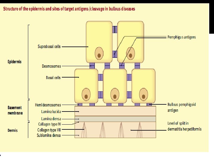
Classification of Immunoblistering Diseases The bullae result from skin cleavage (splitting) splitting that can occur at any level within the skin with subsequent accumulation of fluid. According to the level of skin cleavage, immunoblistering diseases are classified into: 1. Epidermal blistering disorders e. g. Pemphigus group of disorders. 2. Dermal blistering disorders in which splitting occurs within or beneath the basement membrane e. g. Pemphigoid group of disorders, DH, Linear Ig. A disease and CBBDC (Linear Ig. A disease of Children). 6
. B, Suprabasal (as in")
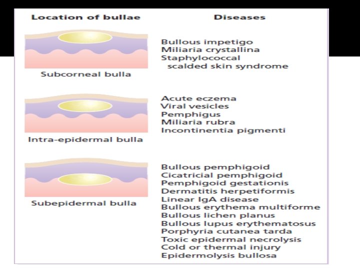
Levels of blister formation A, Subcorneal (as in pemphigus foliaceus). B, Suprabasal (as in pemphigus vulgaris). A, these lesions. C, Subepidermal (as in bullous pemphigoid & dermatitis herpetiformis). Assessment of the levels of epidermal separation forms the basis of the initial differential diagnosis of these lesions.

Pemphigus ﺩﺍﺀ ﺍﻟﻔﻘــــــﺎﻉ Ø An autoimmune intraepidermal blistering disease. Ø Relatively uncommon disease. Ø Often severe and can be life-threatening. Ø It presents as flaccid bullae and painful erosions. Ø According to the level of splitting within the epidermis Pemphigus can be divided into: 1. Superficial types: types characterized by subcorneal cleavage e. g. P. foliaceus and its variant P. erythematosus 2. Deep type: type characterized by suprabasal cleavage e. g. P. vulgaris and its variant P. vegetans Ø A rare variant of Pemphigus which is usually associated with an underlying malignancy (Thymoma or Carcinoma) is called Paraneoplastic Pemphigus 9
. .")
Levels of blister formation A, Subcorneal (as in pemphigus foliaceus). .

Pathogenesis Circulating Antidesmosomal Ig. G autoantibodies + desmosomes Ag-Ab complexes Stimulate Keratinocytes Plasminogen activator Plasminogen Plasmin (Proteolytic enzyme) Acantholysis Bullae 11

Pemphigus vulgaris ﺩﺍﺀ ﺍﻟﻔﻘﺎﻉ ﺍﻟﺨﺒﻴﺚ Clinical Picture Usually affects middle age group [40 -60 years]. Both sexes are equally affected. Incidence is higher among Jews. Presentation Flaccid bullae that easily and rapidly rupture leaving oozy painful erosions and later crusted lesions (bullae may not be seen at presentation because of their flaccid and fragile nature). The bullae are not hemorrhagic. The rash is not pruritic and there is no grouping of lesions. Nikolsky’s sign and Bulla spread sign are positive. 12

+ ve Nikolski’s Sign Nikolsky Sign : Dislodging of epidermis by lateral finger pressure in the vicinity of lesions, which leads to an erosion. Shearing stresses on normal skin cause new erosions to form

Pemphigus vulgaris ﺩﺍﺀ ﺍﻟﻔﻘﺎﻉ ﺍﻟﺨﺒﻴﺚ P. vulgaris usually has generalized distribution, distribution however the predilection sites are the scalp, trunk, itertriginous areas (axillae and groins). Mucous membranes of the mouth, pharynx and larynx may be involved and cause painful oral erosions, dysphagia and hoarseness of the voice respectively. Oral lesions (Painful erosions and ulcerations) are the presenting feature in about 50% of the patient and oral involvement occurs in almost all patients during the whole course of the disease. The course of the disease is usually chronic with a high mortality rate (100% before steroid era and about 10 -30% after corticosteroid introduction), however with careful management many patients can live relatively normal life with occasional exacerbations. 14

P. Vulgaris involving the trunk with painful erosions 15

Bullae are flaccid and fragile in P. vulgaris, they rupture easily and rapidly leaving painful erosions and later crustations. 16

Pemphigus vulgaris: Bullae are transient in this disorder; erosion is more characteristic. 17

Upper lip Lower lip PV of the oral mucous membranes Tongue Inner Cheek 18

Pemphigus vulgaris Suprabasal acantholysis results in an intraepidermal blister containing rounded keratinocytes that are separating from their neighbors. Initially, a single row of basal cells is present on the floor of the blister with dermal papillomatosis (tombstone effect). Follicular involvement by acantholysis is also common.

Binding of Abs to the adhesion molecules loss of cell -cell adhesion acantholysis

Pemphigus vulgaris ﺩﺍﺀ ﺍﻟﻔﻘﺎﻉ ﺍﻟﺨﺒﻴﺚ Complications of P. vulgaris 1. Infections such as septicemia, chest infection… etc. 2. Fluid and electrolyte loss due to widespread denudation of the skin and mucous membranes. 3. Side effects of treatment with corticosteroids and other immunosuppressive agents. These side effects are inevitable and now considered as the leading cause of death in patients with P. vulgaris. 21

Investigations 1. Tzanck Smear: Scrape the floor of an intact blister and stain with Giemsa stain, it shows large rounded acantholytic keratinocytes with hyperchromatic nuclei. 2. Skin Biopsy 3. Immunoflourescence studies: is used to detect tissue-fixed antibodies (local antibodies) and circulating antibodies (serum antibodies) through: A. Direct Immunoflourescence technique (DIF) B. Indirect Immunoflourescence technique (IIF) 22

Investigations Skin Biopsy Take an intact early blister from the skin or oral mm and stain it with H-E stain. Suprabasal acantholysis and intraepidermal bullae containing large round acantholytic cells and some polymorphs & eosinophils P. foliaceus: Subcorneal Acantholysis P. vulgaris: Suprabasilar Acantholysis 23

Investigations DIF Technique: A peri-lesional skin from the patient is stained with a fluorescen-labeled Anti. Ig. G and Anti-C 3 Abs to detect tissue fixed autoantibodies. It shows Ig. G and C 3 deposition in epidermal intercellular spaces around the keratinocytes i. e. Desmosomal Antibodies. 24

Investigations Indirect Immunoflourescence Technique: Ig. G anti-Desmosomal Abs can be detected in the serum of about 90% of patients with P. vulgaris. Technique: Incubate Patient’s serum with monkey’s esophageal mucosa and then stain it with a fluorescenlabeled Anti-Ig. G. Positive test shows Ig. G deposition in epidermal intercellular spaces around the keratinocytes i. e. Desmosomal Antibodies. This technique: 1. Is useful in detecting circulating autoantibodies. 2. Can be used to measure disease activity in P. vulgaris (through measuring Ab titer by serial dilution of the patient serum). It is useful in the follow-up of patient on corticosteroid therapy as the Ab titer correlates with the disease activity in patients with P. vulgaris. 25

Management of P. vulgaris 1. 2. 3. A. ü Admission to the hospital. Give supportive therapy (iv fluids, antibiotics) if needed. Start Immunosuppressive therapy: therapy Corticosteroids: Corticosteroids Considered as the first line treatment unless there is a contraindication. ü Start with 80 -120 mg Prednisolone per day in divided doses (higher doses may be needed in more severe cases). ü Start to taper the dose gradually over weeks when the disease is controlled (Control is reached when no new lesions appear and disappearance of old lesions) until reaching a minimal possible maintenance dose (Usually in the range of 5 -15 mg per day). 26

Management of P. vulgaris B. Cytotoxics: Cytotoxics Used as Steroid-sparing Agents (Used in combination with steroid to decrease the dose of steroid) or as Steroid alternatives in cases of corticosteroid contraindications. These agents act through suppressing immunity and hence decrease autoantibody production. Examples of these agents: • Azothioprine (Immuran®): 150 mg per day • Cyclophosphamide (Cytoxan®): 2 mg per Kg per day • Cyclosporine • Mycophenolate moefetil C. Gold: Gold therapy (oral or parenteral “im”) may be useful in some cases of P. vulgaris. D. Plasmapheresis: Plasmapheresis Rarely used, however it may be reserved for very severe and refractory cases. 27

TREATMENT Systemic steroid ; 2 to 3 mg/kg of prednisolone until cessation of new blister formation and disappearance of Nikolsky sign. Concomitant Immunosuppressive Therapy(steroid sparing agents) such as Azathioprine , 2– 3 mg/kg Methotrexate , either orally or IM at doses of 25 to 35 mg/week. cyclophosphamide or mycophenylate mofetil High-dose intravenous immunoglobulin (HIVIg) ; (2 g/kg every 3– 4 weeks) may help gain quick control whilst waiting for other drugs to work. Rituximab ( Anti-CD 20 monoclonal antibody) has been reported to help multidrug resistance, IV , once a week for 4 weeks. Rx is usually prolong and need regular follow up Dosage should be dropped only when new blisters stop appearing

Bullous Pemphigoid ﺷﺒﻴﻪ ﺩﺍﺀ ﺍﻟﻔﻘﺎﻉ Clinical Picture Ø BP is an autoimmune subepidermal blistering disease. Ø More prevalent in western countries (BP>PV), whereas the reverse occurs in Iraqi people (PV>BP). Ø Usually affects old people (60 -80 years). Ø Both sexes are equally affected. Ø BP is often pruritic. Ø Characterized by tense bullae that may arise from normalappearing skin or from an urticarial or eczematous areas. Ø The bullae are tense (not flaccid because they are subepidermal and covered with an intact epidermis). 29

Bullous Pemphigoid Ø The bullae may be hemorrhagic and may rupture leaving erosions and crustations. Ø The lesions usually heal without scar formation. Ø Nikolsky's sign is negative. Ø Predilection areas: 1. Extremities (esp. inner thighs and flexor aspects of upper arms). 2. Trunk (especially axillae, groins & lower abdomen). Ø Mucous membrane involvement is occasional (2030%) and is rarely being the initial presenting feature of BP. 30

Bullous Pemphigoid Tense bullae and hemorrhagic scabs Thigh 31

Bullous Pemphigoid Tense bulla BP mimicking Pompholyx 32

Bullous Pemphigoid 33

Bullous Pemphigoid Tense bullae 34

Bullous pemphigoid

Levels of blister formation A, these lesions. , Subepidermal (as in bullous pemphigoid

Pathogenesis of BP Ig. G autoantibodies Complement system Produces Binds BP Ag (in LL of BMZ) Ag-Ab complex Subepidermal bullae v ti c A s e at Chemotactic factors + Proteolytic enzymes Separation of LL 37

Investigations 1. Skin Biopsy: Biopsy shows subepidermal unilocular bullae, often filled with eosinophils and some neutrophils. Subepidermal bulla Eosinophils 38

Investigations 2. Immunoflourescent Studies A. DIF: Shows linear Ig. G and C 3 deposition along the Basement Membrane Zone. B. IIF: Circulating Ig. G antibodies can be detected in about 70% of patients. The titer of antibodies dose not correlate with the disease activity i. e. it is not useful in the follow up of the patients. DIF 39

DIF: Linear Ig. G deposits in the BMZ in BP 40

Treatment of BP Treatment 1. Prednisolone: start with 40 -60 mg per day and after control state taper the dose to the minimum possible maintenance dose or even in some cases treatment may stopped. 2. Dapsone: 2 mg per kg per day may be useful in some cases. ü Course of BP is often self-limiting and treatment can often be stopped after one or few years of treatment. ü Remission of BP may be spontaneous or treatmentinduced. ü Complications of BP are similar but milder and to lesser extent than those of PV (less mortality rate). 41

DIFF BTW PEMPHIGUS AND PEMPHIGOID PEMPHIGUS • • • Usually affects the middle age Acute and non itchy Seen on the trunk, flexures and scalp Mouth Blister is common Nature of blister is superficial and flaccid • Circulating Ab is Ig. G to intracellular adhesion proteins • Serum Ab Titer correlate with clinical disease activity. • • Acantholysis Nikolsky sign is positive Elderly patients Chronic and itchy • • • Usually flexural Mouth Blister is Rare • Blister is tense and bloody • Ig. G to BM region • Serum Ab Titer does not correlate with clinical disease activity. • • • No acantholysis Nikolsky sign is negative

- Slides: 43