IMAGING OF HEAD TRAUMA Dr Thanh Binh Nguyen

IMAGING OF HEAD TRAUMA Dr. Thanh Binh Nguyen University of Ottawa, Canada July 2009

OUTLINE n Clinical indications for imaging n Imaging technique n Extraaxial hemorrhage n Intraaxial injury n Brain herniations n Skull fractures

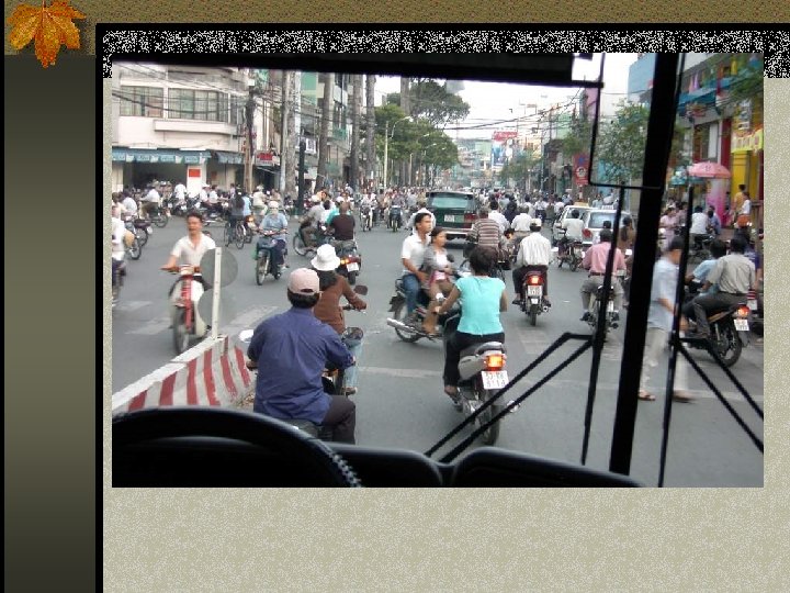
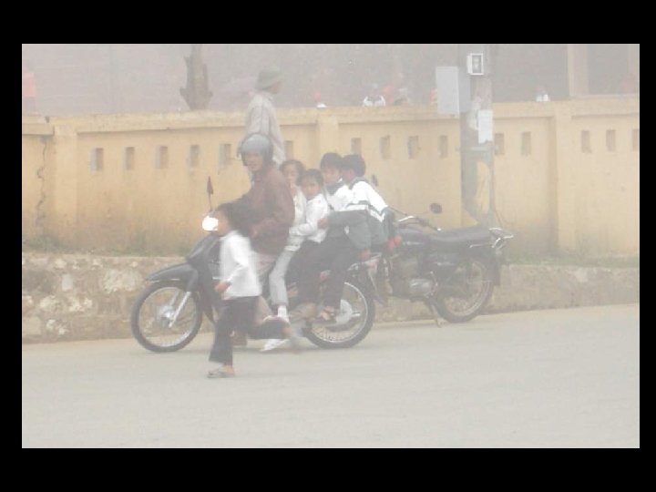
INTRODUCTION n Head trauma is the leading cause of death in people under the age of 30. n Males have 2 -3 x frequency of brain injury than females n Due mainly to motor vehicle accidents and assaults

Classification of TBI n Primary n Injury to scalp, skull fracture n Surface contusion/laceration n Intracranial hematoma n Diffuse axonal injury, diffuse vascular injury n Secondary n Hypoxia-ischemia, swelling/edema, raised intracranial pressure n Meningitis/abscess

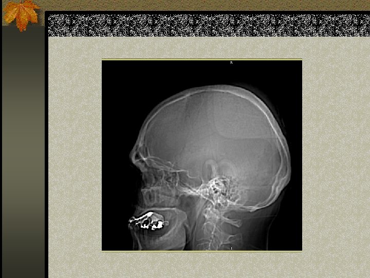
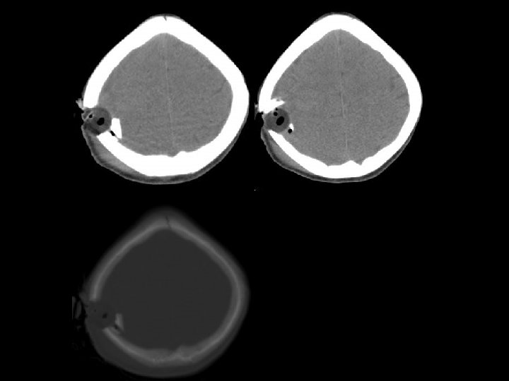
IMAGING TECHNIQUE n The presence of a skull fracture increases the risk of having a posttraumatic intracranial lesion. n However, the absence of a skull fracture does not exclude a brain injury, which is particularly true in pediatric patients due to the capacity of the skull to bend. n NO ROLE FOR PLAIN FILMS IN ACUTE HEAD TRAUMA

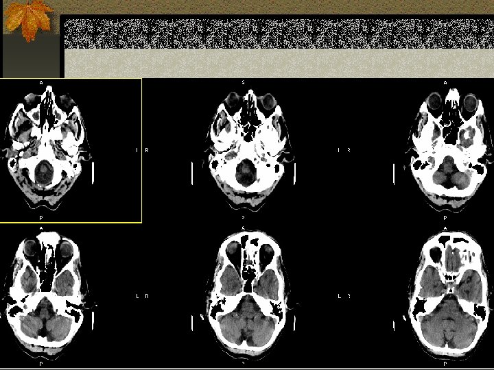
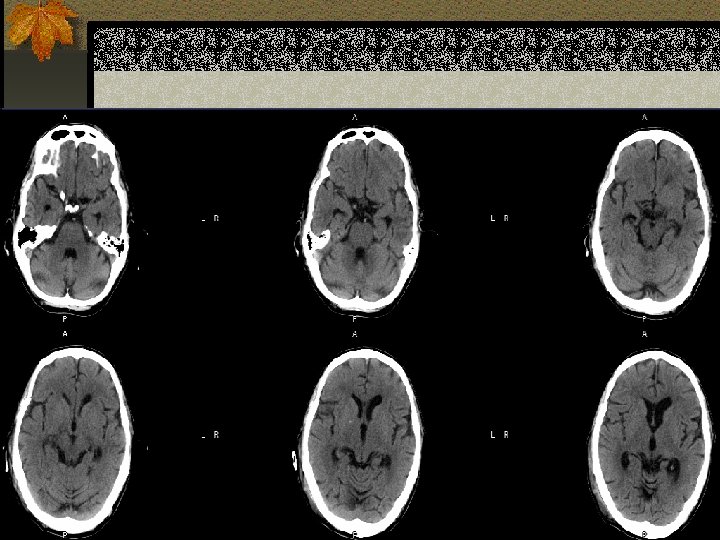
IMAGING TECHNIQUE n CT without contrast is the modality of choice in acute trauma (fast, available, sensitive to acute subarachnoid hemorrhage and skull fractures) n MRI is useful in non-acute head trauma (higher sensitivity than CT for cortical contusions, diffuse axonal injury, posterior fossa abnormalities)

OUR CT PROTOCOLS n “ROUTINE”: posterior fossa and supratentorial region (slice thickness = 5 mm) n “TRAUMA”: posterior fossa (2. 5 mm), supratentorial region (5 mm) n “TEMPORAL BONE”: <1 mm in axial or coronal plane n “ORBITS/FACIAL BONES”: 1. 25 mm axial/coronal orbits

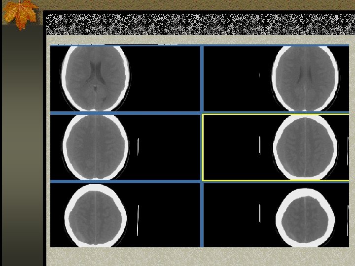
APPROACH TO CT BRAIN n Look at the scout film: ? Fracture of upper n n n cervical spine or skull Look for brain asymmetry Look at sulci, Sylvian fissure and cisterns to exclude subarachnoid hemorrhage Change windows to look for subdural collection Look at bone windows to see fractures Determine if mass is intraaxial (in the brain) or extraaxial (outside)

SCALP INJURY

SCALP INJURY n Cephalohematoma: blood between the bone and periosteum. Cannot cross the suture lines. n Subgaleal hematoma: blood between the periosteum and aponeurosis. Can cross the suture lines. n Caput Succ: swelling across the midline with scalp moulding. Resolves spontaneously.
 n Subdural hematoma(SDH) n Epidural hematoma n Subdural")
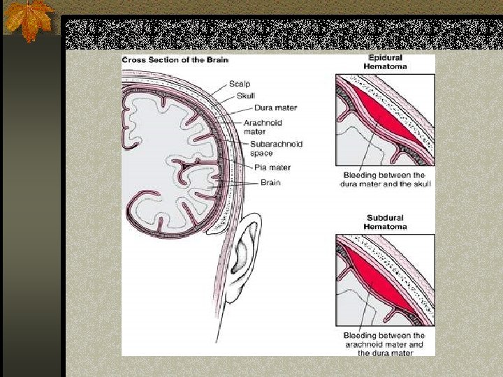
Extraaxial fluid collections n Subarachnoid hemorrhage(SAH) n Subdural hematoma(SDH) n Epidural hematoma n Subdural hygroma n Intraventricular hemorrhage

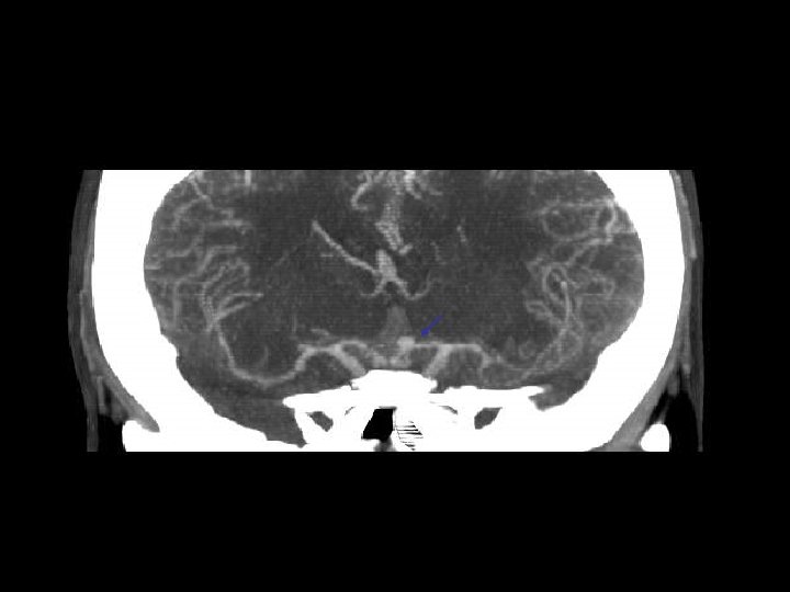
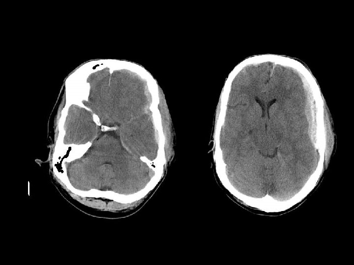
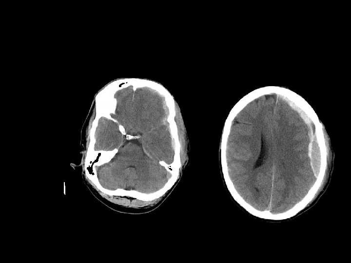
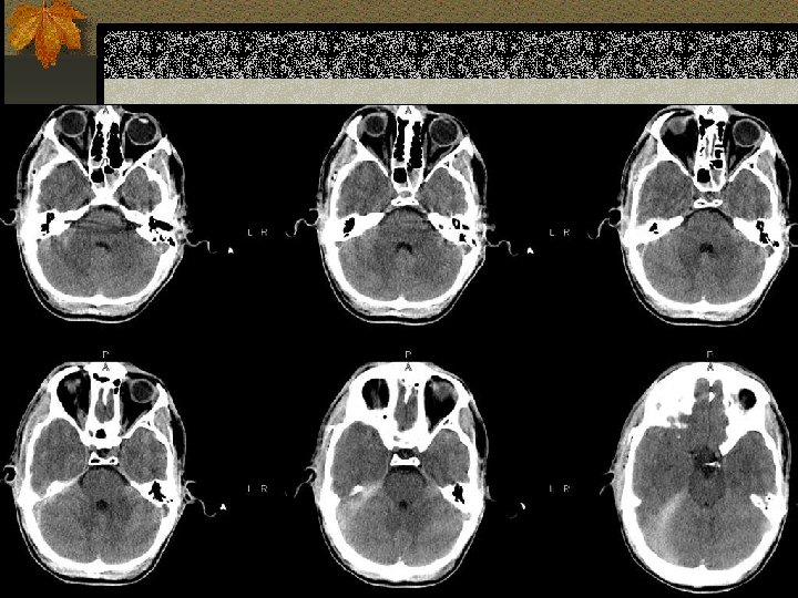
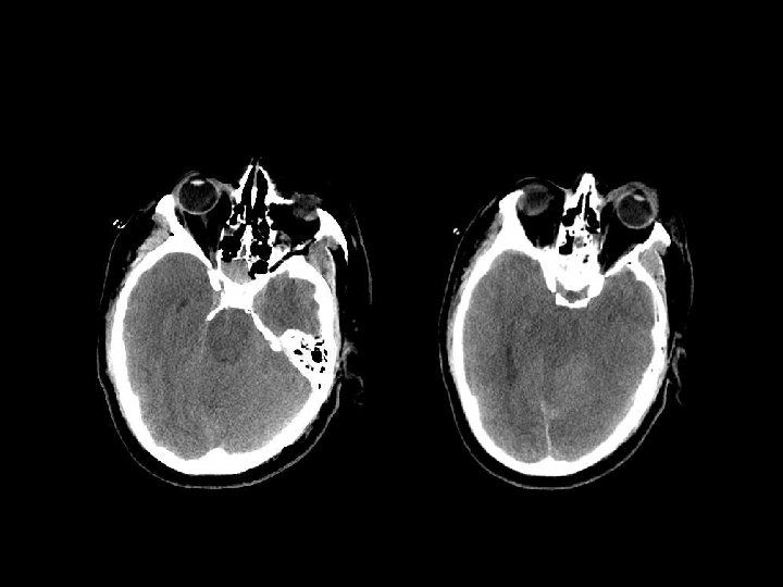
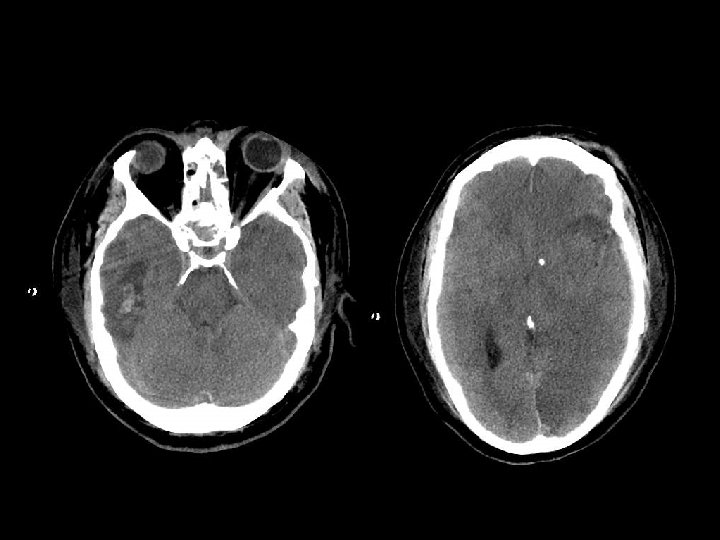
Subarachnoid hemorrage n Can originate from direct vessel injury, contused cortex or intraventricular hemorrhage. n Look in the interpeduncular cistern and Sylvian fissure n Usually focal (but diffuse from aneurysm) n Can lead to communicating hydrocephalus

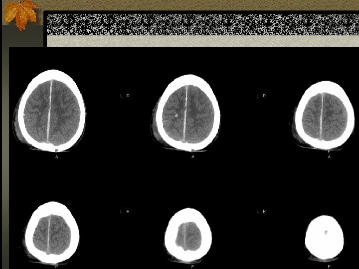
SUBDURAL HEMATOMA n Occurs between the dura and arachnoid n Can cross the sutures but not the dural reflections n Due to disruption of the bridging cortical veins n Hypodense(hyperacute, chronic), isodense(subacute), hyperdense(acute)

W=33 L=41

MANAGEMENT OF a. SDH n Acute SDH with thickness > 10 mm or midline shift > 5 mm should be evacuated n Patient in coma with a decrease in GCS by >2 points with a SDH should undergo surgical evacuation.

EPIDURAL HEMATOMA n Located between the skull and periosteum n Due to laceration of the middle meningeal artery or dural veins n Can cross dural reflections but is limited by suture lines n Lentiform shape (but concave shape in SDH)

MANAGEMENT OF a. EDH n EDH > 30 cm 3 should be evacuated. n EDH < 30 cm 3 and <15 mm thickness and < 5 mm midline shift and GCS >8 may be managed nonoperatively with serial CT

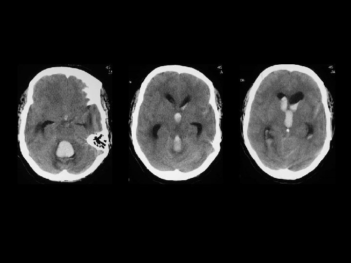
Intraventricular hemorrhage n Most commonly due to rupture of subependymal vessels n Can occur from reflux of SAH or contiguous extension of an intracerebral hemorrhage n Look for blood-cerebrospinal fluid level in occipital horns

INTRA-AXIAL INJURY n Surface contusion/laceration n Intraparenchymal hematoma n White matter shearing injury/diffuse axonal injury n Post-traumatic infarction n Brainstem injury

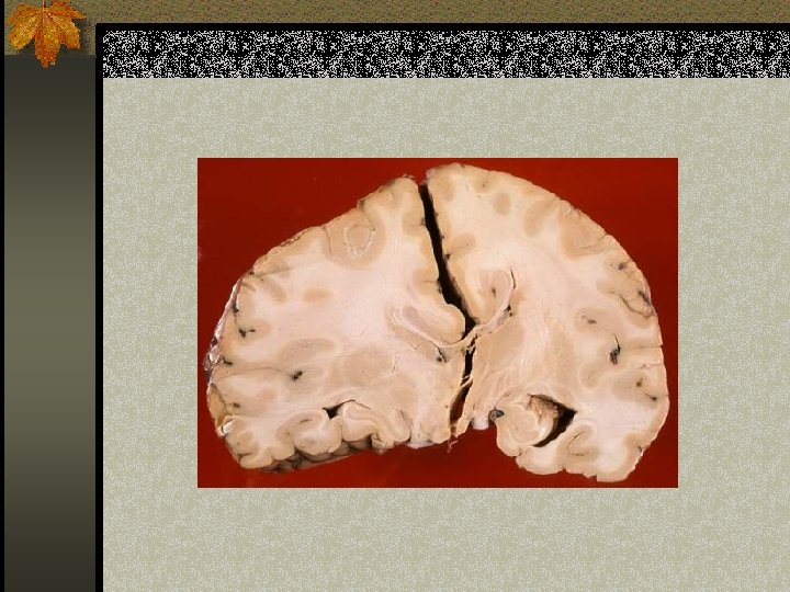
CONTUSION/LACERATIONS n Most common source of traumatic SAH n Contusion: must involve the superficial gray n n matter Laceration: contusion + tear of pia-arachnoid Affects the crests of gyri Hemorrhage present ½ cases and occur at right angles to the cortical surface Located near the irregular bony contours: poles of frontal lobes, temporal lobes, inferior cerebellar hemispheres

From http: //neuropathology. n eoucom. edu/ Dr. Agamanolis

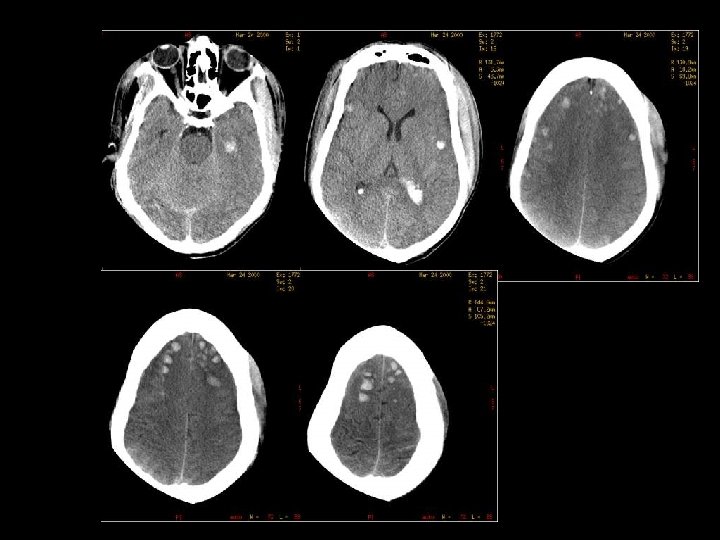
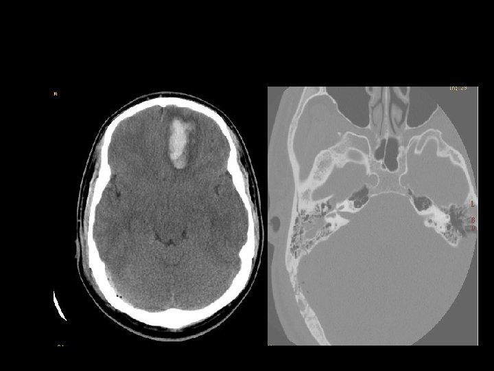
Intraparenchymal hematoma n Focal collections of blood that most commonly arise from shear-strain injury to intraparenchymal vessels. n Usually located in the frontotemporal white matter or basal ganglia n Hematoma within normal brain n DDx: DAI, hemorrhagic contusion

DIFFUSE AXONAL INJURY n Rarely detected on CT ( 20% of DAI lesions are hemorrhagic) n MRI: T 1, T 2 GRE, SWI

DAI n Due to acceleration/deceleration to whtie matter + hypoxia n Patients have severe LOC at impact n Grade 1: axonal damage in WM only 67% n Grade 2: WM + corpus callosum (posterior > anterior) – 21% n Grade 3: WM + CC + brainstem

DAI n Hours: n hemorrhages and tissue tears n Axonal swellings n Axonal bulbs n Days/weeks: clusters of microglia and macrophages, astrocytosis n Months/years: Wallerian degeneration

From http: //neuropathology. neou com. edu/ Dr. Agamanolis

Sagittal T 1 -W images
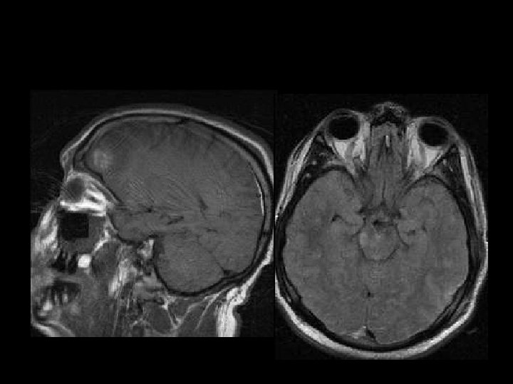

Axial FLAIR images

AXIAL FLAIR

AXIAL T 2 GRADIENT-ECHO

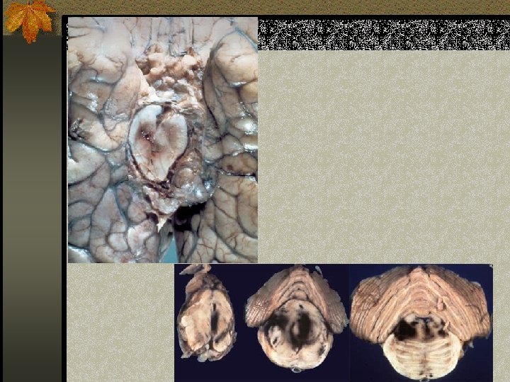
BRAINSTEM INJURY n By direct or indirect forces n Most commonly associated with DAI n Involves the dorsolateral midbrain and upper pons and is usually hemorrhagic n Duret hemorrhage is an example of indirect damage: tearing of the pontine perforators leading to hemorrhage in the setting transtentorial herniation n <20% of brainstem lesions are seen on CT

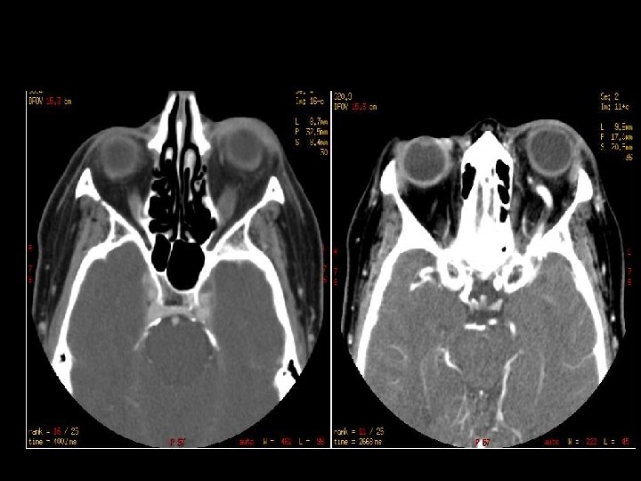
18 biker hit by a car

BRAIN HERNIATIONS

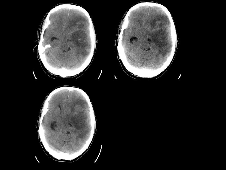
SUBFALCIAL HERNIATION n Subfalcial: displacement of the cingulate gyrus under the free edge of the falx along with the pericallosal arteries. n Can lead to anterior cerebral artery infarction

UNCAL HERNIATION n Displacement of the medial temporal lobe n n n through the tentorial notch Displacement of the midbrain Effacement of the suprasellar cistern Displacement of the contralateral cerebral peduncle against the tentorium Widening of the ipsilateral cerebello pontine angle Compression of the posterior cerebral artery
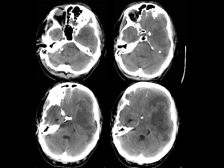

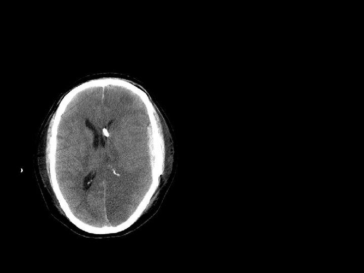
DOWNWARD HERNIATION n Caudal displacement of the thalamus and midbrain n Effacement of the perimensencephalic cistern and 4 th ventricle. n Can cause a 3 rd nerve palsy and disrupt pontine vessels leading to brainstem hemorrhage

UPWARD HERNIATION n Due to posterior fossa mass causing superior displacement of the vermis through the tentorial incisura n Compression of the 4 th ventricle and effacement of the quadrigeminal plate cistern. n Compression of the superior cerebellar artery

TONSILLAR HERNIATION n Inferior displacement of the cerebellar tonsils through the foramen magnum n Can lead to posterior cerebellar artery infarction

EXTERNAL HERNIATION n Due to a defect in the skull in combination with elevated ICP n Venous obstruction can occur at the margins of the defect.

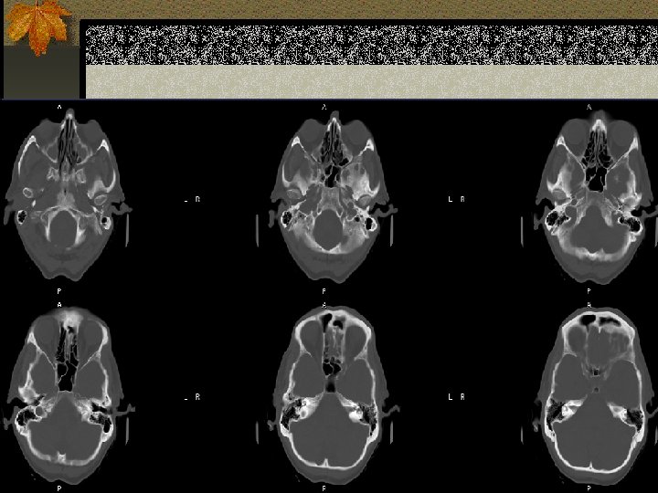
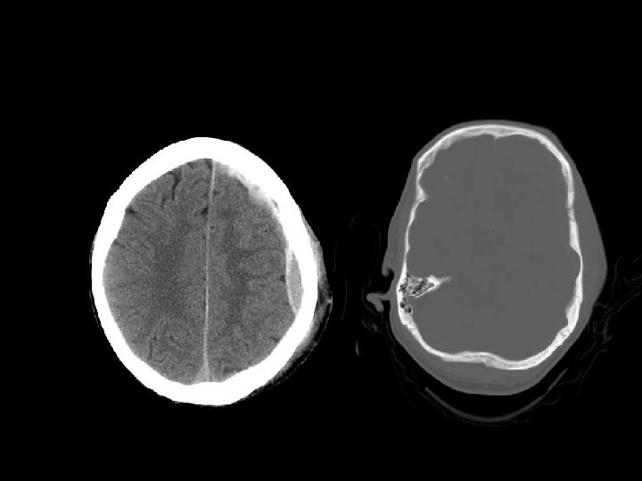
SIGNIFICANT SKULL FRACTURES n “Depressed”: inner table is depressed by the thickness of the skull. n Overlie major venous sinus, motor cortex, middle meningeal artery n Pass through sinuses n Look for sutural diastasis (lambdoid)

TEMPORAL BONE FRACTURES n Look for opacification of the mastoid n Longitudinal: 70%, parallel to long axis of petrous bone, conductive hearing loss (from ossicular dislocation), facial nerve paralysis (20%) n Transverse: 20%, sensorineural hearing loss, facial nerve paralysis (50%) n Complex n Complications: meningitis, abscess
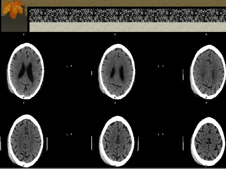
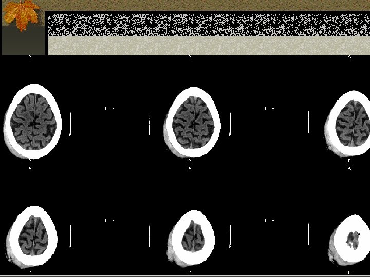
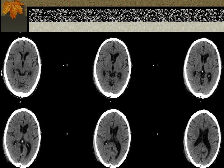
 n Dissection/pseudoaneurysm n Infarction n Atrophy/encephalomalacia n Infection")
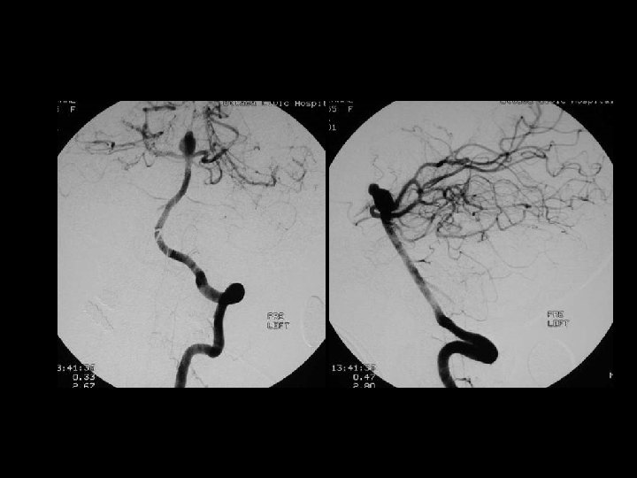
POST TRAUMATIC SEQUELAE n Carotid-cavernous fistula(CCF) n Dissection/pseudoaneurysm n Infarction n Atrophy/encephalomalacia n Infection n Leptomeningeal cyst
- Slides: 82