Imaging of extraocular orbital pathology BY Ali Hekmatnia

Imaging of extraocular orbital pathology BY : Ali Hekmatnia M. D

Imaging indications • Ophthalmologist suspects pathology symptomatically or by sonography not exactly delineated • In cases of trauma (e. g. foreign body, fractures) • Posttreatment

Imaging modalities • US • CT – MDCT often working horse • MRI
, coronal/sagittal reconstructions, soft")
Imaging techniques � CT MDCT, axial, -/+ CM (depending on pathology), coronal/sagittal reconstructions, soft tissue/bone window level � MRI Headcoil/surface coils, axial IR, axial T 1 w. SE -/+ CM, coronal/sagittal T 1 w. SE+CM+ FS, matrix 512, FOV ~20 cm

CT Scan : TECHNIQUE - Axial and coronal images - Axial 3 mm sections - Coronal 5 mm sections - Coronal sections from the lateral orbital rim to the posterior aspect of the optic canals (anterior clinoid or dorsum sellae)

Coronal images : - Extraocular muscles , optic nerve sheath , nasal complex , vessels and globes , Spread of processes from surrounding structures - Windowing : soft tissue as well as bone-oriented window

MRI : - Multiplanar capability , without ionizing radiation and bony artifact(especially in the orbital apex, optic canal and parasellar regions ). Best soft tissue contrast. - Protocol of MRI : coronal and axial T 1 and T 2 W images , coronal T 1 W with fat saturation(before and after contrast injection )

Anatomy of the Orbit Compartimental anatomy • • • Extraconal Conal Intraconal Globe Lacrimal gland

Orbital Anatomy : - Bony cavity , the globe, muscle cone, optic nerve-sheath complex, lacrimal apparatus, orbital fat, vascular and nerve structures, orbital septum and lids

Muscle Cone : - Superior, medial, lateral and inferior recti, Superior and inferior obliques, Levator palpebrae superioris. - Introconal space : Surgical problems - Extraconal space : Medical management - Globe : Cornea, lens, anterior chamber, vitreous, retinal - scleral complex

Optic nerve sheath complex : - Optic nerve , subarachnoid space , fluid between dura and nerve , diameter of complex (4 -6 mm)

Anatomy of the Orbit • • Intraorbital Extraconal. Conal Intraconal Globe

Anatomy of the Orbit Compartimental anatomy • • • Intraorbital Extraconal Conal Intraconal Globe Lacrimal gland

Supraorbital fissure Infraorbital fissure and pterygopalatine fossa

Close relationship to PNS Variant

Anatomy of the orbit Muscles

Anatomy of the orbit Muscles

Close relationship to vascular/nerval structures!!! Orbita may be easily affected!!!

Anatomic regions Fossa pterygopalatina • Close relation Orbit-PNSOropharynx • Nerves III, IV, V, VII • Parasellarregion Maxillary nerve Greater petrosal nerve

Pathology • • • Inflammation orbital-extraorbital Blastoma orbital-extraorbital benign-malignant Trauma Foreign bodies
 MRI (Phlegmone)")
Inflammation of lid CT (Abscess) MRI (Phlegmone)

Harnsberger R: Head and Neck 2004 Subperiostal abscess CT – Spread of infection from ethmoid cells – Compression of optic nerve – Thrombosis

Harnsberger R: Head and Neck 2004 Subperiostal abscess MRI -Spread of infection from ethmoid cells -Compression of optic nerve!! -Thrombosis!!

Dacryoadenitis

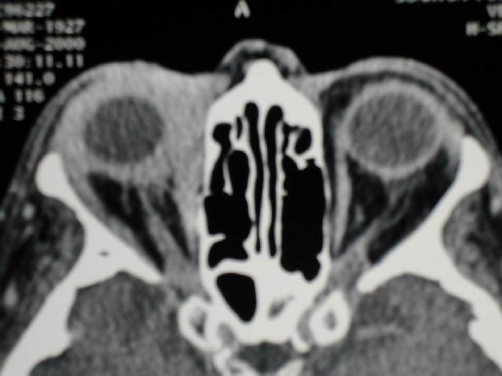
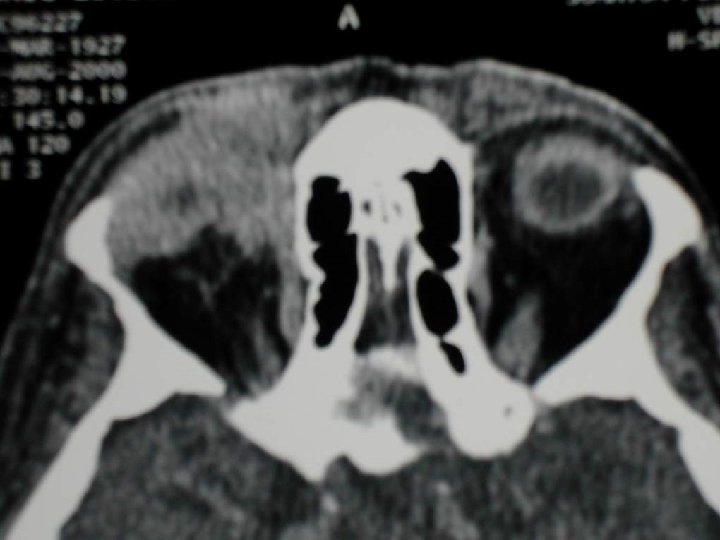
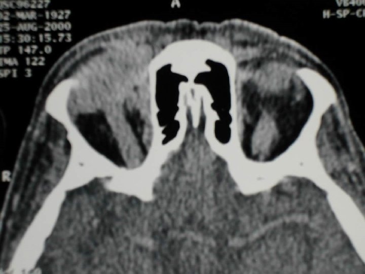
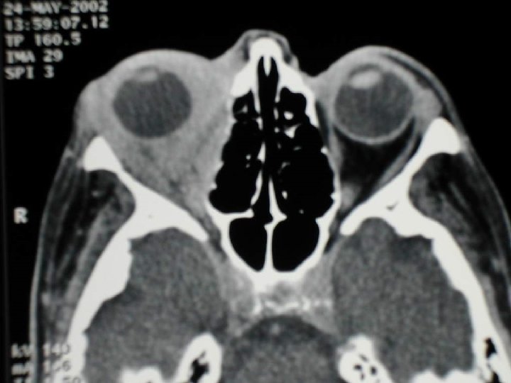
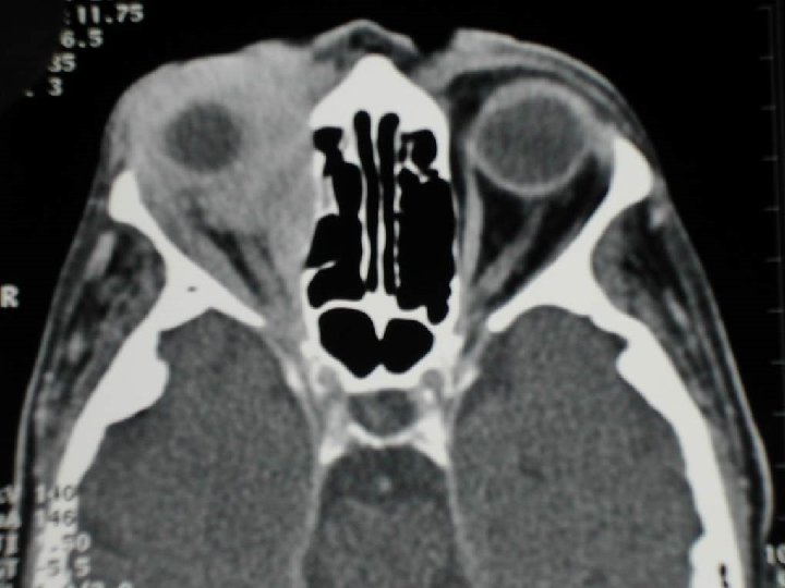
ORBITAL CELLULITIS IN A 13 -YEAR-OLD BOY WITH EXTENSIVE RIGHT ETHMOID SINUS DISEASE The inflammation involved the medial extraconal portions of the right Axial CTorbit scan shows lateral displacement of the medial rectus muscle and infiltration ofthe extraconal fat (arrows) Subperiosteal abscess in a 4 -year-old girl with chronic right ethmoid sinusitis The inflammation involved the preseptal and extraconal portions of the medial right orbit. Axial CT scan shows the slightly displaced and thickened medial rectus muscle and a small focal fluid collection (arrow), which was confirmed as representing a subperiosteal abscess

Orbital pseudotumor Gross mass-like enlargement of the medial rectus muscle, with characteristic hypointense signal on T 1 W (a) and T 2 W (b) sequences. Moderate heterogeneous enhancement is seen in the post gadolinium image (c)
 Diffuse enlargemen t of the lacrimal gland is seen with")
Orbital pseudotumor (different patients) Diffuse enlargemen t of the lacrimal gland is seen with preservatio n of its shape Axial CECT shows a diffuse infiltrative right orbital mass involving the globe and causing marked proptosis There is diffuse thickening of the bilateral medial and lateral rectus muscles including their tendinous insertion (arrows) which is typically spared in thyroid ophthalmopathy
")
Optic nerve neuritis (MS)

Pseudo-inflammation 3 T Orbital pseudotumor No diffuse infiltration

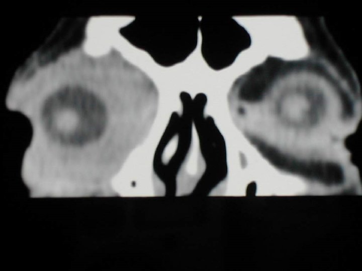
Endocrine orbitopathy CT Graves disease / M. Basedow

Endocrine orbitopathy MR Graves disease / M. Basedow

Endocrine orbitopathy 3 T MR Thickening and hyperintensity of medial and inferior rectus muscle

Small capillary hemangioma

3 T MR Large capillary hemangioma

Hemangiomatosis

3 T MR Large lymphaticvenous malformation

LYMPHANGIOMA IN A 4 -YEAR-OLD BOY WITH SUDDEN SUPRAORBITAL FULLNESS OF THE RIGHT EYE Axial CT scan reveals a multilocular intraconal lymphangioma in the right orbit. Lymphangioma in a patient who experienced sudden proptosis and discoloration about his right eye Axial T 2/W MR image demonstrates hemorrhage into a multilocular lymphangioma. The high-signal-intensity methemoglobin is layering anteriorly in each cyst.

VENOLYMPHATIC MALFORMATION USG reveals a multiseptate cystic mass in the orbit MRI reveals a heterogeneous intraconal mass in the right orbit displacing the optic nerve. Lesion is heterogeneous in signal intensity with a hyperintense area on T 1 W image (a) which shows blood-fluid level on T 2 W sequence (arrows) (b). There is only mild enhancement following contrast administration (c)

Blastoma/Tumor-like � Bone Fibrous dysplasia, Metastasis � Lacrimal gland Adenoma, Dermoid, pleomorphic Adenoma, Lymphoma � Conus � (Globe) (Melanoma, Retinoblastoma) � Nerve Glioma, Grave`s, Hemangioma, Lymphoma, Schwannoma, Pseudotu Meningeoma P. Som Head and Neck Imaging 4 th ed. 2003

Orbital pathology Pathology bony orbit Fibrous dysplasia orbit

Blastoma Melanoma lower lid

Fat Dermoid

3 T MR Pleomorphic adenoma
 Pleomorphic adenoma parotid gland")
Pleomorphic carcinoma with papilla infiltration (II) Pleomorphic adenoma parotid gland

MALT-lymphoma lacrimal gland

Orbital lymphoma diffuse infiltration

Rhabdomyosarcoma

3 T MR Rhabdomyosarcoma M. rectus superior

Rhabdomyosarcoma

Cavernous hemangioma

Orbital varix Axial T 1 W and T 2 W MRI reveal an elongated lesion around the optic nerve which is hypointense on T 1 W and hyperintense on T 2 W sequence. Note the characteristic “club like” configuration of the lesion in the sagittal T 2 W

Orbit Varix : - Large , tortuous vein or a mass like confluence of small veins may markedly enlarge with changes in venous pressure (Valsalva ` maneuver)
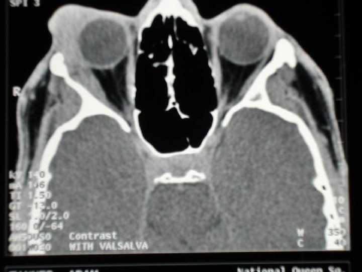
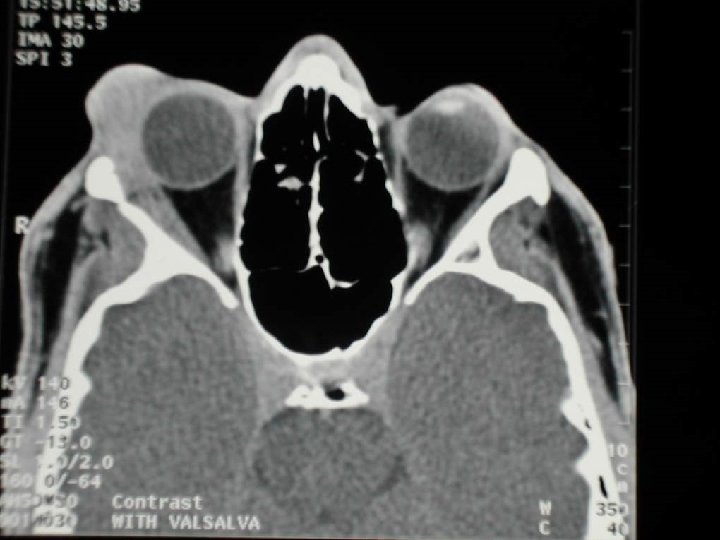
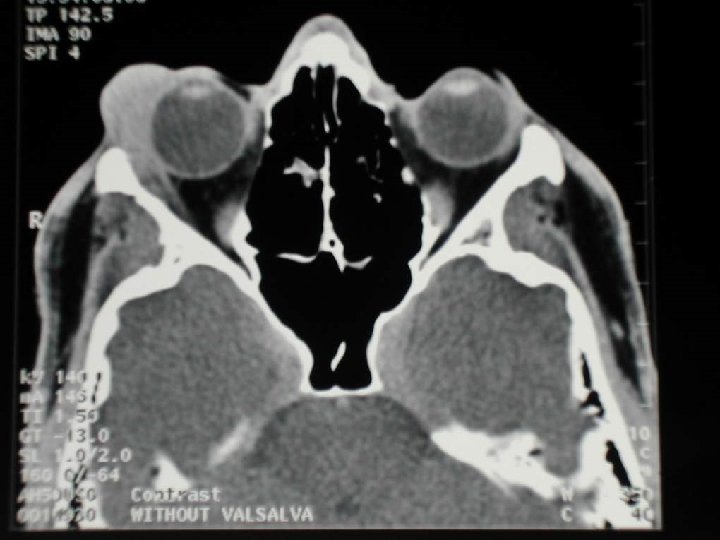
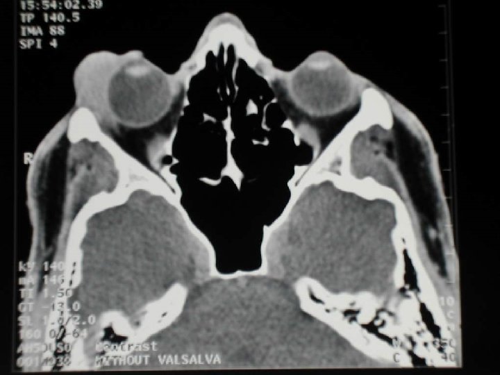


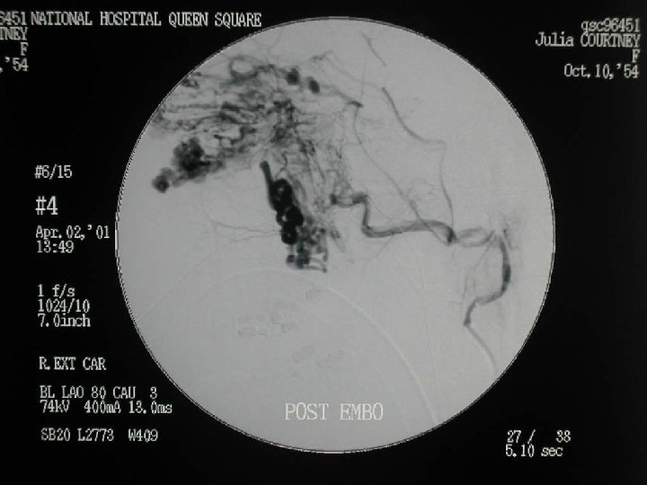

ORBITAL VARIX Axial CECT in a child with intermittent proptosis is almost normal. However, during valsalva maneuver the enhancing mass and the associated tortuous venous channels stand out causing significant proptosis

IN A 5 -MONTH-OLD GIRL WITH DIFFUSELY DILATED CAPILLARIES AND CHEMOSIS OF THE EYELID • Axial and sagittal Ti-weighted MR images demonstrate a capillary hemangioma superficially and preseptally about the left orbit. • Several prominent vessels are noted within the mass.

CAPILLARY HEMANGIOMA Axial CECT shows an intensely enhancing mass in the eyelid and extraconal space of the left orbit causing displacement of the globe

CAVERNOUS HEMANGIOMA IN A 16 -YEAR OLD BOY NTRACONAL ENHANCE GREATLY

CAVERNOUS HEMANGIOMA A homogenous well-defined intraconal mass is seen in the left orbit which is isointense on T 1 W , hyperintense on T 2 W sequence and reveals heterogeneous enhancement. Cavernous hemangiomas are not uncommon in children

Tram-track Optic nerve meningeoma

Glioma II

3 T MR Glioma II
 and coronal Ti-weighted (b) MR images demonstrate extensive involvement of the")
Axial proton-weighted (a) and coronal Ti-weighted (b) MR images demonstrate extensive involvement of the left eyelid and extraconal region by a plexiform neurofibroma LEFT ORBITAL PLEXIFORM NEUROFIBROMA IN A 10 -MONTH-OLD BOY Optic nerve gliomas in a teenage girl with neurofibromatosis Axial Ti-weighted (a) and T 2 -weighted (b) MR images show diffuse bilateral enlargement ofthe optic nerves by gliomas (arrows)
 reveals the characteristic enlarged and “bare” left orbit")
NF-1 Radiograph of the orbit (a) reveals the characteristic enlarged and “bare” left orbit in a child with NF 1 Axial CECT shows the dysplastic left greater wing of sphenoid with anterior herniation of the temporal lobe and an ill-defined infiltrative mass in the temporal fossa invading the orbit suggestive of a plexiform neurofibroma

Multifocal meningioma in an 18 -year-old male adolescent with neurofibromatosis Coronal T 1/W MR image demonstrates bilateral isointense intraventricular meningiomas Axial CT scan shows a calcified meningioma of the right optic nerve

BILATERAL OPTIC NERVE MENINGIOMAS IN A 15 -YEAR-OLD GIRL WITH NO OTHER FINDINGS OF NEUROFIBROMATOSIS Axial CT scan reveals bilateral calcified meningiomas ofthe optic sheath Optic nerve glioma in a young boy without neurofibromatosis Axial CT scan shows diffuse involvement of the right optic nerve by a glioma. Pediatric optic nerve gliomas are frequently associated with neurofibromatosis

Extraorbital pathology Schwannoma III

Schwannoma V 2 with elevation of rectus inf. muscle

Angiofibroma with orbital infiltration

SCC

TRAUMA CT Le Fort II

Orbital floor-fx with herniation of fat

Hematoma

Orbital floor fracture, Motility disturbance

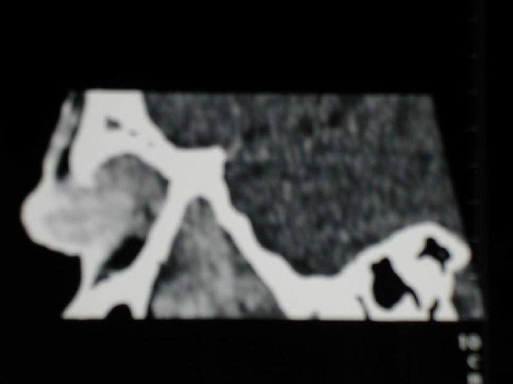
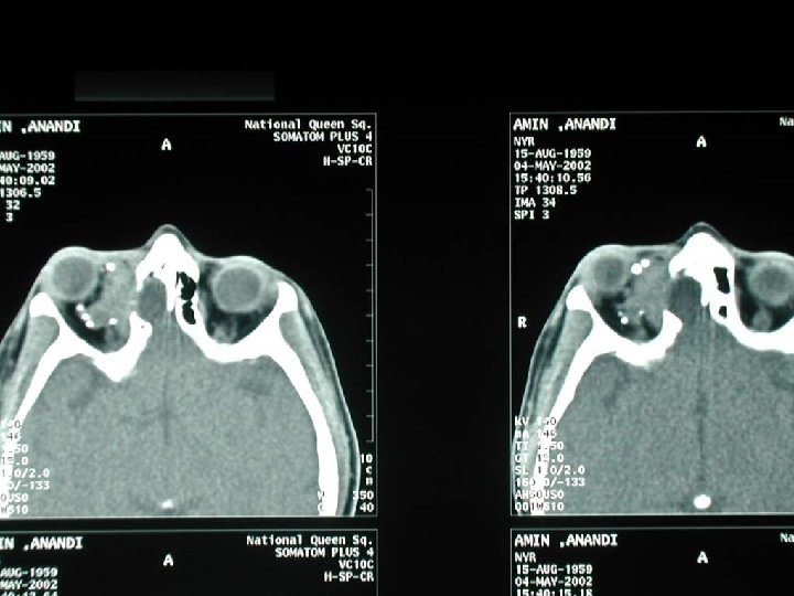
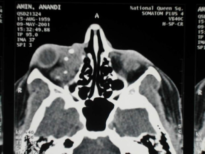
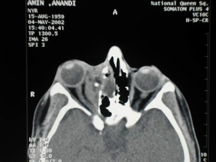
Foreign bodies No MRI metallic !!! Foreign bodies extraconal intraorbital

Foreign bodies Foreign body sclera

Foreign bodies Perforating intraocular fb

Take home points • • Remind anatomic situation Imaging technique and characteristics and localisation of pathology (intraorbital compartments) Involvement of adjacent structures Careful analysis DIAGNOSIS

Intraocular disorders

Calcified retinoblastoma Axial CT scan demonstrates a calcified mass in the left globe, accompanied by some increased attenuation of the vitreous.

Bilateral retinoblastoma üCoronal CT scan and T 1 -w axial MR image demonstrate bilateral calcified retinoblastomas üThe increased signal intensity of the right globe is likely secondary to hemorrhage üThe calcifications so prominent on the CT scan are poorly visualized on the MR image

Trilateral retinoblastoma

Medulloepithelioma
 in a 3 -year-old boy Axial contrast")
Persistent hyperplastic primary vitreous ( PHPV ) in a 3 -year-old boy Axial contrast material-enhanced CT scan shows a coneshaped, noncalcified, central retrolental area of increased attenuation in the right eye Coronal T 2/W MR image better depicts this abnormality. The increased signal intensity in the right globe is due to hemorrhage

PHPV Transverse color Doppler USG shows an echogenic retrolental structure with a vascular channel within, suggestive of PHPV

RETROLENTAL FIBROPLASIA WITH BILATERAL MEDIAL RETINAL DETACHMENTS IN AN 1 1 -MONTH-OLD GIRL WITH BILATERAL LEUKOKORIA The infant, born prematurely, had received oxygen therapy for respiratory distress syndrome. Axial CT scan clearly shows the high-attenuation detached retinas (arrows). Sclerosing endophthalmitis Axial CT scan shows a uniform increased attenuation throughout the right globe. The linear area of high attenuation seen in the middle to lateral aspects of the globe is a detached retina. A classic nematode infection was confirmed at the histopathologic analysis. The lack of a focal mass and of calcification helps differentiate sclerosing endophthalmitis from retinoblastoma.

Coat’s disease Color doppler USG shows a large retinal detachment with hypoechoic subretinal exudates CT shows diffuse increase in the intraocular density

Orbital rhabdomyosarcoma in a young child A large superior right orbital mass compressed and displaced the globe anteriorly and inferiorly. The mass is hypointense on the coronal T 1/W image and hyperintense on the T 2/W image

Orbital rhabdomyosarcoma Axial CECT show a homogeneous multicompartmental soft tissue density mass causing orbital expansion and destruction of the medial orbital wall
 Axial CECT : A welldefined fat-containing lesion is")
Dermoid and Epidermoid cyst (different patients) Axial CECT : A welldefined fat-containing lesion is seen near the outer canthus Axial CECT : The large wellcircumscribed cystic lesion at the inner canthus is suggestive of an epidermoid cyst. The adjacent bone is remodelled

Leukemic involvement of the orbit in a 6 -month-old boy with acute lymphocytic leukemia Axial CT scan shows preseptal swelling and involvement the left orbit, diffuse scleral thickening with enhancement, and a poorly defined intraconal mass that envelops the optic nerve. There is resultant proptosis.

NEUROBLASTOMA IN A 14 -YEAR-OLD BOY Axial CT scan reveals diffuse sclerotic bone involvement from metastatic neuroblastoma. In addition, there is extraconal involvement about the right orbit with resultant proptosis. Midline granuloma Coronal CT scan demonstrates a midline mass with encroachment into the left orbit.

Langerhan’s cell histiocytosis Axial CECT shows destruction of lateral orbital walls and the greater wing of the sphenoid bone on both sides with associated complex soft tissue mass encroaching on the orbits. The skull radiograph in the same patient reveals multiple well-defined lytic lesions in the cranial vault with typical bevelled edges
 1953 Lichtenstein Bone or bone marrow lesions")
Langerhans ‘cell Histiocytosis ( Orbital LCH ) 1953 Lichtenstein Bone or bone marrow lesions ( Overall incidence 23% ) Most commonly in frontal bone ( superior or superolateral wall of orbit )

CT and MRI findings : - An osteolytic lesion or multiple lesions - Well defined or diffuse soft tissue mass , encroaching lacrimal gland , lateral rectus or even the globe

Differential diagnosis : - Rhabdomyosarcoma - Juvenile fibrosarcoma - Aggressive fibromatosis - Lacrimal gland tumor - Leukemic infiltration - Metastasis ( Neuroblastoma , Wilm's , Ewing's)

Bilateral Anophthalmia

MICROPHTHALMOS IN A 13 -YEAR-OLD BOY ü CT scan shows the small right globe ü The presence of the radiopaque lens differentiates microphthalmos from an orbital Posterior optic coloboma in a 2 -year-old girl with seizures ü Sagittal T 1/W small cyst insertion Microphthalmos

Bilateral coloboma Axial CECT shows defect of the right globe at the optic nerve head via which the vitreous projects posteriorly (arrow). The left globe is small, with a retrobulbar (colobomatous) cyst (arrow) and dystrophic calcification at the site of coloboma

Duplication cyst

Congenital cystic eye ü ü Axial CECT shows a large right orbital cyst with absence of the globe. Also note the microphthalmia with coloboma and dysplastic lens on the left side

Septo-optic dysplasia in a 22 -month-old deaf, mute, and blind boy Axial T 1/W MR image shows bilaterally small globes (left larger than the right), with hemorrhage in the left globe. Since no trauma had occurred, the presence of hemorrhage may indicate PHPV is present Sagittal midline T 1/W MR image demonstrates agenesis of the corpus callosum and a prominent anterior inferior recess ofthe third ventricle (arrow).

Septo - optic dysplasia : - A part of a spectrum of developmental and congenital brain abnormalities - Absence of the septum pellucidum and pituitary hypothalamic endocrine dysfunction and hypoplasia of the optic nerves

Trigonocephaly secondary to metopic suture synostosis Axial CT scan obtained at the level of the orbit roofs demonstrates a marked frontal and rbital asymmetry secondary to cranial synostosis

Apert syndrome in a 3 -week-old boy Anterior radiograph of the skull demonstrates the typical features Axial CT scan demonstrates the proptosis and shallow orbits

Orbital encephalocele in a newborn Axial CT scan demonstrates an encephalocele protruding through the left orbit. A left globe was not seen, although retinal remnants were found within the encephalocele at autopsy Coronal CT scan demonstrates a prominent encephalocele that has ballooned inferiorly into the nasal cavity and medially into both orbits (arrows).

Metastatic disease : - Most common primary tumor is breast , then lung - In any part of the orbit , bone or soft tissue - Findings may be subtle , with small focal areas of thickening of the globe to large destructive lesion
- Slides: 137