Imaging Interpretation for the Comprehensive Eye Care Professional


 • Event Analysis (EA): change that")


 20")


- Slides: 67
Imaging Interpretation for the Comprehensive Eye Care Professional Blair Lonsberry, MS, OD, MEd. , FAAO Professor of Optometry Pacific University College of Optometry blonsberry@pacificu. edu 1
Disclosures • I have received honoraria from the following companies: – Alcon – Carl Zeiss Meditec – Optovue – Shire – Sun
Glaucoma Statistics • High percent of undiagnosed cases – Over 50% of open‐angle glaucoma cases in the USA – Many undiagnosed patients have undergone prior eye exams – Worsening problem as the proportion of older Americans increases • January 22 nd, 2016: Project blindness projects significant increase in glaucoma – By 2032 there will be a 50% increase in glaucoma cases
Challenges of Glaucoma • Risk factors are not widely known among patients – Many do not know it runs in families or is more common in African and Hispanic ancestry • The structural changes in early glaucoma can be difficult to distinguish – Wide variation of optic disc size in both normal and glaucoma patients • Patients can’t tell that they have it – Most do notice loss of function until they are nearly blind
Structural / Functional Relationship in Glaucoma as the Disease Progresses VF • The RNFL usually changes before both the visual fields and optic disc % Loss Moderate FL • The Optic disc often changes before visual fields Early c RN • Visual Field changes occur late in the disease Dis Severe Time
Glaucoma Progression • “Once the diagnosis of glaucoma has been made, the MOST IMPORTANT remaining question is whether the disease is stable and therapy/compliance are sufficient, or whether the disease is progressive and therapy in relation to the life expectancy has to be intensified. ” Progression of Glaucoma, World Glaucoma Association, 2011 Kugler Publications
Progression of Glaucoma “Although most glaucoma patients will show some evidence of progression if followed long enough, the rate of deterioration can be highly variable among them. While most patients progress slowly, others have aggressive disease with fast deterioration which can eventually result in blindness or substantial impairment unless appropriate interventions take place”
WGA Consensus Statements on Structure & Function • Both ON structure and function should be evaluated for detection of progression • Currently, no specific test can be regarded as the perfect standard for determination of progression • Progression detected by functional means will not always be corroborated using structural tests, and vice‐versa
WGA Consensus Statements • A statistically significant change in structure and/or function is not always clinically relevant. • Life expectancy should be considered when evaluating the clinical relevance of a structural and/or functional change in glaucoma. • Structural and/or functional testing should be conducted throughout the duration of the disease. – Detection of progression is more difficult in eyes with advanced disease
Detection & Measurement of Change (Structure & Function) • Event Analysis (EA): change that exceeds a certain predefined threshold compared to the baseline value; generally determined by measurement reproducibility • Trend Analysis: change over a designated time period using regression analysis. Generally takes more exams to obtain a reliable slope
Structural Changes ‐ WGA • The agreement for progression among ONH, RNFL, and macular parameters is poor • The rates of change vary considering within and between glaucoma patients • Differences in technologies and scan protocols could influence the detection of progression
Clinical Exam of the Optic Nerve Head Utility and Limitations • Disc exam at the first visit – normal or abnormal? – Disc exams are subjective, or at best semiquantitative – The wide variety of disc appearances requires long experience and expert judgment to separate normal from abnormal – Disc diameter must be taken into account
Case 72 year old glaucoma suspect Cup-to-disc ratio asymmetry Highest untreated IOP 24 mm Hg OU Anterior chamber angles open OU Clear visual fields • Central corneal thickness – 533 microns OD – 545 microns OS • No prior ocular history or surgery • No family history of glaucoma • Good general health except for elevated cholesterol
Case: Optic Nerves OD OS
Cirrus OCT: The Basics 15
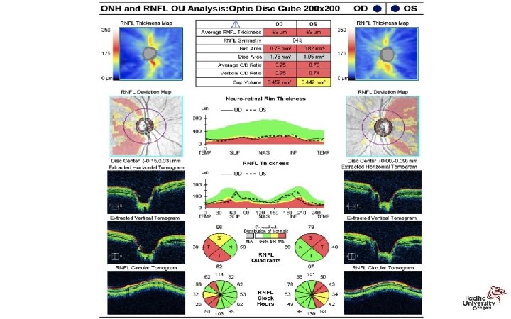
Cirrus RNFL Analysis OPTIC DISC CUBE SCAN The 6 mm x 6 mm cube is captured with 200 A-scans per B-scan, 200 B-scans. CALCULATION CIRCLE Auto. Center™ function automatically centers the 1. 73 mm radius peripapillary calculation circle around the disc for precise placement and repeatable registration.
RNFL/ONH Analysis RNFL THICKNESS MAP shows the patterns and thickness of the nerve fiber layer within the full 6 mm x 6 mm area ONH Analysis: rim/disc area, average C/D ratio, vertical C/D ratio and cup volume RNFL DEVIATION MAP, overlaid on the OCT fundus image, illustrates precisely where RNFL thickness deviates from the normal range. Data points that are not within normal limits are indicated in red and yellow. RNFL THICKNESS AND COMPARISON TO NORMATIVE DATABASE is shown in circle, quadrants and clock hour display RNFL THICKNESS along the calculation circle is displayed in graphic format and compared to age-matched normative data
Cirrus RNFL and ONH Analysis Elements Optic Nerve Head calculations are presented in a combined report with RNFL thickness data. Key parameters are compared to normative data and displayed in table format • RNFL Peripapillary Thickness profile, OU • compared to normative data Neuro-retinal Rim Thickness profile, OU - compared to normative data
Cirrus HD‐OCT GPA Analysis Image Progression Map Baseline §Two baseline exams are required § Third exam is compared to the two baseline exams § Sub pixel map demonstrates change from baseline: § Yellow pixels denote change from both baseline exams § Third and fourth exams are compared to both baselines: § yellow pixels denote change from both baselines § change identified in three of the four comparisons is indicated by red pixels Change refers to statistically significant change, defined as change that exceeds the known variability of a given pixel based on population studies
Page 2 Guided Progression Analysis (GPA™) 20
Ganglion Cell Analysis • Measures thickness for the sum of the ganglion cell layer and inner plexiform layer (GCL + IPL layers) – RNFL distribution in the macula depends on individual anatomy, while the GCL+IPL appears regular and elliptical for most normals – Deviations from normal are more easily appreciated in the thickness map by the practitioner, and arcuate defects seen in the deviation map may be less likely to be due to anatomical variations. Carl Zeiss Meditec, Inc Cirrus 6. 0 Speaker Slide Set CIR. 3992 Rev B 01/2012
Ganglion Cell Analysis 22 Carl Zeiss Meditec, Inc Cirrus 6. 0 Speaker Slide Set CIR. 3992 Rev B 01/2012
Be Careful: Red Disease FALSE POSITIVES • A red OCT that is believed to be glaucoma but may be indicative of another disease or just red as a result of poor imaging quality • Anatomic anomalies: – Tilted disc – PPA • Media opacities – Reduce image quality • ONH masqueraders – Optic neuritis, AION etc 23
Be Careful: Green is Clean FALSE NEGATIVE • A green OCT that is believed to be normal but actually has clinically detectable evidence of glaucoma found by methods of testing other than just looking at the colors on the OCT The CCT should match the: • clinical optic nerve evaluation • clinical retinal nerve fiber layer evaluation • visual field 24
Cases
Case 1: Case History • 55 black male complaining about decreased vision at near • Positive medical history of hypertension for 10 years, currently medicated. • Mother has glaucoma
Entrance Skills Va’s: 20/20 OD, OS Pupils: PERRL EOM’s: FROM CVF: full to finger count • IOP’s: 16, 16 OD, OS • • §Pachy: 540, 550 OD, OS §Fundus eval: see photos OD: c/d 0. 45/0. 45 OS: c/d 0. 6/0. 6 §HVF: see photos
OD OS
OS OD
Consider the below PSD plots. OS OD Predict what TSNIT graphs you would obtain for this patient.
OS
What we did. • We discussed with the patient: – appears he has early glaucomatous changes • early nasal step OS, • reduced NFL OS – positive family history – educated patient that we could monitor him very closely every 3 months and watch for further change, and then begin treatment at that time – or he could begin treatment
Treatment/Follow Up • Patient chose to begin treatment • We started him on Travatan Z qhs in the left eye – felt this would be the best medication for lowering his eye pressure without significant side effects related to his hypertension • Patient returned 2 weeks later for a follow up and his IOP had decreased from 16 to 12 in the left eye. • Patient asked whethere was potential to have glaucoma in his right eye – said it was possible and he decided he wanted to initiate treatment in his right eye as well.
Vesta: 61 y/o Hatian Female • • • GL suspect 2001 – suspicious ON’s NTG since 2006 Meds: Alphagan P bid OU, latanoprost qhs OU Medical Hx: HTN, HIV (+) for > 15 yrs VA: 6/6 (20/20) TA for the past 3 or 4 yrs: 9‐ 13 mm. Hg OU – Last 2 visits 9 mm. Hg – today 13 – Pachs: 450 microns Case Courtesy of Dr. Mark Dunbar
2010 What’s This? ? ? 2012 OD OSCase Courtesy of Dr. Mark Dunbar
OD OS RE 2010 2011 2012 Case Courtesy of Dr. Mark Dunbar
GPA Progression Analysis OD
GPA Progression Analysis OS
Vesta: 61 y/o Hatian Female • NTG OU with thin corneas • OS: – Optic Nerve and HVF show trend towards progression…. • OCT shows no change Case Courtesy Dr. Mark Dunbar
Vesta: 61 y/o Hatian Female • How do you manage this patient? – Currently on latanoprost and alphagan OU • This is what was done…. – Stopped Alphagan P – Switch to Combigan bid OU – Continue with latanoprost qhs OU – RTC 1 mo Case Courtesy of Dr. Mark Dunbar
RETINA 42
Question Which OCT goes with a patient undergoing hydroxychloroquine toxicity? 1 2 3 4
Revised Recommendations on Screening for Hydroxychloroquine Retinopathy • 2002 recommendations for screening were published by Ophthalmology • Revised recommendations on screening published in Ophthalmology 2011; 118: 415 -42 – Significant changes in light of new data on the prevalence of retinal toxicity and sensitivity of new diagnostic techniques – Risk of toxicity after years of use is higher than previously believed • Risk of toxicity approaches 1% for patients who exceed 5 years of exposure
“New” New Recommendations • Recommendations on Screening for Chloroquine and Hydroxychloroquine Retinopathy – Ophthalmology 2016; 123: 1386‐ 1394 – Released March 2016 from American Academy of Ophthalmology – revised in light of new information about the prevalence of toxicity, risk factors, fundus distribution, and effectiveness of screening tools.
2016 Recommendations • maximum daily HCQ use of 5. 0 mg/kg real weight, which correlates better with risk than ideal weight. • risk of toxicity is dependent on daily dose and duration of use. – at recommended doses: • risk of toxicity up to 5 years is under 1% • up to 10 years is under 2% • rises to almost 20% after 20 years. However, even after 20 years, a patient without toxicity has only a 4% risk of converting in the subsequent year.
2016 Recommendations • High dose and long duration of use are the most significant risks. – Other major factors are concomitant renal disease, or use of tamoxifen • A baseline fundus examination should be performed to rule out preexisting maculopathy. • Begin annual screening after 5 years for patients on acceptable doses and without major risk factors.
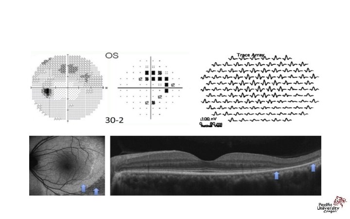
2016 Recommendations • primary screening tests are automated visual fields plus spectral‐domain optical coherence tomography (SD OCT) • most patients of Asian descent will show initial damage in a more peripheral extramacular distribution near the arcades (require a 24‐ 2 as opposed to 10‐ 2 and OCT scans need to be analyzed further out)
Revised Recommendations on Screening for Retinopathy • Parafoveal loss of visual sensitivity may appear before changes are seen on fundus evaluation • Many instances where retinopathy was unrecognized for years as field changes were dismissed as “nonspecific” until the damage was severe • 10 -2 VF should always be repeated promptly when central or parafoveal changes are observed to determine if they are repeatable • Advanced toxicity shows well-developed paracentral scotoma
Paracentral Scotomas
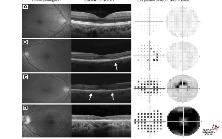
Normal Retina: VF/OCT/ERG TD-OCT Outer Nuclear Layer PIL SD-OCT Rodriguez-Padilla, J. A. et al. Arch Ophthalmol 2007; 125: 775 PIL=PR Integrity Line 780. Copyright restrictions may apply.
Mild Maculopathy Normal Foveal Peak Paracentral Scotomas Thinned Outer Nuclear Layer PIL Rodriguez-Padilla, J. A. et al. Arch Ophthalmol 2007; 125: 775780. Copyright restrictions may apply.
Bull’s Eye Maculopathy Flattened Foveal Peak Dense Para/Central Defects RPE Atrophy Remnant of PIL Rodriguez-Padilla, J. A. et al. Arch Ophthalmol 2007; 125: 775780. Copyright restrictions may apply.
Major Risk Factors
Screening Recommendations
Macular hole • Unilateral, decreased vision – Often in 60‐ 80 year old women – Anyone w/ a history of trauma • Symptoms: – Decreased vision, metamorphopsia • 20/200 for full thickness holes • Signs: – Red hole in the macula – (+) Watzke‐Allen sign
• Stages Macular hole – Stage 1 a ‐> impending hole. Normal foveal depression with yellow spot/dot in fovea. – Stage 1 b ‐> Abnormal foveal depression with yellow ring. Stage 1 b macular hole
• Stages Macular hole – Stage 2 ‐> Small full‐thickness hole. 20/80 ‐ 20/400. – Stage 3 ‐> Full‐thickness hole w/ cuff of SRF. No PVD – Stage 4 ‐> Full‐thickness hole with cuff of SRF, with complete PVD. Stage 2 macular hole
• Stages Macular hole – Stage 2 ‐> Small full‐thickness hole. 20/80 ‐ 20/400. – Stage 3 ‐> Full‐thickness hole w/ cuff of SRF. No PVD – Stage 4 ‐> Full‐thickness hole with cuff of SRF, with complete PVD. Stage 3 Macular hole Stage 4 macular hole →
New Macular Hole Staging
New Macular Hole Staging
New Macular Hole Staging Small FTMH w/o traction 154 microns 237 microns
New Macular Hole Staging Medium FTMH w/o traction 250 -400 microns
New Macular Hole Staging Large FTMH with traction > 400 microns
Thank You! blonsberry@pacificu. edu 67