Image Quality Part II 1 Image Quality Photographic


- Slides: 39
Image Quality Part II 1
Image Quality Photographic Quality Geometric Quality Image Quality 2
The films or images have different levels of density – different shades of gray X-rays show different features of the body in various shades of gray. The gray is darkest in those areas that do not absorb X-rays well – and allow it to pass through The images are lighter in dense areas (like bones) that absorb more of the X-rays. Why you see what you see… 3
Terminology Density, contrast Detail Distortion Image Sharpness spatial resolution Lp/mm Blur Motion SID OID Focal spot size Image receptor Magnification Penumbra Size distortion Shape distortion Focal Spot size
Density Contrast Detail Distortion Image quality 5
detail Resolution, ability to distinguish shapes, ability to distinguish lines on image 6
RECORDED DETAIL The degree of sharpness in an object’s borders and structural details. How “clear” the object looks on the radiograph 7
Recorded detail Image sharpness Spatial resolution Smallest separation of two lines or edges Measured by lp/mm 8
RESOLUTION TEST TOOLS LINE PAIRS/ MM Depicts how well you can see the differences in structures More lines=more detail 9
10
11
Recorded Detail Factors that affect detail SID Focal spot size OID Image receptor type Motion
Focal Spot Geometry Angle of anode size
Focal Spot Geometry Focal spot is not really a single point Dependent on filament size and anode angle.
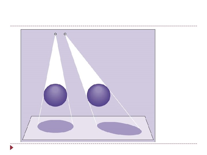
SID 15 Shine a flashlight on a 3 -D object, shadow borders will appear “fuzzy” -On a radiograph called penumbra Penumbra (fuzziness) obscures true border Farther the flashlight from object = sharper borders. Same with radiography.
Recorded Detail: Penumbra and SID
Recorded Detail: Penumbra and Focal Spot Size
Recorded Detail: Penumbra and OID
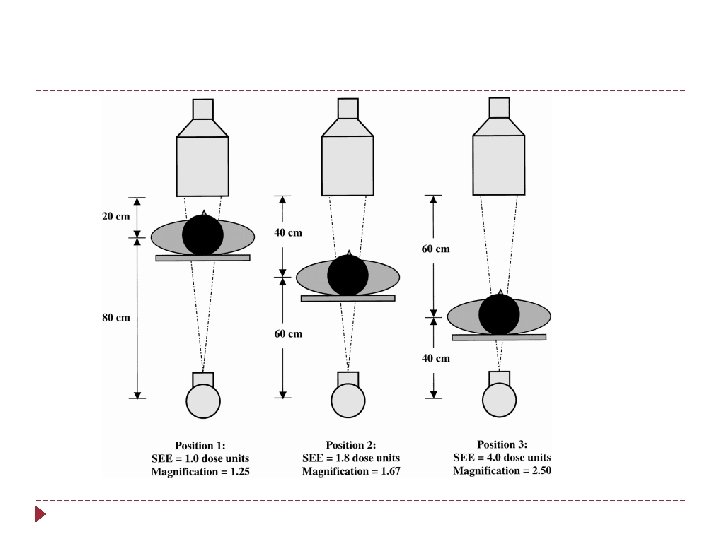
The position of the structure in the body will influence how magnified it will be seen on the image The farther away – the more magnified 19
Recorded Detail: OID and Penumbra The closer the object to the film, the sharper the detail. OID , penumbra , sharpness Structures located deep in the body, radiographer must know how to position to get the object closest to the film. 20
Recorded Detail: Image Receptor Type Film/Screen Imaging excellent spatial resolution-smallest detail that can be detected in an image Computed Radiography (cassettes) Digital Radiography (cassette-less) improved contrast resolution
Motion 22 Can be voluntary or involuntary Best controlled by short exposure times Use of careful instructions to the patient Suspension of patient respiration Immobilization devices
Blurring of image due to patient movement during exposure. 23
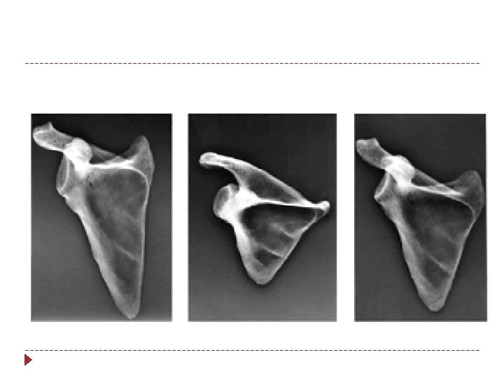
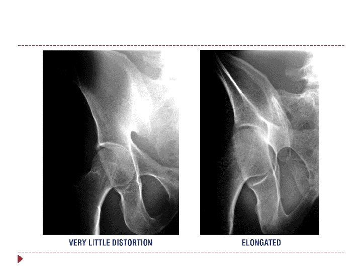
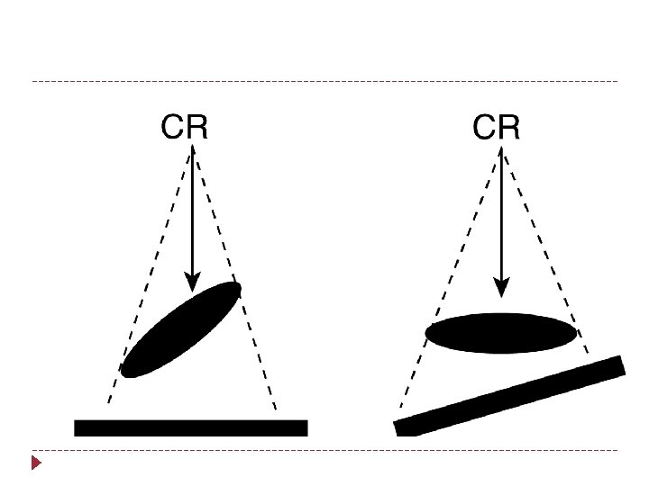
distortion Misrepresentation of size or shape of anatomic part; when part is distorted, detail is reduced 24
Distortion An increase of decrease in the size of an object : magnification or reduction Three types: size, shape, placement of part in body
Types of Distortion FACTOR INFLUENCING DISTORTION SID-size distortion OID-size distortion Beam Angulation-shape distortion Body Part-Beam alignment-shape distortion
Distortion: SID
40” SID VS 72” SID Which one is which? 28
Which one was taken at 72”? 29
Distortion: OID
31
Elongation 37 Foreshortened Normal
Which is normal? 38