Il laboratorio di fisiopatologia respiratoria Riccardo Pistelli Universit

Il laboratorio di fisiopatologia respiratoria Riccardo Pistelli Università Cattolica - Roma

Una lista condivisibile • • • • 1. Indications for Pulmonary Function Testing 2. Spirometry 3. Diffusing Capacity Tests 4. Lung Volumes, Airway Resistance and Gas Distribution Tests 5. Ventilation and Ventilatory Control Tests 6. Blood Gases and Related Tests 7. Cardiopulmonary Exercise Testing 8. Pediatric Pulmonary Function Tests 9. Bronchoprovocation Testing 10. Specialized Test Regimens 11. Pulmonary Function Test Equipment 12. Quality Systems in the Pulmonary Function Laboratory 13. Reference Values and Interpretation Strategies

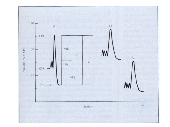
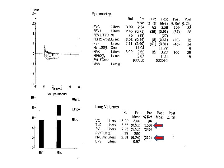
Cosa si intende comunemente per prove di funzionalità respiratoria • Misura dei volumi polmonari • Misura della capacità ventilatoria • Misura dell’efficienza di trasferimento dei gas

Un algoritmo per interpretare le PFT ERS 2005

L’invenzione della spirometria

Mr Hutchinson

Lo spirometro di Mr Hutchinson

Lo spirometro a campana

La prima misura di Volume Residuo

Le prime misure in circuito aperto dell’aria residua


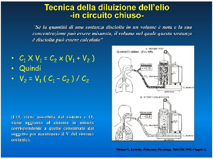
Equilibrazione di un gas traccia non solubile

La funzione di equilibrazione di un gas traccia: wash-in

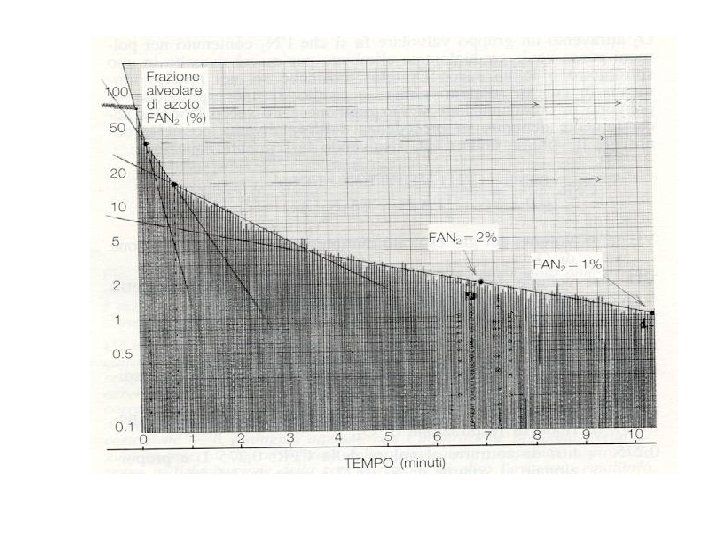
La funzione di lavaggio di un gas traccia: wash-out

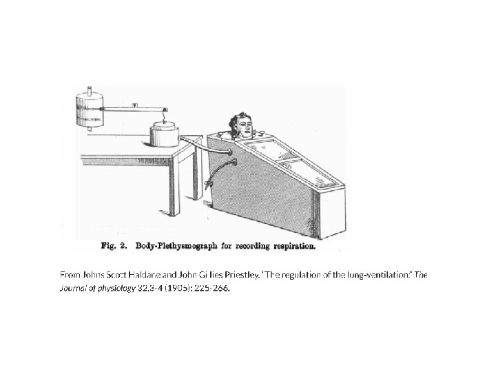
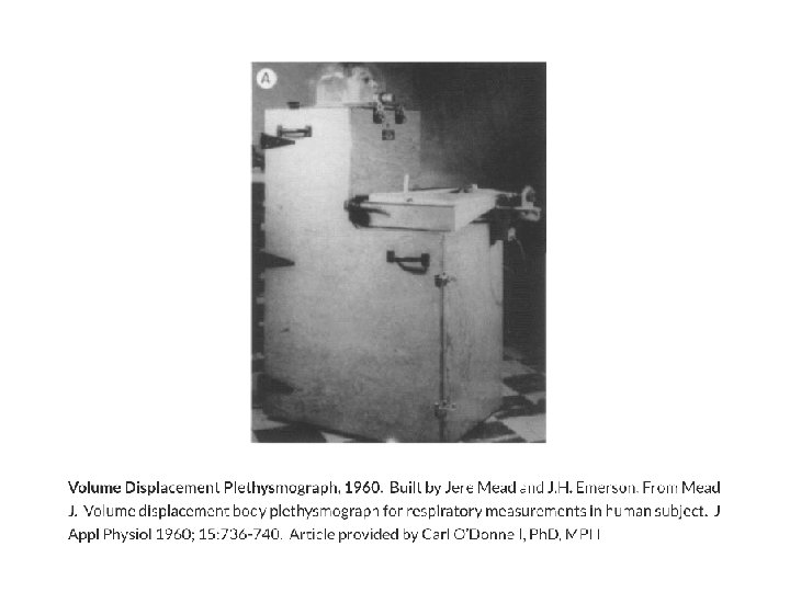
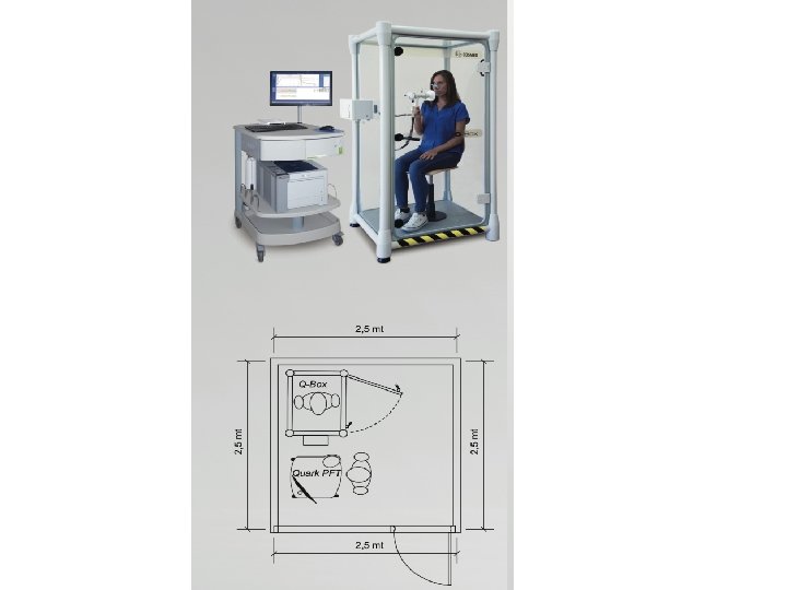
Il metodo pletismografico
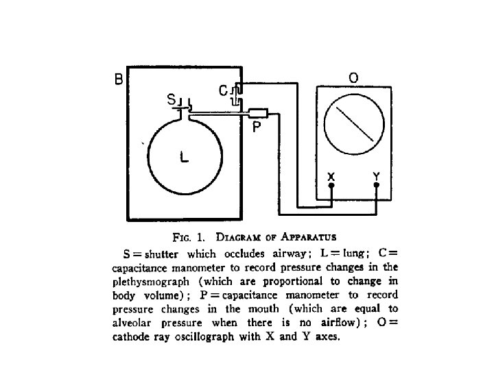

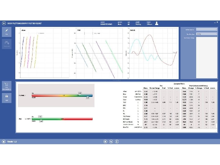

1 2 3

BMJ. MAY 10. , 1958

Iperinflazione dinamica negli asmatici R. Pellegrino, V. Brusasco. Eur Respir J 1997; 10: 468– 475


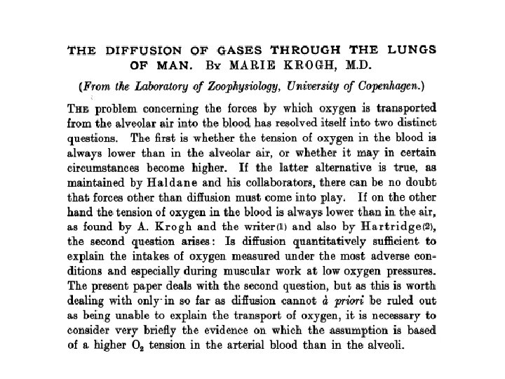
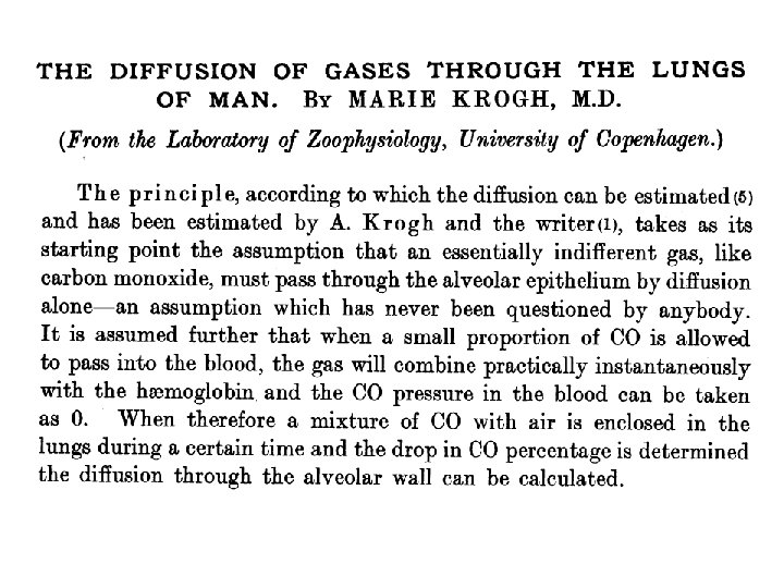
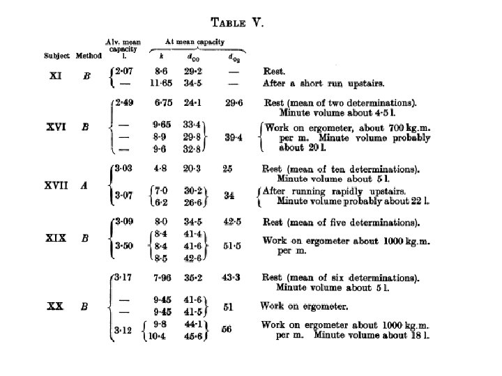
Diffusion properties of the lung

John Scott Haldane
 1. Respiro tranquillo")
Misura della DLCO col metodo del respiro singolo con apnea (DLCOsb) 1. Respiro tranquillo 2. Espirazione non forzata fino a RV, per non più di 6 sec 3. Rapida inspirazione a TLC in < 4 sec (volume inspirato > 85% EVC) 4. Tempo di breath-holding di 10 ± 2 sec (evitare manovre di Valsalva o Mueller) 5. Espirazione in < 4 sec Durante l’inspirazione il soggetto inala una miscela composta da CO allo 0. 3%, da un gas inerte normalmente assente (ad esempio elio, 10 -14%), da ossigeno (1821%) e azoto, in percentuali variabili in base alla miscela standard utilizzata Macintyre N et al. Standardisation of the single-breath determination of carbon monoxide uptake in the lung. Eur Respir J 2005; 26: 720 -735.

Interpretazione della DLCO • Il test di Diffusione del CO misura la Capacità di trasferimento di un gas non inerte dall’aria alveolare al sangue. • I fattori fisiologici che influiscono sul trasferimento del CO sono molteplici e analizzabili in vivo soltanto sulla base di modelli interpretativi semplici. • In condizioni di patologia, si aggiungono molteplici fattori, non accuratamente identificabili per qualità ed entità, che possono influire sul trasferimento del CO. • Da queste considerazioni di deduce che il test di Diffusione del CO può essere utilizzato per – Indagare con elevata sensibilità l’insorgere di patologie per le quali preesista un sospetto clinico – Valutare l’evoluzione di una patologia nota, specialmente nella sua fase iniziale – Contribuire alla diagnosi differenziale di patologie polmonari, quando sia inserito in un panel di esami funzionali, clinici e radiologici.

Note all’algoritmo interpretativo. A simplified algorithm that may be used to assess lung function in clinical practice. It presents classic patterns for various pulmonary disorders. As in any such diagram, patients may or may not present with the classic patterns, depending on their illnesses, severity and lung function prior to the disease onset (e. g. did they start with a vital capacity (VC) close to the upper or lower limits of normal (LLN)). The decisions about how far to follow this diagram are clinical, and will vary depending on the questions being asked and the clinical information available at the time of testing. The forced expiratory volume in one second (FEV 1)/ VC ratio and VC should be considered first. Total lung capacity (TLC) is necessary to confirm or exclude the presence of a restrictive defect when VC is below the LLN. The algorithm also includes diffusing capacity for carbon monoxide (DL, CO) measurement with the predicted value adjusted for haemoglobin. In the mixed defect group, the DLCO patterns are the same as those for restriction and obstruction. This flow chart is not suitable for assessing the severity of upper airway obstruction. Pellegrino R et al. Eur Respir J 2005
- Slides: 38