Idiopathic Thrombocytopenic Purpura ITP During Pregnancy Dr Muhammad
 During Pregnancy Dr Muhammad El Hennawy Ob/gyn specialist Dumyat Specialised")
Idiopathic Thrombocytopenic Purpura (ITP) During Pregnancy Dr Muhammad El Hennawy Ob/gyn specialist Dumyat Specialised Hospital and Rass El Barr Central Hospital www. geocities. com/mmhennawy

Platelet structure and physiology • • • • • The Normal Platelet are small, disc-shaped cells without a nucleus, normally measuring 1 to 2 um in diameter and 0. 5 to 1. 0 um in thickness with a volume of about 6 ul. The mean platelet count in normal children and adults is about 250 x 109/L, usually ranging from 150 to 400 x 109/L. Platelets are derived from the cytoplasm of megakaryocytes, primarily located in the bone marrow. Normally, a platelet is released to the bloodstream and circulates for about 10 days before its removal, largely by the spleen. Platelets circulate freely without adhesion to the vessel wall or aggregation with other platelet. If stimulated, platelets become spherical, extend pseudopods, and adhere to vessel walls and to each other. It participates with the blood vessel, coagulation factors, and other platelets in the initiation of hemostasis A phospholipid bilayer lies within a glycoprotein rich outer coat. Glycoproteins (GP’s) have essential roles in platelet activity, for example GP Ib and GP IIb-IIIa are important in platelet activation. Internally, a platelet contains an open canicular network, through which granule contents are secreted, and a dense tubular system, in which intracellular calcium is stored. Dense bodies, alpha granules and lysosomal granules are all visible within the inactive platelet, in addition to glycogen granules and mitochondria. During activation, the platelet contracts using actin and myosin. Alpha granules contain von Willebrand factor Platelet factor 4 - inhibits anticoagulants (and heparin) (PDGF) promotes fibroblast activity in repairing vessel wall damage Factor V Fibrinogen Fibronectin Dense bodies contain ADP - initiates platelet activation and recruitment Serotonin - vasoconstrictor -
platelets")
Platelet formation megakaryocyte formation of demarcation membranes platelets (pro)platelets

Approach to thrombocytopenia THROMBOCYTOPE NIA rule out pseudothrombocytopenia SEQUESTRATION â PRODUCTION á DESTRUCTIO N look for underlying disorders look for splenomegaly bone marrow investigation review meds Causes of splenomegaly • infection • inflammation • congestion • maligancy • red cell disorders • storage diseases • aplasia • infiltration • ineffective megakaryopoiesis eg. MDS • selective impairment of platelet production Also Artefactual (false) or pseudothrombocytopenia –Clot in the sample. - Platelets clumped. Congenital thrombocytopenia –Rare inherited disorders (eg May Hegglin Anomaly). review meds • immune auto-immune (ITP, SLE drugs infections allo-immune • non-immune sepsis DIC, TTP, HUS hypertensive disorders of pregn
: the primary mediator of platelet adhesion.")
Platelet receptors • • The glycoprotein Ib-IX(CD 42): the primary mediator of platelet adhesion. In the presence of flow (arterial shear stress), adhesion is accomplished by the interaction of the von Willebrand factor(v. WF) with subendothelial collagen and platelet GPIb-IX. When the platelet glycoprotein Ib-IX complex interacts with thrombin, at lease a partial internal translocation of the Ib-IX complex occurs, presumably mediated by the cytoskeleton. Therefore, the number of Ib-IX complexes on the platelet surfaces activation-sensitive and may vary considerably. There is no conformational activation of the GPIb-IX complex. Glycoprotein IIb-IIIa: the primary mediator of platelet aggregation. Activation of the complex by strong agonists results in a poorly understood conformational change resulting in the expression of a fibrinogen binding site. This includes a binding site on GPIIIa for the two Arg-Gly-Asp(RGD) sequences in the fibrinogen a chain as well as a separate dodecapeptide binding site on GPIIb. A single molecule of fibrinogen is bound for each IIb-IIIa "active site. " Fibrinogen binding to two contiguous platelets serves to bridge the two platelets, resulting in the essential step in development of platelet aggregates. There are 50, 000 to 60, 000 GPIIb-IIIa sites per platelet that remain relatively constant. Following activation and aggregation, platelet contractility is mediated by the IIb-IIIa complex. This complex may be linked to the platelet cytoskeleton, perhaps via talin.
 Adhesion")
The reactions of platelets that enable them to carry out this function (1) Adhesion The process by which platelets adhere to the basement membrane at sites of vascular injury. von Willebrand factor functions as the glue which sticks the platelet to the basement membrane collagen. Platelet adhesion is defective in von Willebrand disease (2) Secretion (a) fibrinogen - which will be used to stabilise the clot. (b) platelet factor 4 ( anti heparin) (c) inositol triphosphate (controls Ca release). Ca is used to activate some of the clotting factors of the cascade system. (3) Aggregation (4) Pro coagulant activity-PDFG induces proliferation of fibroblasts, microglia, and smooth muscle. It is stored in platelet granules and is released following platelet aggregation. PDGF may also serve as a chemotactic agent for inflammatory cells.

Causes of Thrombocytopenia During Pregnancy • • • • Common causes Immune thrombocytopenic purpura ( ITP ) (3. 8%), Gestational thrombocytopenia (73. 6%), Severe preeclampsia (21%), HELLP syndrome Disseminated intravascular coagulation Rare causes Lupus anticoagulant/antiphospholipid antibody syndrome Systemic lupus erythematosus Thrombotic thrombocytopenic purpura Hemolytic uremic syndrome Type 2 b von Willebrand's syndrome Folic acid deficiency Human immunodeficiency virus infection Hematologic malignancies May-Hegglin syndrome (congenital thrombocytopenia)
")
Immune Thrombocytopenic Purpura • Maternal thrombocytopenia (platelet count of less than 150, 000/m. L) • Normal platelet range for a person without ITP is 150, 000 to 400, 000 platelets per microliter. • It occurs in 5– 7% of all pregnancies. • The most common clinical signs are • petechiae, easy bruising, epistaxis, gingival bleeding, and hematuria. • A patient with a platelet count of more than 20, 000/m. L is at low risk for bleeding, but the risk increases as the platelet count drops below 20, 000/m. L.

the platelet count • • • The normal range of the platelet count in nonpregnant individuals is 150, 000 -400, 000/m. L. thrombocytopenia is defined as any platelet value less than 150, 000/m. L, with counts of 100, 000 -150, 000/m. L indicative of mild thrombocytopenia, 50, 000 -100, 000/m. L indicative of moderate thrombocytopenia, less than 50, 000/m. L indicative of severe thrombocytopenia. Clinically significant bleeding usually is limited to patients with platelet counts less than 10, 000/m. L. Serious bleeding complications are rare, even in those with severe thrombocytopenia Excessive bleeding associated with trauma or surgery is uncommon unless the patient's platelet count is less than 50, 000/m. L. The mean platelet count in pregnant women is lower than in nonpregnant individuals
 • 30, 000")
Clinical Signs • > 50, 000 - Asymptomatic (rarely following trauma) • 30, 000 - 50, 000 - bruising on trauma. • 10, 000 - 30, 000 - spontaneous bruising, menorrhagia, - prolonged bleeding after trauma. • <10, 000 - mucosal bleeding (epistaxis, gastrointestinal and genitourinary) and severe bleeding (includes life-threatening bleeding such as CNS bleeding)

Thrombocytopenic bleeding ? Risk of bleeding ! platelet count ! cause of thrombocytopenia ! comorbid disease ! drugs ? Clinical manifestations ! petechiae ! purpura, ecchymoses ! mucosal bleeding ! menorrhagia ! intracranial bleeding
 shows low platelet count (platelet count <150, 000/m.")
Tests • Complete blood count (CBC) shows low platelet count (platelet count <150, 000/m. L). • Bone marrow aspiration or biopsy appears normal or increased numbers of megakaryocytes. . • PTT (coagulation studies) is normal. • Platelet associated antibodies may be detected Assays for these antibodies are commercially available but should not be routinely performed because they are nonspecific, poorly standardized, and subject to a large degree of interlaboratory variation. Furthermore, levels of antiplatelet antibodies do not correlate well with the degree of fetal thrombocytopenia

Platelet counts • Platelet counts use a freshly-collected blood specimen to which a chemical called EDTA has been added to prevent clotting before the test begins. • About 5 m. L of blood are drawn from a vein in the patient's inner elbow region. • Blood drawn from a vein helps to produce a more accurate count than blood drawn from a fingertip. Collection of the sample takes only a few minutes • Counting methods • Platelets can be observed in a direct blood smear for • approximate quantity and shape. • Hemacytometer counting • Electronic counting of platelets is the most common method. There are two types of electronic counting, voltage-pulse and electro-optical counting systems
 platelets")
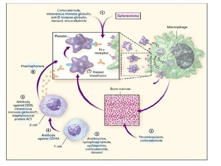
The pathophysiology • Lymphocytes produce antiplatelet antibodies directed at platelet surface glycoproteins. (a) platelets are taken up and internally degraded by antigen-presenting cells (APCs). (b) APCs present platelet antigen in association with major histocompatibility complex (MHC) class II to T helper (Th) cells, which become activated and secrete the Th 1 cytokines interleukin (IL)-2 and interferon (IFN)-g. (c) Th 1 cytokines activate and drive autoreactive B cells to differentiate into autoantibody-producing cells. • The immunoglobulin G (Ig. G)–coated platelets are cleared by splenic macrophages, which results in thrombocytopenia. • The course of ITP is affected by pregnancy (relapse during pregnancy after remission worse if active ). • Placental transfer of the Ig. G platelet antibodies can result in fetal or neonatal thrombocytopenia.

The Complications of ATP Maternal • ATP may lead to complications in pregnancy, • the most serious of which is maternal hemorrhage around the time of delivery. • No maternal deaths from ATP in pregnancy have been recorded since the early 1980 s, • peripartum bleeding may result in serious morbidity. Fetal and Neonatal • the placenta selectively transports maternal Ig. G antiplatelet antibodies into the fetal circulation, • fetal thrombocytopenia may also occur, sometimes leading to purpura, ecchymosis, or melena. Intracranial hemorrhage is only rarely reported and appears to be unrelated to the mode of delivery • levels of antiplatelet antibodies do not correlate well with the degree of fetal thrombocytopenia

Aim Of Treatment • Treatment of pregnant women with ATP are aimed at preventing bleeding • by maintenance of the platelet count above 20, 000 per mm 3 in the antepartum period • and over 50, 000 per mm 3 for delivery • No direct evidence indicates that any of these treatments reduce bleeding complications or mortality from ITP.

Methods Of Treatment • Glucocorticoids • Intravenous Immunoglobulin • Platelet Transfusions • Splenectomy • Accessory Splenectomy • intravenous anti-Rho (D) • aminocaproic acid • • • Azathioprine cyclophosphamide Vinca alkaloids danazol ascorbic acid, colchicine protein A immunoadsorption cyclosporine Plasmapheresis plasma exchange

Therapy for Adults with Refractory Chronic Immune Thrombocytopenic Purpura • Level 1 Therapy Corticosteroids - Vinca Alkaloids- Danazol -Colchicine -Dapsone • Level 2 Therapy Staphylococcal-A Immunoadsorption - Cyclophosphamide Azathioprine • Level 3 Therapy Combination Chemotherapy --High-Dose Cyclophosphamide • Level 4 Therapy Interferon - Gammaglobulin. Vinblastine - Cyclosporine

Glucocorticoids • Glucocorticoid drugs are the cornerstones of therapy for ATP in pregnancy. • Prednisone (1– 2 mg/kg/d in divided doses) for 2 to 3 weeks is the most typical regimen. • An increase in platelet count to more than 50, 000 per m. L, accompanied by a decrease in clinical bleeding, is usually achieved within 21 days. • More than 70% of patients have some response and complete remission occurs in up to 25%. • The prednisone dose is tapered by 10% to 20% decrements at 2 -week intervals to a dose that maintains the platelet count above 50, 000 per mm 3. • Dexamethasone and betamethasone also cause an increase in platelet count but both readily cross the placenta and have harmful fetal effects. • The side effects of glucocorticoids in pregnancy include steroidinduced moon facies, gestational diabetes mellitus, psychosis, adrenocortical insufficiency, osteoporosis, aseptic necrosis, hypertension, and uteroplacental insufficiency

Intravenous Immunoglobulin • • • Given at high doses (i. e. , 400 mg/kg/d for 5 days), IVIG usually induces a peak in platelet count within 7 to 9 days. More than 80% of patients achieve a platelet count greater than 50, 000 per mm 3, and the response lasts for more than 30 days in 30% of patients. • Only 2 to 3 days of IVIG therapy may be needed in some patients, and doses greater than 800 mg or 1 g per kg may suffice as a single or double infusion. • Although expensive, IVIG therapy initiated 1 to 2 weeks before delivery or surgery may be useful in some obstetric patients who must undergo operative procedures or who develop bleeding problems and require emergency treatment. The exact mechanism of action of IVIG is unclear, • but may be related to decreased antiplatelet antibody production, • interference with antibody attachment to platelets, • inhibition of macrophage receptor-mediated immune complex clearance, • or interference with platelet receptor mechanisms in the reticuloendothelial system. • Ig. G is selectively transported across the placenta and the amount transferred increases with gestational age and dose so that after 32 weeks of gestation, • maternally infused Ig. G sometimes has a beneficial effect on the fetal platelet count. • No cases of human immunodeficiency virus (HIV) transmission have been reported with the use of IVIG, • but adverse effects include thrombosis, alopecia, liver function disturbances, transient neutropenia, chills, nausea, flushing, tightness of the chest, wheezing, and anaphylactic reactions in patients with immunoglobulin A (Ig. A) antibodies

Platelet Transfusions • Platelet transfusions are used only as a temporizing measure to control life-threatening hemorrhage or to prepare a patient for splenectomy or cesarean section. • The survival of transfused platelets is decreased in patients with ATP because antiplatelet antibodies also bind to donor platelets. • In addition, patients with ATP do not respond as well as normal individuals to platelet transfusions but 6 to 10 U is usually sufficient to temporarily control hemostasis • Whenever possible the plasma of a platelet component should be ABO compatible with the recipient's red cells, • Calculation of CCI (corrected count increment ): CCI = Post-txn count - Pre-txn count x Body Surface Area (M 2) Platelets given x 1011 One unit of platelet concentrate averages 0. 7 x 10¹¹ platelets. One unit of single donor platelets averages 4. 0 x 1011 platelets

Aminocaproic Acid • The bleeding complications of some forms of thrombocytopenia are difficult to control. • Many patients become refractory to platelet transfusions even when HLA-matched. • Aminocaproic acid has proved to be a valuable agent in the management of patients with amegakaryocytic thrombocytopenia, especially in decreasing the need for platelet transfusions • Some studies -- Low dose bolus aminocaproic acid: an alternative to platelet transfusion in thrombocytopenia?

Splenectomy • Splenectomy serves to remove the site of destruction of damaged platelets as well as the major source of antibody production. • During pregnancy, it is used only for patients with ATP who are refractory to or cannot tolerate glucocorticoids and IVIG. • A complete remission is obtained in 80% of patients. • The postsplenectomy platelet count increases rapidly and often is normal within 1 to 2 weeks. The surgery is associated with a modest risk of spontaneous abortion or preterm labor and is technically more difficult late in gestation. • If splenectomy is unavoidable, it is best performed in the second trimester; • it has also been combined safely with cesarean section at term. • Splenectomy does not always protect the fetus from thrombocytopenia because antibodies to platelets are also produced in other lymphoid tissues
")
Accessory Splenectomy • Not all patients who undergo splenectomy for idiopathic thrombocytopenic purpura (ITP) respond to operation. • This may be due to the presence of an accessory spleen. • patients who underwent successful accessory splenectomy after relapse of their disease. • Patients initially experienced a complete response to splenectomy, • but recurrence occurred months to years later. • All of the patients had persistent Howell-Jolly bodies on peripheral blood smear, yet 2/3 of the patients had complete and sustained remissions of their disease after accessory splenectomy. • Intraoperative localization of an accessory spleen may be exceedingly difficult in the patient who has undergone a previous operation. • However, localization of accessory splenic tissue is greatly facilitated by use of a sterile isotopic detector probe intraoperatively after the injection of technetium-99 m-labeled red blood cells. • Given the morbidity and mortality rates of refractory ITP, patients who have a relapse after, or who fail to respond to, splenectomy should be evaluated for the presence of an accessory spleen, even if Howell-Jolly bodies are present on peripheral blood smear.
 • IV anti-(Rh)D, also known as IV Rh immune globulin (IG),")
Intravenous Anti-Rho (D) • IV anti-(Rh)D, also known as IV Rh immune globulin (IG), was not recommended by the 1996 American Society of Hematology practice guidelines. • However, recent studies using higher dosages of IV Rh. IG in acute ITP in children and adults show platelet count increases at 24 hours faster than medicating with steroids and at 72 hours similar to IVIg. • Although generally less toxic than IV steroids, • IV Rh. IG is more expensive than IV steroids. • Studies in children with chronic ITP show that escalating or elevated doses of IV Rh. IG have comparable responses to those of high-dose IVIg therapy in children. • This therapy is not appropriate for patients who have undergone splenectomy. • Acute intravascular hemolysis after infusing IV Rh. IG has been reported, with an estimated incidence of 1 in 1115 patients

Other agents • Other agents have been used with some success in patients who are refractory to glucocorticoids, IVIG, and splenectomy. • Those most commonly used, such as azathioprine, cyclophosphamide, Vinca alkaloids, and danazol, ascorbic acid, colchicine, , protein A immunoadsorption, cyclosporine are to be avoided in pregnancy because of their toxicity and potential adverse effects on the fetus. • Plasmapheresis , plasma exchange has also been tried, but the results of this treatment are variable level V evidence
 is suggested because occasional patients achieve")
Vinca Alkaloids Vincristine (1 to 2 mg/wk intravenously) is suggested because occasional patients achieve complete remission. If a permanent response is not achieved, vincristine therapy should be discontinued after 4 to 6 doses before peripheral neuropathy becomes a problem. Danazol • • • Danazol is given at a dose of 200 mg orally four times daily for at least 6 months because responses are often slow Liver function should be checked monthly. If a response occurs, doses should be continued at full levels for at least 1 year and then tapered by 200 mg/d every 4 months Colchicine • • • In patients who do not respond to vinca alkaloids and danazol, 0. 6 mg of colchicine should be administered orally three times daily for at least 2 months If a response occurs, the dose may be tapered to the lowest level that results in safe platelet counts; withdrawal causes relapse. Diarrhea may require a reduction in dose and may be minimized with diphenoxylate or loperamide. Dapsone • • The dose for dapsone therapy should be 75 mg orally per day; responses occur within 2 months Patients should be screened for erythrocyte glucose-6 -phosphate dehydrogenase because persons with low values can have serious hemolysis Staphylococcal-A Immunoadsorption • • Patients receiving staphylococcal-A immunoadsorption should be treated 3 times per week for 2 weeks, according to manufacturer's instructions; responses may occur after 2 to 3 cycles of therapy If no response occurs, therapy should be discontinued.

Cyclophosphamide • • The dosage for treatment with cyclophosphamide should begin with 150 mg/d orally and should be adjusted with the aim of maintaining mild neutropenia Responses occur within 8 weeks. If the platelet count becomes normal, patients should receive the full dose for 3 additional months, and then therapy should be stopped. If relapse occurs, such long-term risks as secondary malignancy must be weighed against the benefits of resuming therapy. During treatment, patients should drink at least 2 L of liquid daily to prevent hemorrhagic cystitis, and the blood count should be checked at least weekly Azathioprine • • • Responses occur slowly over 3 to 6 months with azathioprine therapy, and many physicians stop therapy before giving it a fair trial The dosage should begin at 150 mg/d orally and should be adjusted to result in mild neutropenia. If a response occurs, the full dose should be continued for 18 months, and therapy should be discontinued thereafter. Many patients who have relapse respond if therapy is repeated. The decision to continue therapy for the long term (>18 months) is determined by weighing the risk–benefit ratio. Becauseneutropenia is the major complication, serial blood counts should be monitored Interferon • • • The recommended dose is 3 million U subcutaneously three times per week for 4 weeks One report suggests that more prolonged therapy may be beneficial. Interferon may suppress platelet production, and this suppression was thought to have contributed to a patient's death in at least one case Vinblastine • In patients who respond to vinca alkaloids, vinblastine (5 to 10 mg intravenously every 1 to 4 weeks) can occasionally be used effectively for long intervals Cyclosporine • • • The suggested dose for cyclosporine is 1. 25 to 2. 5 mg/kg orally twice daily (total daily dose, 2. 5 to 5. 0 mg/kg). Serum creatinine and cyclosporine levels must be measured periodically, and the dose of cyclosporine should be adjusted as needed. In view of its serious side effects, this drug should be used cautiously and only when no good alternatives are available

Treatment in pregnant women • >50, 000 -no routine treatment is required • 30, 000 -50, 000 -no treatment in the 1 st and 2 nd - 3 rd trimester - CDOG trimester. (Conventional dose oral glucocorticoids ) • 10, 000 -30, 000 - 2 nd and 3 rd trimester - No bleeding - CDOG - Bleeding - IV Ig. G. • < 1 0, 000 -No bleeding - CDOG - Bleeding or 3 rd trimester - IV Ig. G • Splenectomy - < 1 0, 000 - who are bleeding - in whom CDOG and IV Ig. G have failed. - Appropriate in the 2 nd trimester

At labour and delivery. >10, 000 -----platelet transfusions • 10, 000 -30, 000 -----? platelet transfusions • 30, 000 - 50, 000 ------ CDOG (Conventional dose oral glucocorticoids ) • > 50, 000 ----safe, no treatment is indicated

After delivery • Infants platelet count should be monitored during the first week after delivery. • For severe thrombocytopaenia or mucosal bleeding --- IV Ig. G is the treatment of choice and Platelet transfusions can be added

Obstetric Management of Autoimmune Thrombocytopenia

Antenatal Management • • • Patients with ITP may experience greater morbidity from therapeutic regimens used to treat the disease than from the disease itself. The goal of therapy is to raise the platelet count to a safe level (more than 20, 000– 30, 000/µL) with the least amount of intervention possible; it is important to remember that a safe platelet count is not necessarily a normal platelet count Mothers with ATP do not require substantial alterations in prenatal care. Serial platelet counts should be obtained during the pregnancy. If the platelet count is less than 50, 000 per m. L in the weeks preceding delivery, patients with ATP should be treated with glucocorticoids or IVIG. Women requiring chronic glucocorticoid therapy during pregnancy should be carefully monitored for the development of gestational diabetes and should have serial ultrasounds to assess fetal growth When the maternal platelet count falls below 20, 000– 30, 000/µL, treatment is initiated ( cortisone , IV globulin ) Splenectomy rarely is indicated during pregnancy. Immunosuppressive therapy is controversial and usually not pursued. Although the efficacy of these treatments in increasing maternal platelet count is well established, they are potentially harmful to the developing fetus. Women with prior splenectomy should be monitored for the development of infection.

Intrapartum Management • • • Intrapartum management. For years, the assumption that a fetus with a platelet count lower than 50, 000/µL is at significant risk for intracranial hemorrhage, coupled with the belief that cesarean delivery is less traumatic than spontaneous vaginal delivery, led to the recommendation of cesarean delivery for severe fetal thrombocytopenia in ITP patients. Fetal platelet counts were determined either by scalp sampling or by cordocentesis. There is currently no recommendation to assess fetal platelet counts to determine route of delivery, however, in light of the evidence that intracranial hemorrhage is a neonatal event, not an intrapartum event. In a retrospective study, the incidence of neonatal intracranial hemorrhage was lower with vaginal delivery than with cesarean (0. 5% versus 2. 0%). The investigators, however, did not subdivide the cesarean section group into those with elective surgeries and those with surgeries performed after the onset of labor. Other reviews found equal morbidity in thrombocytopenic neonates after cesarean or vaginal delivery. Without evidence that cesarean section provides fetal benefit, we believe spontaneous vaginal delivery without antepartum or intrapartum fetal platelet determination to be the most reasonable method of delivery for women with ITP Delivery is best accomplished in a setting in which platelets, fresh frozen plasma, and IVIG are available. A neonatologist or pediatrician familiar with the disorder should be present to promptly treat any hemorrhagic complications in the neonate.

• An Individual Management Of Delivery For Parturient Patients With ITP Is Recommended • Management of pregnancies complicated by ATP is controversial largely • because of uncertainties regarding the actual risk of fetal thrombocytopenia. • some authorities recommended cesarean delivery was advocated in all women with ATP because of anecdotal reports of intracranial hemorrhage associated with vaginal delivery. Prompted by the fact that clinically significant bleeding is extremely unlikely in fetuses with platelet counts more than 50, 000 per m. L, • some authorities recommended cesarean delivery only if the fetal platelet count was less than 50, 000 per m. L. They advise cordocentesis after 37 weeks if the mother's bleeding time is normal and her platelet count is greater than 50 x 109/L considered for vaginal delivery • some authorities recommended delivery on the basis of obstetric indications and that sampling of fetal blood (cordocentesis )is not indicated.

Surgery • Excessive surgical bleeding is rare when the platelet count is more than 50, 000/µL. • Platelet transfusions may be used to elevate the platelet count to this level before surgery. • Platelet transfusions are available both as platelet concentrates and as platelet-enriched plasma. • Each unit of platelets transfused increases the platelet count 5000– 10, 000/µL. • In the presence of clotting abnormalities, use of epidural anesthesia can increase the patient's risk of intraspinal hematoma. • When the platelet count is more than 100, 000/µL, the patient is a candidate for regional anesthesia. • If the platelet count is between 50, 000 and 100, 000/µL, however, measurement of bleeding time might help to determine whether the patient is at increased risk for intraspinal bleeding. If the bleeding time is prolonged, epidural anesthesia should be avoided.
 should be")
Postnatal Management • In the puerperium, salicylates and nonsteroidal antiinflammatory drugs (NSAIDs) should be avoided. • Though breast-feeding may theoretically induce neonatal thrombocytopenia because of the passage of antiplatelet antibodies in the colostrum, • it is considered safe and reasonable by most pediatricians

Neonatal Thrombocytopenia • Samuels et al reported a neonatal morbidity rate of 278 per 1, 000 infants born to mothers with true ITP. The sample size, however, was too small to determine if mode of delivery or degree of neonatal thrombocytopenia made an impact on this morbidity. This rate may be overly high, as these were patients referred to two large tertiary care centers. Nonetheless, this study does point out that ITP does not always carry a benign course for the neonate. The neonatal morbidities included intraventricular hemorrhage, hemopericardium, gastrointestinal bleeding, and extensive cutaneous manifestations of bleeding. Regardless of the complication rates in profoundly thrombocytopenic fetuses, cordocentesis is rarely indicated in patients with ITP

• As labor and delivery approach, women with ITP do not need testing for maternal platelet antibodies. Percutaneous umbilical blood sampling or fetal scalp vein sampling to measure the fetal platelet count and predict risk for neonatal bleeding are not necessarily required. • Percutaneous umbilical blood sampling and fetal scalp vein sampling are unnecessary in pregnant women without known ITP, even if they have platelet counts of 40 to 75 x 109/L at term.
![Fetal Scalp Sampling • In 1980, Scott et al] were the first to institute](http://slidetodoc.com/presentation_image_h2/926cce40559a4bf9de6c0b15e44cbcbc/image-41.jpg "Fetal Scalp Sampling • In 1980, Scott et al] were the first to institute")
Fetal Scalp Sampling • In 1980, Scott et al] were the first to institute direct fetal platelet determination in a series of women with ITP by utilizing fetal scalp sampling. • This procedure, however, requires operator skill, an engaged fetal vertex, a dilated cervix, ruptured membranes, and the ability to obtain a pure sample of fetal blood without any contamination with maternal blood or amniotic fluid. • The procedure has proven to be technically difficult in the hands of many practicing obstetricians who do not perform fetal scalp sampling on a regular basis. • In many cases, amniotic fluid in the vagina contaminates the specimen. Amniotic fluid contains procoagulants, which cause fetal platelet clumping and spurious thrombocytopenia

Cordocentesis • With the development and increased use of ultrasound-guided cordocentesis in the mid-1980 s, • accurate in utero sampling of fetal platelets has become feasible. • Some authors advocate routine use of this technique in mothers with ITP • Some maternal-fetal specialists believe that there is minimal risk involved with the cordocentesis. Ghidini and colleagues • however, reviewed cordocentesis complications at medical centers where more than 100 procedures had been performed. They found a 1. 4 percent risk of perinatal death in low-risk fetuses at more than 28 weeks' gestation undergoing the procedure. • The complication rate may be appreciably higher when larger numbers of severely thrombocytopenic neonates have been studied. • Bleeding may occur in up to 41 percent of cases, but most stop in less than 60 seconds. • Complication rates will probably fall as experience increases and imaging, including color Doppler, improves. • However, cordocentesis is expensive when including the price of the procedure, the physician consultation fee, the ultrasound guidance, and the fetal monitoring that must accompany the procedure. • Indeed, the risks, associated costs, and low yield with which profoundly thrombocytopenic infants are identified do not justify the routine use of cordocentesis in all thrombocytopenic mothers

Alloimmune Fetal or neonatal thrombocytopenia • • • Alloimmune thrombocytopenia is the result of a fetal-maternal platelet incompatibility analogous to the incompatibility that causes Rh hemolytic disease. In alloimmune throm. Qbocytopenia, the fetal platelets carry a specific paternal antigen that is not present on maternal platelets. These fetal platelets can traverse the placenta and immunize the mother. The Ig. G maternal antiplatelet antibodies cross to the fetal circulation and cause fetal thrombocytopenia. Unlike in ITP, the maternal platelet count is normal in alloimmune thrombocytopenia; it is only the fetus who becomes thrombocytopenic. Incidence and complications. Unlike with Rh disease, 20– 59% of diagnosed cases of neonatal alloimmune thrombocytopenia occur in primiparous women. In approximately 80% of cases, the thrombocytopenia is a benign, selflimited condition of 1– 16 weeks' duration postpartum. Twenty percent of affected offspring have intracranial hemorrhage, half of which cases occur in utero. Ninety percent of subsequent pregnancies are likely to be equally or more severely affected. If alloimmune thrombocytopenia has complicated a previous pregnancy, appropriate prenatal management entails determination of fetal platelet genotype so that antenatal treatment can be initiated if the fetus is at risk.

• • • • Five major human platelet antigen systems have been described: PLA I, Bak, Br, Ko, and Pen (also known as HPA 1– 5). The platelet antigens are localized on platelet membrane glycoprotein complexes. All are implicated in alloimmune thrombocytopenia. These human antigen systems are biallelic and are inherited as autosomal codominant traits. Parental genotypes determine whether the fetus is potentially at risk. When a father is homozygous for a platelet antigen allele lacking in the mother, all of the offspring are at risk for alloimmune thrombocytopenia. When the father is heterozygous, however, only half of the fetuses are at risk. When the father is heterozygous, amniocentesis can be performed to determine whether the fetus is at risk. . Management. Optimal management of pregnant patients at risk of alloimmune thrombocytopenia is still evolving. Therapeutic options include maternal administration of steroids alone, maternal administration of IVIG with or without steroids, and fetal platelet transfusions using washed, irradiated maternal platelets. Fetal blood sampling is often performed at 20– 22 weeks' gestation, after which therapy is initiated based on the initial fetal platelet count. If a previous offspring was severely affected, therapy is initiated as early as 12 weeks in subsequent pregnancies. More than one fetal blood sampling procedure may be necessary, particularly if failure of maternal therapy necessitates serial fetal platelet transfusions. At the time of labor, fetal blood sampling can be offered to the patient before allowing vaginal delivery. The patient also might choose an elective cesarean near term.

Gestational Thrombocytopenia • Gestational thrombocytopenia is a benign condition that occurs in 4– 8% of pregnancies • It is not merely due to dilution of platelets with increasing blood volume. • It appears to be due to an acceleration of the normal increase in platelet destruction that occurs during pregnancy • This is demonstrated by the fact that the mean platelet volume (MPV) is increased in patients with gestational thrombocytopenia. • The increase in platelet-associated Ig. G seen in these patients may merely reflect immune complexes adhering to the platelet surface rather than specific antiplatelet antibodies. • Diagnosis. Gestational thrombocytopenia is usually a diagnosis of exclusion, for which the following three cardinal criteria are present: mild thrombocytopenia (70, 000– 150, 000/µL), no prior history of thrombocytopenia, and no bleeding symptoms • Pregnant women who have gestational thrombocytopenia do not require any special therapy during the puerperium unless their platelet counts fall below 20, 000/mm 3 or if there is clinical bleeding. • These complications, however, are rare, and it is difficult to determine whether these patients, with profound thrombocytopenia, have gestational thrombocytopenia or new onset of immune thrombocytopenic purpura • Gestational thrombocytopenia usually resolves by 6 weeks postpartum and can recur in subsequent pregnancies

Thrombocytopenic Purpura and Hemolytic Uremic Syndrome • These two conditions are characterized by microangiopathic hemolytic anemia and severe thrombocytopenia. • Pregnancy does not predispose a patient to these conditions, • but these conditions should be considered when evaluating the gravida with severe thrombocytopenia
 • • It is characterized by a pentad of findings,")
Thrombotic thrombocytopic purpura (TTP) • • It is characterized by a pentad of findings, which are shown in the box "Pentad of Findings in TTP. " The complete pentad only occurs in approximately 40 percent of patients, but approximately 75 percent present with a triad of microangiopathic hemolytic anemia, thrombocytopenia, and neurologic changes Pathologically, these patients have thrombotic occlusion of arterioles and capillaries. These occur in multiple organs, and there is no specific clinical manifestation for the disease. The clinical picture will reflect the organs that are involved. The pathophysiology of TTP remains elusive, but diffuse endothelial damage and impaired fibrinolytic activity are hallmarks of this disorder. Weiner has published the most extensive literature review concerning TTP. In this series of 45 patients, 40 developed the disease antepartum, with 58 percent occurring before 24 weeks' gestation. The mean gestational age at onset of symptoms was 23. 5 weeks.
 1")
• • • • Pentad of Findings in Thrombotic Thrombocytopenic Purpura (TTP) 1 - Microangiopathic hemolytic anemia 2 -Thrombocytopenia 3 -Neurologic abnormalitiesconfusion, headache, paresis, visual hallucinations, seizures 4 -Fever 5 -Renal dysfunction *The classic pentad is only found in 40% of patients. These three findings are present in 74% of patients This finding may be helpful when trying to distinguish TTP from other causes of thrombocytopenia and microangiopathic hemolytic anemia occurring during gestation. In Weiner's review, the fetal and maternal mortality rates were 80 and 44 percent, respectively These mortality rates are overly pessimistic, as this series included many patients who contracted the disease before plasma infusion/exchange therapy was used for treating TTP. This disorder may be confused with rarely occurring early-onset severe preeclampsia. In preeclampsia, antithrombin III levels are frequently low, and this is not the case with TTP This test, therefore, may be a useful discriminator between these two disorders
 • It has many features in common with TTP,")
The Hemolytic Uremic Syndrome (HUS) • It has many features in common with TTP, it usually has its onset in the postpartum period. • Patients with HUS display a triad of microangiopathic hemolytic anemia, acute nephropathy, and thrombocytopenia. • HUS is rare in adults, and the thrombocytopenia is usually milder than that seen in TTP, with only 50 percent of patients having a platelet count less than 100, 000/mm 3 at time of diagnosis. • The thrombocytopenia worsens as the disease progresses • A major difference between TTP and HUS is that 15 to 25 percent of patients with the latter develop chronic renal disease • ] HUS often follows infections with verotoxin-producing enteric bacteria • Cyclosporine therapy, cytoxic drugs, and oral contraceptives may predispose adults to develop HUS • The majority of cases of HUS occurring in pregnancy develop at least 2 days after delivery • In fact, in one series, only 9 of 62 cases (6. 9 percent) of pregnancyassociated HUS occurred antepartum • Four of those nine developed symptoms on the day of delivery. • The mean time from delivery to development of HUS in patients in this series was 26. 6 days. • Maternal mortality may exceed 50 percent in postpartum HUS.
 syndrome is the most")
HELLP • HELLP (hemolysis, elevated liver enzymes, and low platelet) syndrome is the most common pathologic cause of maternal throm. Qbocytopenia. • It occurs in approximately 10% of women who have severe preeclampsia. • HELLP syndrome results from increased platelet turnover related to either endothelial damage or consumptive coagulopathy. • Symptoms spontaneously resolve by the fifth postpartum day. • Around 0. 4% of the offspring of mothers with HELLP syndrome have mild thrombocytopenia, mainly as a consequence of prematurity. • Preeclampsia may be difficult to distinguish from thrombotic thrombocytopenic purpura, especially when thrombocytopenia, microangiopathic hemolysis, and renal failure are present. • Measurement of antithrombin III levels may help make the diagnosis, as antithrombin III activity is decreased in severe preeclampsia but normal in thrombotic thrombocytopenic purpura and hemolytic uremic syndrome

Congenital Inherited Thrombocytopenia • The Bernard-Soulier syndrome • is characterized by lack of platelet membrane glycoprotein (GPIb/IX) and severe dysfunction. Maternal antibodies against fetal GPIb/IX antigen cause isoimmune fetal thrombocytopenia. Peng and colleagues (1991) described an affected woman who during four pregnancies had episodes of postpartum hemorrhage, gastrointestinal hemorrhage, and fetal thrombocytopenia. Fujimori and associates (1999) described a similarly affected woman whose neonate died from intracranial hemorrhage from thrombocytopenia. • Chatwani and associates (1992) • described a woman with autosomally dominant May-Hegglin anomaly. Because fetal inheritance could not be excluded, they performed cesarean section at term. The infant was not affected. Urato and Repke (1998) described a women diagnosed 2 years before pregnancy. Vaginal delivery was allowed, and despite a platelet count of 16, 000/u. L, she did not bleed excessively. The neonate inherited the anomaly, but also had no bleeding despite a platelet count of 35, 000/u. L.

Pseudothrombocytopenia • It can result from laboratory artifacts such as platelet clumping induced by ethylenediaminetetraacetic acid (EDTA) in the collection tube, blood clotting related to techniques of blood withdrawal, and an inadequate amount of anticoagulant. • These factors can be confirmed by examining a stained peripheral maternal blood smear. • Once the diagnosis of pseudothrombocytopenia is established, no further treatment is needed for mother or infant
 and levels of recommendations (A-C) are")
RECOMMENDATIONS MAJOR RECOMMENDATIONS The grades of evidence (I-III) and levels of recommendations (A-C) are defined at the end of "Major Recommendations" field
 Neonatal")
The following recommendations are based on good and consistent scientific evidence (Level A) Neonatal alloimmune thrombocytopenia should be treated with intravenous immune globulin (IVIG) as the initial approach when fetal thrombocytopenia is documented.
 •")
The following recommendations are based on limited or inconsistent scientific evidence (Level B) • The mode of delivery in pregnancies complicated by immune thrombocytopenic purpura (ITP) should be chosen based on obstetric considerations alone. • Prophylactic cesarean delivery does not appear to reduce the risk of fetal or neonatal hemorrhage. • Epidural anesthesia is safe in patients with platelet counts greater than 100, 000/microliter. • Mild maternal thrombocytopenia (>70, 000/microliter) in asymptomatic pregnant women with no history of bleeding problems is usually benign gestational thrombocytopenia. • These women should receive routine prenatal care with periodic repeat platelet counts (monthly to bimonthly).
: •")
The following recommendations are based primarily on consensus and expert opinion (Level C): • Platelet counts of at least 50, 000/microliter rarely require treatment. • Neonatal alloimmune thrombocytopenia should be suspected in cases of otherwise unexplained fetal or neonatal thrombocytopenia, hemorrhage, or porencephaly. • Prior to initiating any plan of treatment for a woman based on thrombocytopenia in her fetus, consultation should be sought from a physician with experience dealing with that problem. • Laboratory testing for neonatal alloimmune thrombocytopenia should be performed in a regional laboratory with special interest and expertise in dealing with the problem

AMG 531 is being investigated as a new approach to treat ITP, and other platelet deficiencies, by directly increasing platelet production to outpace platelet destruction by the immune system. • AMG 531 is a first-in-class investigational protein called a peptibody, which contains two component regions. • AMG 531 works similarly to thrombopoietin (TPO), a natural protein in the body. • The active peptide component of AMG 531 stimulates the TPO receptor, or "on-off switch, " which is necessary for growth and maturation of bone marrow cells and plays a very important role in platelet production. • The carrier component contains a portion of natural immunoglobulin called the constant or Fc component, which increases the half-life of AMG 531. • In 2004, the U. S. Food and Drug Administration (FDA) granted fast track designation for AMG 531. Phase 3 clinical trials for ITP were initiated in 2005.
- Slides: 57