
Identification of Microplastics Simon Nunn Ph D Thermo


- Slides: 24
Identification of Microplastics Simon Nunn, Ph. D. Thermo Fisher Scientific, Madison, WI The world leader in serving science
Webinar Outline • Overview of Microplastics • What are they? • Why are they important? • Identification of Microplastics • Choices of Analytical Technique • Review of Resources 2
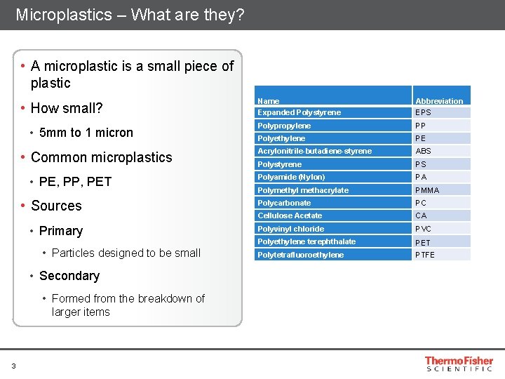
Microplastics – What are they? • A microplastic is a small piece of plastic • How small? • 5 mm to 1 micron • Common microplastics • PE, PP, PET • Sources • Primary • Particles designed to be small • Secondary • Formed from the breakdown of larger items 3 Name Expanded Polystyrene Abbreviation EPS Polypropylene PP Polyethylene PE Acrylonitrile-butadiene-styrene ABS Polystyrene PS Polyamide (Nylon) PA Polymethyl methacrylate PMMA Polycarbonate PC Cellulose Acetate CA Polyvinyl chloride PVC Polyethylene terephthalate PET Polytetrafluoroethylene PTFE
Why the concern? • Ubiquitous • Found in many places • Environmental • Aquatic life • Food Chain • Water Supply • Bottled Water • Fish & Seafood 4
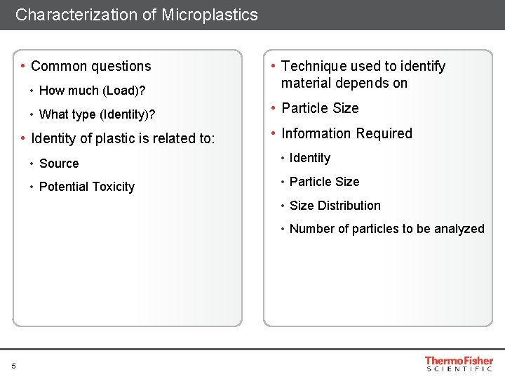
Characterization of Microplastics • Common questions • How much (Load)? • What type (Identity)? • Identity of plastic is related to: • Technique used to identify material depends on • Particle Size • Information Required • Source • Identity • Potential Toxicity • Particle Size • Size Distribution • Number of particles to be analyzed 5
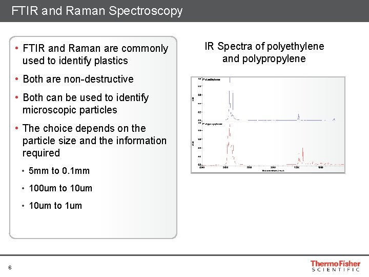
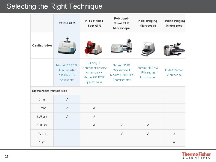
FTIR and Raman Spectroscopy • FTIR and Raman are commonly used to identify plastics • Both are non-destructive • Both can be used to identify microscopic particles • The choice depends on the particle size and the information required • 5 mm to 0. 1 mm • 100 um to 10 um • 10 um to 1 um 6 IR Spectra of polyethylene and polypropylene
Particle Size 5 mm to 0. 1 mm The world leader in serving science
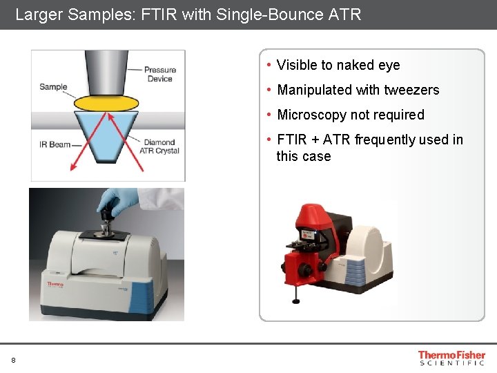
Larger Samples: FTIR with Single-Bounce ATR • Visible to naked eye • Manipulated with tweezers • Microscopy not required • FTIR + ATR frequently used in this case 8
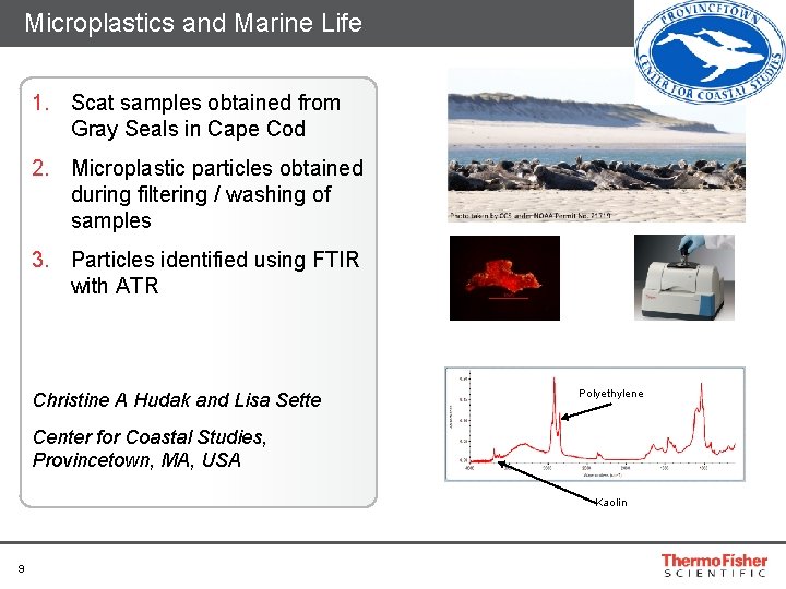
Microplastics and Marine Life 1. Scat samples obtained from Gray Seals in Cape Cod 2. Microplastic particles obtained during filtering / washing of samples 3. Particles identified using FTIR with ATR Christine A Hudak and Lisa Sette Polyethylene Center for Coastal Studies, Provincetown, MA, USA Kaolin 9
Particles 100 microns to 10 microns The world leader in serving science
Micro spectroscopy • Particle sizes below 100 microns require micro spectroscopy • To visualize the sample • To focus the spectrometer beam to a suitably small size • Microscopy has two benefits • The ability to measure small particles • The ability measure multiple particles automatically • Both FTIR and Raman are available in microscope configurations 11
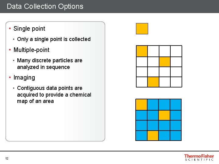
Data Collection Options • Single point • Only a single point is collected • Multiple-point • Many discrete particles are analyzed in sequence • Imaging • Contiguous data points are acquired to provide a chemical map of an area 12
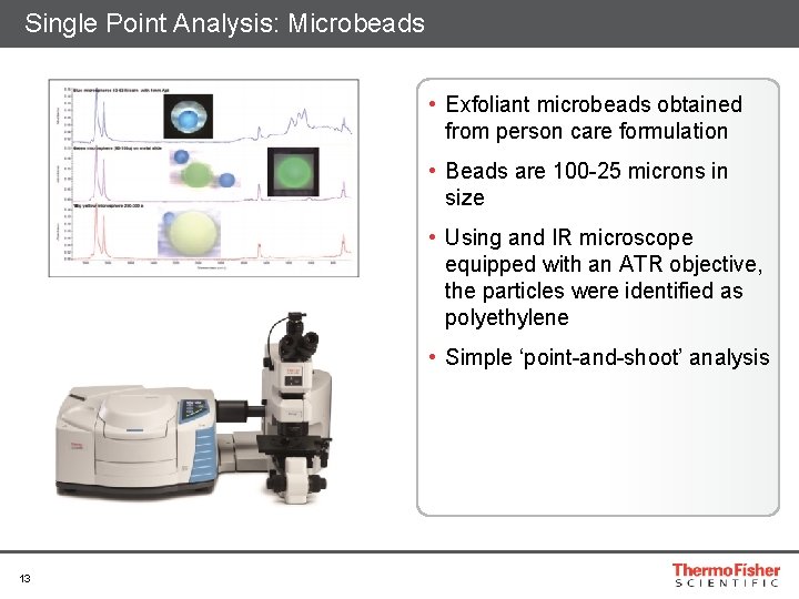
Single Point Analysis: Microbeads • Exfoliant microbeads obtained from person care formulation • Beads are 100 -25 microns in size • Using and IR microscope equipped with an ATR objective, the particles were identified as polyethylene • Simple ‘point-and-shoot’ analysis 13
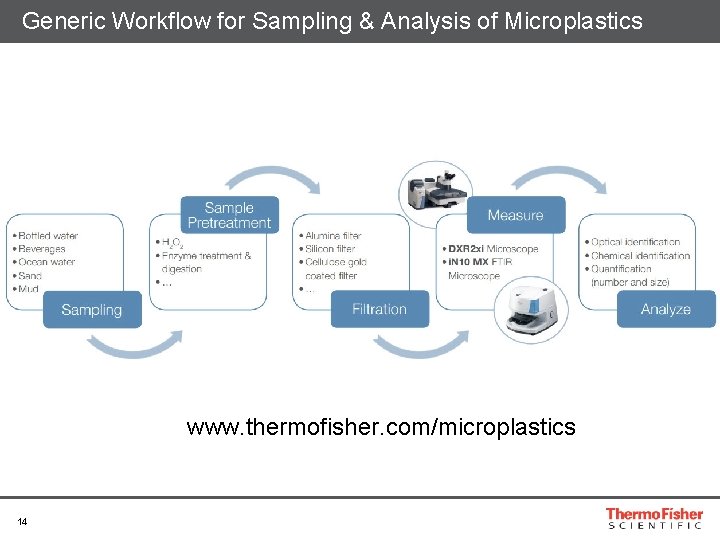
Generic Workflow for Sampling & Analysis of Microplastics www. thermofisher. com/microplastics 14
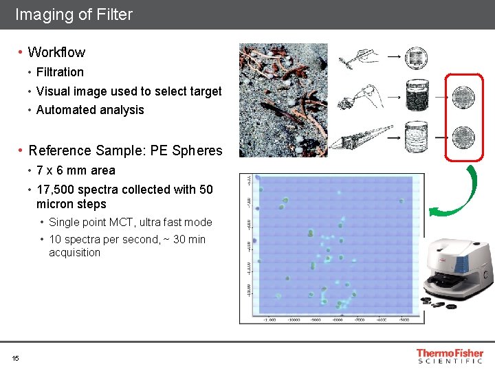
Imaging of Filter • Workflow • Filtration • Visual image used to select target • Automated analysis • Reference Sample: PE Spheres • 7 x 6 mm area • 17, 500 spectra collected with 50 micron steps • Single point MCT, ultra fast mode • 10 spectra per second, ~ 30 min acquisition 15
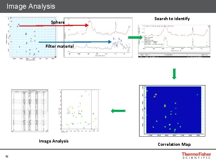
Image Analysis Sphere Search to identify Filter material Image Analysis 16 Correlation Map
Particles 10 to 1 micron The world leader in serving science
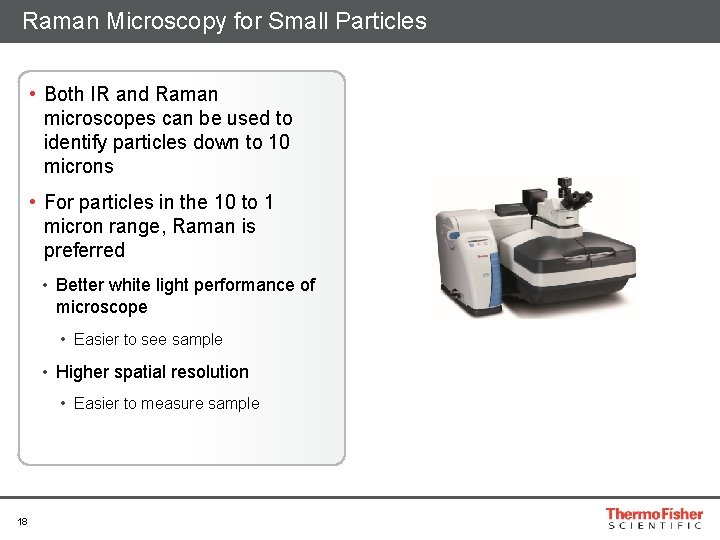
Raman Microscopy for Small Particles • Both IR and Raman microscopes can be used to identify particles down to 10 microns • For particles in the 10 to 1 micron range, Raman is preferred • Better white light performance of microscope • Easier to see sample • Higher spatial resolution • Easier to measure sample 18
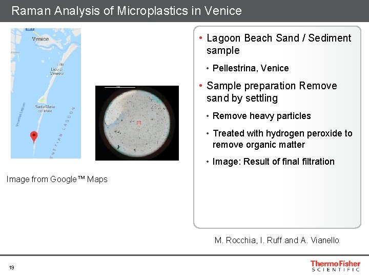
Raman Analysis of Microplastics in Venice • Lagoon Beach Sand / Sediment sample • Pellestrina, Venice • Sample preparation Remove sand by settling • Remove heavy particles • Treated with hydrogen peroxide to remove organic matter • Image: Result of final filtration Image from Google™ Maps M. Rocchia, I. Ruff and A. Vianello 19
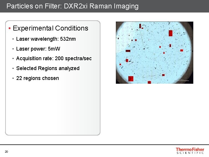
Particles on Filter: DXR 2 xi Raman Imaging • Experimental Conditions • Laser wavelength: 532 nm • Laser power: 5 m. W • Acquisition rate: 200 spectra/sec • Selected Regions analyzed • 22 regions chosen 20
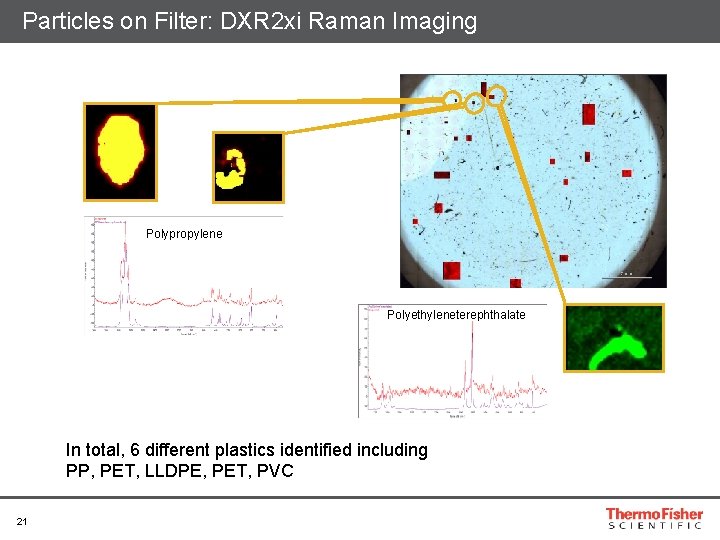
Particles on Filter: DXR 2 xi Raman Imaging Polypropylene Polyethyleneterephthalate In total, 6 different plastics identified including PP, PET, LLDPE, PET, PVC 21
Selecting the Right Technique 22
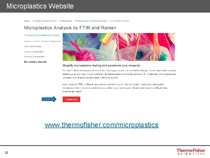
Microplastics Website www. thermofisher. com/microplastics 23
Acknowledgments • Christine A Hudak • Lisa Sette • Center for Coastal Studies, Provincetown, MA • Prof. Lorena M. Rios Mendoza • University of Wisconsin – Superior • Alvise Vianello • Aalborg University, Denmark • • • Michael Bradley Suja Sukumaran Massimiliano Rocchia Ines Ruff Steve Lowry • Thermo Fisher Scientific www. thermofisher. com/microplastics 24