ICU Curriculum Liberation from Mechanical Ventilation Clinical Case

ICU Curriculum Liberation from Mechanical Ventilation

Clinical Case 68 year old female with COPD presented to the emergency department with 3 days of cough, dyspnea and fevers. She was intubated and transferred to the ICU due to hypercapneic respiratory failure. She has been treated for pneumonia and a COPD exacerbation. Is she ready for extubation?

Complications related to MV VAP Sinusitis Pneumothorax Hypotension Stress gastropathy, gastric hypomotility Need for sedatives Oral, laryngeal injuries

Expediting Liberation from MV Fix the primary condition. Did the patient need the ventilator, an artificial airway, or both? What was the mechanism of respiratory failure? Is airway resistance improved? Is compliance improved? Is minute ventilation requirement reasonable?

Key Points Gas exchange or circulatory disturbance needs to have improved prior to considering weaning. > 50% of critically ill patients can be weaned the first day after the precipitating factor has resolved. There is a difference between weaning and extubation. Gradual reduction of IMV breaths prolongs weaning Daily SBT in patients off pressors and with adequate oxygenation
 Peak Inspiratory Flow Normal")
Resistance Formula Resistance. AW = (Peak inspiratory pressure – Pplateau) Peak Inspiratory Flow Normal Resistance. AW in an intubated patient is 4 -6 cm H 2 O/l/sec
 Normal Compliancestatic intubated = 0.")
Compliance Formula Compliancestatic = Tidal volume (Pplateau – PEEP) Normal Compliancestatic intubated = 0. 05 -0. 07 L/cm. H 2 O
 with resultant hypoxemia Increased venous return")
Physiological effects of weaning Increased atelectasis (no PEEP) with resultant hypoxemia Increased venous return can precipitate pulmonary edema Increased LV afterload Risk of hypoventilation (residual sedatives)

Expediting Liberation from MV Weaning parameters No parameter can be used exclusively to make decisions. The best parameter is performance on a SBT. Patients should be hemodynamically stable, no myocardial ischemia, have adequate oxygenation (Pa. O 2 > 60 on Fi. O 2 , 50%) with PEEP less than 5 cm H 2 O Discontinue sedatives & narcotics

Cause of respiratory failure improved Hemodynamically stable Awake, alert and co-operative Pa. O 2/Fi. O 2 >120 on PEEP < 5 cm. H 2 O SBT PS < 7 cm. H 2 O, CPAP = 5 cm H 2 O or T-piece trial for 0. 5 -1 hour on < 50% Fi. O 2 RR < 30 /min HR increase < 20 SBP increase < 20 mm. Hg RSBI < 100 Acceptable gas exchange Yes Cough, secretions, mental status all OK No Extubate Define Mechanism of failure and correct if possible

SBT Failure Clinical Diagnosis Rapid shallow breathing is a sign of impending respiratory failure Accessory muscle use, retractions, paradoxical motion of the abdomen Tachycardia Hypertension Distress Do not prolong trials that are exhausting the patient.
 : bronchodilators, steroids Compliance: (pulmonary edema, abdominal distention): diuresis,")
Correctable Factors Resistance (airway obstruction) : bronchodilators, steroids Compliance: (pulmonary edema, abdominal distention): diuresis, tap ascites, time Minute ventilation: (fever, sepsis, PE) Malnutrition, overfeeding Electrolyte abnormalities ICU Myopathy Metabolic alkalosis Atelectasis Sedation Secretions Endotracheal tube
 347 (76%) extubated successfully the first day after resolution of")
456 patients (medical/surgical) 347 (76%) extubated successfully the first day after resolution of acute illness 109 who failed initial SBT were randomized to weaning by: T-piece trials IMV with gradual rate reduction PSV with gradual reduction of PSV down to 8 cm. H 2 O Brochard L, et al. AJRCCM 1994; 150: 896

PSV had the shortest weaning time compared to T-piece and SIMV. PSV was not compared directly to the T-piece group but was compared to the cohort of T-piece and SIMV. Brochard L, et al. AJRCCM 1994; 150: 896
 extubated on the first day 130")
546 medical / surgical patients 416 (76%) extubated on the first day 130 remaining patients randomized to: Once a day T-piece trial Two or more T-piece or CPAP trials per day PSV with reduction of 4 cm H 2 O per day SIMV with rate reduction Esteban A, et al. NEJM 1995, 332: 345

Once daily T-piece trials were significantly better than PSV and SIMV Twice daily T-piece trials were not significantly better. Median duration of weaning: 3 days T-piece 4 days PSV 5 days SIMV Esteban A, et al. NEJM 1995, 332: 345

Liberation is not Extubation To extubate, the patient has to be liberated from ventilatory support (pass a SBT) and meet the following criteria: 1) have a good cough 2) be awake and alert to protect their airway 3) not require frequent suctioning or have copious secretions

Post extubation Stridor Need to be around when patients get extubated Due to edema of the upper airway These patients can rapidly develop pulmonary edema due to the generation of negative intrathoracic pressure and increased LV afterload. Treat with steroids, and possibly reintubation.
 NIPPV was")
Multicenter trial of 221 patients failing extubation (only 10% had COPD) NIPPV was ineffective in preventing re-intubation and did not reduce mortality. Esteban A, et al. NEJM 2004; 350: 2452

Weaning parameters The ideal parameter is an objective measure that decreases dependence on the skill and wisdom of the clinician.

Weaning parameters Minute Ventilation < 10 l/min High rate of false negative and false positive results.
 PImax value of -30 cm. H 2 O")
Weaning Parameters Maximal Inspiratory Pressure (NIF) PImax value of -30 cm. H 2 O has classically been taught as the threshold for successful weaning. PImax value of -20 cm. H 2 O has been used to predict failure. However, not very sensitive or specific as it does not take into account the demands being placed on the muscles.
 of respiratory frequency to tidal volume")
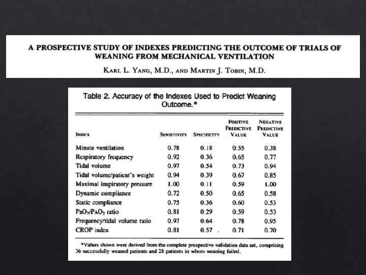
Weaning Parameters Rapid Ratio Shallow Breathing Index (RSBI) of respiratory frequency to tidal volume f/VT Useful tool if performed properly.

f/VT was measured using a hand held spirometer over one minute of spontaneous breathing after the patient was disconnected from the ventilator circuit. RSBI was not validated during PSV trials RSBI had a PPV of 0. 78 and a NPV of 0. 95 Yang K, Tobin MJ, NEJM 1991; 324: 1445

ICU Protocol Nursing/RT driven Plan for SBT starts several hours before Nurse weans sedation to RASS -1 to +1 Analgesia/sedation stopped before SBT Criteria to start SBT Adequate mental status, “acceptable” oxygenation/PEEP, temp < 38. 5, low dose/off vasopressors, physician order to wean, RR <30, minute ventilation < 15 L/min.

ICU Protocol SBT should start between 04: 30 -06: 30 (or whenever patient is ready) Record baseline data, HOB >30 degrees, suction. PSV 5/5 (higher pressure support if ETT <7) SBT for 30 minutes (never > 60 minutes; if they’re failing, and if they’re passing) Resume full support if: RR >35, Sp. O 2 <90%, HR >140 (or new arrhythmia), HR >20% change, new hypo/hypertension, agitation, diaphoresis, anxiety, increased ETCO 2. ABG is not necessary as routine

ICU Protocol Notify physician pass/fail If decision to extubate, physician order Extubate to nasal cannula (can decide NIPPV) If fails, resume sedation at half dose and analgesia at full dose once agitated.
 Daily systematic scrutiny of readiness for weaning")
Summary Protocol driven weaning teams (RT driven) Daily systematic scrutiny of readiness for weaning Examine the patient before and during weaning trials Many patients don’t need to be weaned If apneic, hold sedation and repeat once awake

Clinical question Is our patient with hypercapneic respiratory failure ready for extubation? Awake, alert, and following commands Off vasopressors, without tachycardia Minimal secretions, strong cough PEEP of 5, Fi 02 of 40% RSBI of 60 on PSV of 5 for 30 minutes AM ABG – p. H 7. 38| p. CO 2 55 | p. O 2 90
- Slides: 30