
ICRP Publication 121 Radiological Protection in Paediatric Diagnostic
ICRP Publication 121 Radiological Protection in Paediatric Diagnostic and Interventional Radiology Authors on behalf of ICRP P-L. Khong, H. Ringertz, V. Donoghue, D. Frush, M. Rehani, K. Applegate, R. Sanchez
Increased risk in Infants and Children Diagnostic radiological examinations carry higher risk per unit of radiation dose, on average, for the development of cancer in infants and children compared to adults. The higher risk is explained by the longer life expectancy in children for any harmful effects of radiation to manifest and the fact that developing organs and tissues are more sensitive to the effects of radiation. Moreover, the average risk is higher in infants and young children compared with older children ICRP 121, Introduction, Pg 15
General Aspects of Radiological Protection Justification the use of the radiological examination in question will do more good than harm to the patient the specific radiological examination when required for a specific disease and age group has a specified objective and this will usually improve the diagnosis or treatment or will provide necessary information about the exposed individuals the examination is required for that individual patient. Optimization ALARA Radiological equipment for children Adequacy of equipment and technical parameters Diagnostic reference level (DRL) Quality criteria implementation and audit ICRP Publication 103
Justification Imaging procedure is reliable, i. e. reproducible, and of sufficient sensitivity, specificity, accuracy Radiologist able to make accurate interpretation Performed by qualified radiographer/technologist with appropriate monitoring for quality and safety by medical physicists Accountability by single person, usually radiologist Imaging techniques that do not employ the use of ionizing radiation should always be considered as an possible alternative.
Some examples of radiographic examinations not routinely justified • Skull radiograph in an infant or child with epilepsy • Skull radiograph in an infant or child with headaches • Sinus radiograph in an infant or child under 6 years • • suspected of having sinusitis Cervical spine radiograph in an infant or child with torticollis without trauma Radiographs of the opposite side for comparison in limb injury Scaphoid radiographs in children under 6 years Nasal bone radiographs in children under 3 years ICRP 121, Pg 22
Optimisation: ALARA Optimisation of radiological protection in such a way that the required image is obtained with least radiation dose and net benefit is maximised ALARA (as low as reasonably achievable) principle should be adhered to for every examination
Radiological Equipment and adequacy of radiological equipment and technical parameters At procurement, installation and commissioning Radiographic equipment should have broadest range of settings to optimise the dose to the size of the child Modifications of parameters may be necessary both at installation and later in the use of the equipment ICRP 121, Pg 24
Conventional Radiography and Fluoroscopy: Equipment Special consideration to availability of dose reduction methods e. g. Virtual collimation Low attenuation table tops Removable grids Pulsed fluoroscopy and last image hold Spectral filters and adaptive technologies to minimise blooming Adding a copper filter in addition to the aluminium filtration should be considered
Diagnostic reference levels (DRLs) Assist in optimisation process If value is exceeded regularly, practice should be investigated. The upper DRL is often taken as the third quartile value, i. e. the value below which the measurements for three quarters of the institutions lie. Measurements taken in any institution should lie below the upper DRL, and if above, it should be possible to reduce exposures below the DRL without loss of clinical information. ICRP Publication 103
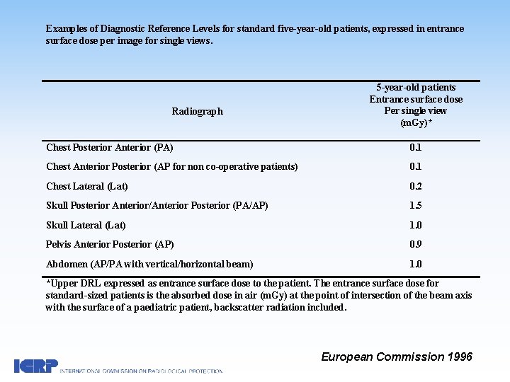
Examples of Diagnostic Reference Levels for standard five-year-old patients, expressed in entrance surface dose per image for single views. Radiograph 5 -year-old patients Entrance surface dose Per single view (m. Gy)* Chest Posterior Anterior (PA) 0. 1 Chest Anterior Posterior (AP for non co-operative patients) 0. 1 Chest Lateral (Lat) 0. 2 Skull Posterior Anterior/Anterior Posterior (PA/AP) 1. 5 Skull Lateral (Lat) 1. 0 Pelvis Anterior Posterior (AP) 0. 9 Abdomen (AP/PA with vertical/horizontal beam) 1. 0 *Upper DRL expressed as entrance surface dose to the patient. The entrance surface dose for standard-sized patients is the absorbed dose in air (m. Gy) at the point of intersection of the beam axis with the surface of a paediatric patient, backscatter radiation included. European Commission 1996
Quality criteria implementation and audit Need for follow-up and regular audits after implementation of quality criteria Audits of referral criteria, image quality, and imaging technique in paediatric radiology practices have found that better results are obtained for paediatric specialist centers compared with non-specialist centers. Thus, sharing of good practice by paediatric specialist centers is important for improving practices and patient outcomes. ICRP 121, Pg 27
Conventional Radiography and Fluoroscopy Proper patient positioning and applying immobilization which is often required in infants and young children Appropriate field size and correct X-ray beam limitation Use collimation to expose only the area intended for examination ICRP 121, Pg 29
Protective Shielding Breasts, gonads, and/or thyroid < 5 cm to the primary beam should be protected whenever possible without impairing the necessary diagnostic information Male gonads Not to be included within the primary radiation field Female gonads Gonad protection may not be possible; e. g. for the indications of trauma, incontinence, abdominal pain, misplaced shielding may mask important pathology. Lens SXR: PA rather than AP Breasts CXR and Spine X-Ray: PA rather than AP
Radiographic exposure conditions Knowledge and correct use of appropriate radiographic factors e. g. focal spot size, filtration, focus to image plane distance, tube currentexposure time product Exposure times Short exposure times required due to motion Higher speed classes of screen film system Peripheral skeleton; speed class of 200 -400 Others; speed classes of 400 -800 ICRP 121, Pg 31
Radiographic exposure conditions Additional filtration Anti-scatter grid In neonates, infants and younger children, antiscatter grid often not necessary because of relatively low scatter radiation Removable grids for fluoroscopy Automatic exposure control (AEC) Due to the wide range in size of paediatric patients, there is need to optimise AEC devices for handling patients accordingly Exposure times should be short because children do not generally cooperate and are difficult to restrain, so equipment should work and provide reproducibility in the shortest time range
Fluoroscopy Grid-controlled pulsed fluoroscopy systems Keep fluoroscopy table as far from X-ray source as possible and image intensifier as close to patient as possible Minimize scatter radiation with hanging lead drapes Use pulsed fluoroscopy for monitoring procedures (3 -8 pulses/sec adequate) Image intensifier positioned over area of interest before fluoroscopy is commenced Use virtual collimation to help positioning Apply tight collimation ICRP 121, Pg 36
Fluoroscopy Angle beam away from radiosensitive areas Limit use of magnification Use still images from last-image hold or archive digital images to review findings Consider alarm bells for fluoroscopy beyond a certain time Record radiation dose (air kerma-area-product) AAPM, 1998
Interventional Radiology Increasing complexity and length of procedures, thus overall radiation dose to the patient may be greater. Increased dose to patients due to large size of image intensifier compared to patient and increased need to use magnification (must use collimation). ICRP 121, Pg 39
Interventional Radiology A second, specific level of training in radiation protection is desirable, and in some countries mandatory. Major paediatric interventional procedures, particularly in small infants, should be performed by experienced paediatric interventional operators. Consider ultrasound guidance whenever possible ICRP Publication 85
Reducing unnecessary dose to staff Paediatric procedures may take a longer time Frequently necessary to come close to or enter the beam due to small size of child Angulation of beam off the hands and apply strict collimation Use protective devices (however, caution with gloves as it can reduce dexterity) Power injector instead of hand injecting contrast material ICRP 121, Pg 39
Image acquisition during DSA Each run should be necessary Use fewest number of frames/sec Images obtained using lower magnification (magnify using post-processing instead) Tight collimation to include only area of interest Use last image hold, image capture and archiving of runs ICRP 121, Pg 41
CT: Justification Imaging techniques that do not employ the use of ionizing radiation should always be considered as an possible alternative. Especially in children with chronic illness who require repeated imaging evaluation. Ultrasound should be first line imaging modality for the abdomen e. g. acute appendicitis, intussusception MRI is often best for evaluation of detailed information of soft tissues, nervous system or bone marrow ICRP 121, Pg 46
CT: Justification Chronic illness and malignancy: Follow up CT scans should not be done not too early Alternative modalities should be considered for follow up Consider reducing CT scan volume and adjusting parameters Multi-phase CT scans: Justification for every additional phase needed ICRP 121, Pg 46
Optimisation: CT Equipment Special consideration should be given to dose reduction measures when purchasing new equipment for paediatric use Medical physicist advice for procurement, commissioning, quality control tests etc. ICRP 121, Pg 47
CT software and hardware for dose reduction Tube current modulation and automatic exposure control (AEC): ‘one-size does not fit all’ Organ-based dose modulation Auto k. V technology X-ray detectors of improved efficiency Iterative reconstruction Methods to block unnecessary radiation from ‘helical overdosing’; dynamic or adaptive collimation Filters for control of irradiation beam e. g. bow-tie filters ICRP 121, Pg 48
Optimisation: adjustment of scan parameters Image quality: noise vs contrast Adjustment of scan parameters (m. As, k. Vp, pitch) to weight or age Use of automatic exposure control (AEC) techniques • Noisier images, if sufficient for radiological diagnosis, should be accepted
Image quality and Study quality Depends on structure and region examined More noise acceptable in skeleton or lung compared to brain and abdominal examinations Tube current-time product (m. As) can be significantly reduced in children. e. g. 50 m. As for CT chest 80 k. Vp suggested for infants under 5 kg Depends on clinical indication for the study More noise maybe tolerated for follow-up study
Study Quality and Display Motion artefact Selective use of sedation Adjustment of scan time and pitch, and dual-source CT Meticulous use of IV contrast Post-processing: planar reformation and 3 D reconstruction Display of images Optimise monitors Ambient environment
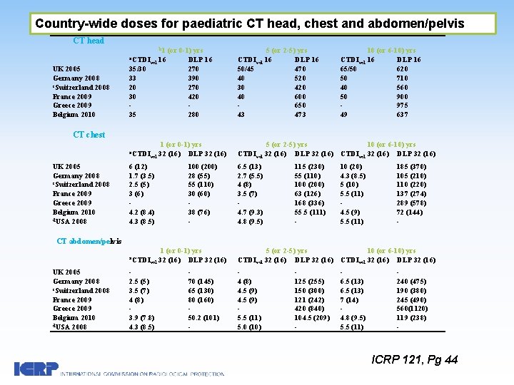
Country-wide doses for paediatric CT head, chest and abdomen/pelvis CT head b 1 (or 0 -1) yrs a. CTDI UK 2005 Germany 2008 c. Switzerland 2008 France 2009 Greece 2009 Belgium 2010 35/30 33 20 30 35 vol 16 DLP 16 270 390 270 420 280 5 (or 2 -5) yrs CTDIvol 16 DLP 16 50/45 470 40 520 30 420 40 600 650 43 473 10 (or 6 -10) yrs CTDIvol 16 DLP 16 65/50 620 50 710 40 560 50 900 975 49 637 CT chest a. CTDI UK 2005 Germany 2008 c. Switzerland 2008 France 2009 Greece 2009 Belgium 2010 d. USA 2008 1 (or 0 -1) yrs 32 (16) DLP 32 (16) vol 6 (12) 1. 7 (3. 5) 2. 5 (5) 3 (6) 4. 2 (8. 4) 4. 3 (8. 5) 100 (200) 28 (55) 55 (110) 30 (60) 38 (76) - 5 (or 2 -5) yrs 10 (or 6 -10) yrs CTDIvol 32 (16) DLP 32 (16) 6. 5 (13) 2. 7 (5. 5) 4 (8) 3. 5 (7) 4. 7 (9. 3) 4. 8 (9. 5) 115 (230) 55 (110) 100 (200) 63 (126) 168 (336) 55. 5 (111) - 10 (20) 4. 3 (8. 5) 5 (10) 5. 5 (11) 4. 5 (9) 5. 5 (11) 185 (370) 105 (210) 110 (220) 137 (274) 289 (578) 72 (144) - CT abdomen/pelvis a. CTDI UK 2005 Germany 2008 c. Switzerland 2008 France 2009 Greece 2009 Belgium 2010 d. USA 2008 1 (or 0 -1) yrs 32 (16) DLP 32 (16) vol 2. 5 (5) 3. 5 (7) 4 (8) 3. 9 (7. 8) 4. 3 (8. 5) 70 (145) 65 (130) 80 (160) 50. 2 (101) - 5 (or 2 -5) yrs 10 (or 6 -10) yrs CTDIvol 32 (16) DLP 32 (16) 4 (8) 4. 5 (9) 5. 5 (11) 5. 0 (10) 125 (255) 150 (300) 121 (242) 420 (840) 104. 5 (209) - 6. 5 (13) 7 (14) 4. 8 (9. 5) 5. 5 (11) 240 (475) 190 (380) 245 (490) 560(1120) 119 (238) - ICRP 121, Pg 44
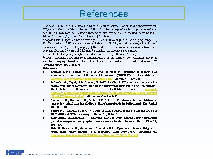
References a. For head CT, CTDI and DLP values refer to 16 -cm phantom. For chest and abdomen/pelvis CT, values refer to the 32 -cm phantom, followed by the corresponding 16 -cm phantom value in parentheses. Data have been adapted from the original publications, expressed according to the 16 -cm phantom [1, 3, 5], the 32 -cm phantom [4] or both [2]. b. Proposed DRLs expressed for children ages 1, 5 and 10 years [1, 4, 5] or using age ranges [2, 3]. Most pediatric DRL surveys do not include a specific 15 -year-old category, although some include an 11 - to 15 -year-old group [2, 3]; the adult DRL in that country, or a value intermediate between adult and 10 -year-old DRL may be considered appropriate for teenagers. c. Switzerland subsequently adopted the values from the larger German [2] study. d. Values calculated according to recommendations of the Alliance for Radiation Safety in Pediatric Imaging, based on the future French DRL values for adult abdominal CT recommended by IRSN in 2008. References: 1. Shrimpton, P. C. , Hillier, M. S. , et al. , 2005. Doses from computed tomography (CT) examinations in the UK – 2003 review (NRPB-67). Available via www. hpa. org. uk/radiation. publication/index. htm. Accessed 10 Jan 2011. 2. Galanski, M. , Nagal, H. D. , Stamm, G. 2007. Paediatric CT exposure practice in the federal republic of Germany. Results of a nationwide survey in 2005/6. Medizinishe Hochschule Hannover. Available via www. mhhannover. de/fileadmin/kliniken/diagnostische_radiologie/download/Report_German _Paed-CT Survey_2 --5_06. pdf. Accessed 9 Jan 2011. 3. Verdun, F. R. , Gutierrez, D. , Vader, J. P. , 2008. CT radiation dose in children: a survey to establish age-based diagnostic reference levels in Switzerland. Eur Radiol 18, 1980 -1986. 4. Brisse, H. J. , Aubert, B. , 2009. CT exposure from pediatric MDCT: results from the 2007 -2008 SFIPP/ISRN survey. J Radiol 90, 207 -215. 5. Yakoumakis, E. , Karlatira, M. , Gialousis, G. , et al. , 2009. Effective dose variation in pediatric computed tomography : dose reference levels in Greece. Health Phys 97, 595 -603. 6. Buls, N. , Bosmans, H. , Mommaert, C. , et al. , 2010. CT paediatric doses in Belgium: a multi-center study: results of a dosimetry audit 2007 -2009. Available via http: //www. fanc. fgov. be/CWS/GED/pop_View. aspx? LG=1&ID=2449
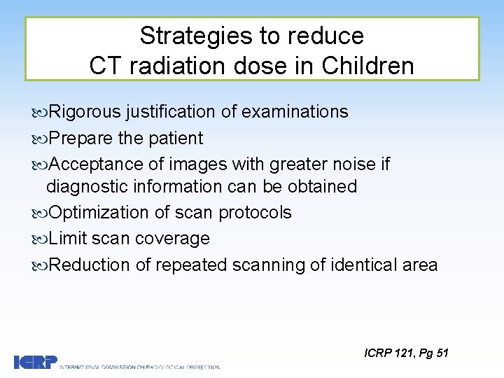
Strategies to reduce CT radiation dose in Children Rigorous justification of examinations Prepare the patient Acceptance of images with greater noise if diagnostic information can be obtained Optimization of scan protocols Limit scan coverage Reduction of repeated scanning of identical area ICRP 121, Pg 51
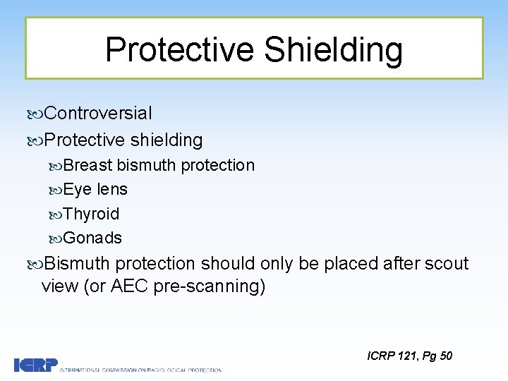
Protective Shielding Controversial Protective shielding Breast bismuth protection Eye lens Thyroid Gonads Bismuth protection should only be placed after scout view (or AEC pre-scanning) ICRP 121, Pg 50
Protective Shielding may increase radiation dose or affect image quality if not placed optimally AEC and dose-modulation systems increasingly available, and beam shielding interferes with these systems Use of proper field size limitation and appropriate parameters ICRP 121, Pg 50
Protective Shielding Studies on paediatric patients and phantoms have shown dose reduction of up to >50% Suggested techniques to reduce streak artifacts: Shield needs to be appropriately placed with enough distance, > 2 cm offset Smoothly placed over the surface Protocols should be tested specifically for the scanner
Summary and Recommendations Justification of every examination involving ionising radiation, followed by optimisation of radiological protection is important in every patient, and especially in paediatric patients in view of the higher risk of adverse effects per unit of radiation dose compared to adults. According to the justification principle, if a diagnostic imaging examination is indicated and justified, this implies that the risk to the patient of not doing the examination is greater than the risk of potential radiation induced harm to the patient. Imaging techniques that do not employ the use of ionising radiation should always be considered as a possible alternative. Optimisation of radiological protection involves optimised functioning of radiological equipment and quality control, ensuring radiological equipment and technical parameters are adequately tailored for paediatric patients and the implementation of diagnostic reference levels (DRL) to assist in the optimisation process. Quality criteria implementation and regular audits should be instituted as part of the radiological protection culture in the institution.
Attention should be paid to good radiographic technique including positioning and immobilisation of paediatric patients, field size and protective shielding, and radiographic exposure parameters should be specially tailored for patient size and age. As most imaging equipment and vendor specified protocols are often structured for adults, modifications of equipment and exposure parameters may be necessary for paediatric use. Advice of medical physicists should be sought if possible to assist with installation, setting imaging protocols and optimisation. Interventional procedures should be performed by experienced paediatric interventional staff due to the potential for high patient radiation dose exposure, and additional training in radiological protection to protect both patient and staff is recommended. For CT, dose reduction should be optimised by adjustment of scan parameters (m. A, k. Vp and pitch) according to patient weight or age, and weight-adapted CT protocols have been suggested and published. For the purpose of minimising radiation exposure, noisier images, if sufficient for radiological diagnosis, should be accepted. Optimised study quality also depends on region scanned and study indication. Other dose reduction strategies include restricting multiphase examination protocols, avoiding overlapping of scan regions, and scanning only the area in question. Furthermore, study quality may be improved by image post-processing to facilitate radiological diagnoses and interpretation.
- Slides: 36