ICD9 CM Coding Proposals Retinopathy of Prematurity Necrotizing


- Slides: 21

ICD-9 -CM Coding Proposals Retinopathy of Prematurity Necrotizing Enterocolitis Disruption of Postoperative Wound

Presented by Patrick Romano, MD, MPH Professor of Medicine and Pediatrics University of California Davis On behalf of the Agency for Healthcare Research and Quality September 28, 2007

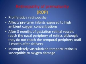
Retinopathy of prematurity • Retinopathy of prematurity (ROP): – A vascular proliferative disorder of the retina in premature infants – Common cause of childhood blindness: 500 -700 children annually – Sequelae affect another 2100: myopia, strabismus, retinal detachment – Incidence and severity decrease with increasing gestational age and birthweight – Incidence of all stages of ROP >50% in preterm infants with birthweight < 1250 g – Incidence of stage 3 or greater ROP is 6% in preterm infants with birthweight <1250 g

ROP developmental biology • Before 16 weeks, the embryonic retina has no vessels. Blood flow is supplied by vessels that are usually reabsorbed by 34 weeks. • Vascularization of retina begins between 15 -18 weeks, and is usually complete by 36 weeks (nasal retina) and 40 weeks (temporal retina) • ROP is thought to develop when new vessels are injured, disrupting normal angiogenesis. • Risk factors include prematurity, low birth weight, and severe respiratory disease with/without hyperoxia

ROP staging • Stages – 0: Immature retina without vascular changes – 1: Flat line demarcates the vascular and avascular areas – 2: Fibrous tissue protrudes into the vitreous between the vascular and avascular retina – 3: New blood vessels and fibrous tissue along ridge or extending into the vitreous – 4: Partial retinal detachment – 5: Total retinal detachment

ROP Copyright © 1995 -2007 Royal National Institute of Blind People

ROP treatment • Threshold ROP: Five contiguous clock hours or eight total clock hours of stage 3 and plus disease (posterior pole vascular dilation and tortuosity) in zone 1 or 2 • Follow-up required for early disease, continuing until ROP regresses or vasculature matures • Retinal ablative therapy is indicated for threshold disease and higher risk prethreshold disease. • More severe cases of ROP can progress to cicatricial disease (scarring)

ROP coding • Current coding lumps together all stages and acute/cicatricial disease into one code – 362. 21 Retrolental fibroplasia • Providing more granularity allows: – Separation between cicatricial (chronic) disease and acute disease – Separation between minor ROP (requiring observation but no treatment) and severe ROP (requiring treatment)

ROP coding rationale • ROP rates are currently tracked by several organizations – Child Healthcare Corporation of America – National Perinatal Information Center • Overall incidence has been stable over the past 20 years, but incidence varies widely across NICUs, and infants delivered at subspecialty perinatal centers have lower rates of ROP than those born elsewhere • AHRQ Clinical panel suggested more granularity in coding would aid in research and surveillance • Best practices are unknown, but careful oxygen management to avoid hyperoxia and repeated episodes of hypoxia-hyperoxia shows promise – RCT of infants with prethreshold disease (p=0. 035)

Necrotizing enterocolitis • Necrotizing enterocolitis (NEC) = Syndrome of intestinal necrosis • Gastrointestinal emergency – Acute: Sepsis, infection, shock, respiratory failure, hypoglycemia, metabolic acidosis – Long term: Short bowel syndrome – Death (20 -30%) • Occurs in 2 -10% of infants with birthweight <1500 g • Incidence decreases with gestational age, but about 13% of cases occur in term infants • Etiologic factors include bowel injury and intestinal mucosal disruption with enteric feedings, immature immune responses, and possibly pathogenic organisms – 10% of cases develop before feeding

NEC staging • Suspected NEC (stage I): nonspecific systemic signs including lethargy, abdominal distention, blood in stool. Radiograph normal or shows dilation of bowel • Proven NEC (Stage II): Absent bowel sounds. Radiograph shows intestinal dilation, ileus and pneumatosis. May have ascites. • Advanced NEC (Stage III): Critically ill. May have hypotension/shock, bradycardia, apnea, peritonitis. May have bowel perforation (free air).

NEC radiography

NEC treatment • Treatment includes supportive care – Cardiovascular and respiratory support if required – May include discontinuation of feedings, GI decompression with nasogastric suction, fluid replacement, parenteral nutrition, antibiotics – Surgical intervention may be required for abdominal mass, ascites/peritonitis, or intestinal obstruction. May include intestinal resection or peritoneal drainage.

NEC coding • Current coding lumps together all NEC into one code – 777. 5 Necrotizing enterocolitis in fetus or newborn • Providing more granularity allows: – Separation between NEC, mild NEC requiring medical therapy (e. g. , pneumatosis), and severe NEC that often requires surgical therapy (e. g. , perforation) • Diagnosis of NEC may vary, especially in early stages. Findings of pneumatosis and perforation provide standards for coding.

NEC coding rationale • NEC rates are currently tracked by several organizations – Child Healthcare Corporation of America – National Perinatal Information Center • Incidence varies across NICUs, and cases occur in clusters suggesting a transmissible agent. • AHRQ Clinical panel suggested more granularity would aid in research and surveillance • Best practices are unknown, but several interventions show promise – Antenatal corticosteroids reduce NEC by 54% (8 RCTs) – Feeding donor human milk (versus formula) reduces NEC by 66% to 75% (4 RCTs) – Proper velocity of feeding volume advancement is controversial – Implementation of any standardized feeding regimen has been associated with an 87% reduction in the risk of NEC (6 quasi-experimental studies)

Disruption of Operative Wound • Failure of operative wound to heal results in full or partial dehiscence (separation) • Disruption can be superficial or extend into the fascia or muscle Anatomy of abdominal wall

Disruption: wound healing • Wound healing occurs over time: initial inflammatory response (days), epithelialization (days), fibroplasia (weeks), maturation (weeks months) • 80% of full strength obtained by 6 weeks • Wound disruption incidence is ~1% of surgeries, 10% in presence of surgical site infection • Disruption occurs when pressure or tension overcomes tissue or suture strength or knot security.

Disruption: risk factors/depth • Risk factors include: age, poor nutrition, comorbid disease, obesity, tissue weakness due to radiation or chemotherapy, infection, ascites • Treatment depends on depth of dehiscence • Complete dehiscence mortality ~10% • Partial dehiscence extending to fascia may be a surgical emergency • Superficial dehiscence may heal without surgical intervention.

Disruption: prevention • Prevention – Proper stitching and surgical technique – Use of appropriate suture material for tissue – Prevention of surgical site infection – Accounting for special situations (e. g. obesity)

Disruption: coding • History of code – FY 2003: 998. 3, “Disruption of operation wound” split into two codes • 998. 31: Disruption of internal operation wound • 998. 32: Disruption of external operation wound • Physicians tend to document the tissues and/or layers involved, rather than using the terms “internal” and “external” • “Disruption of operation wound NOS” is coded to 998. 32 (“external”)

Disruption: coding rationale • Surgical wound disruption rates are currently tracked by several organizations – – AHRQ Patient Safety Indicator AHRQ Pediatric Quality Indicator AHRQ National Healthcare Quality/Disparities Reports National Association of Children’s Hospitals and Related Institutions (NACHRI), etc. • Providing additional guidance, and separating NOS cases, would standardize coding and focus attention on the more severe cases • Best practices are unknown, but considerable interest in prevention: – Surgical approach (size and location of incision) – Continuous versus interrupted sutures
 Rop stages
 Eibschitz-tsimhoni
 Axial coding
 Contoh open coding, axial coding selective coding
 Indication for exploratory laparotomy
 What is nec in babies
 Necrotizing ulcerative gingivitis
 Necrotizing fasciitis hemorrhagic bullae
 Causes of prematurity
 Rickets x ray findings
 Icd9
Icd9 Icd9 dysphagia
 Coding dna and non coding dna
 History of qualitative research
 Diabetic retinopathy
 Drcr net
 Solar retinopathy
 Diabetic retinopathy clinical research network
 Diabetic retinopathy clinical research network
 Albany diabetic retinopathy
 Diabetic retinopathy clinical research network
 Keith wegner classification
