IB BIOLOGY Topic 1 Cell Biology 1 3

IB BIOLOGY Topic 1 Cell Biology 1. 3 -1. 6

Membrane Structure • 1915 - scientists knew structure of membranes included proteins and lipids. • Davson and Danielli Model 1935 – Lipid bilayer model – Covered on both sides with thin layer of globular protein • Singer and Nicholson 1972 – Proteins are inserted into phospholipid bilayer, not covering it • Current model– Fluid mosaic model

Phospholipids • Composed of: – 3 C glycerol – 2 glycerol carbons have fatty acid tails – 3 rd C attached to highly polar organic alcohol bound to a phosphate – Hydrophobic and hydrophylic regions – Fatty acid tails not strongly attracted to each otherallows for fluidity – Structure maintained by hydrogen bonding among H 2 O molecules

Cholesterol • Membranes consistency of olive oil • Various locations in fatty acid tail region, cholesterol molecules are embedded • Determine fluidity of cell, temperature dependent • Allows function during wide temperature range

Proteins • Create extreme diversity in membrane function • Embedded in bilayer • Two Types: – Integral protein • Amphipathic- has both hydrophobic and hydrophylic portions – Peripheral protein • Bound to membrane surface • Can be anchored to integral

Membrane Protein Functions • Hormone binding- specific shapes • Enzymatic action- catalyze chemical reactions • Cell adhesion-permanent or temporary connection • Cell-cell communication-many have carbs attached • Channels for passive transportdown the concentration gradient • Pumps for active transportneeds ATP, up the concentration gradient

Membrane Transport • Passive transport – No ATP used – Goes with concentration gradient (Hi to lo) • Diffusion: membrane – Gases: O 2 diffuses into a cell, CO 2 diffuses out • Facilitated diffusion: – Uses carrier proteins • Osmosis: – H 2 O across semi-permeable membrane – Solute and solvent ratio important

Membrane Transport • Size and Charge of Molecules: – Small, non-polar cross easily • CO 2, N 2, H 2 O, glycerol – Large, polar cross with difficulty • Cl-, K+, Na+, glucose, sucrose

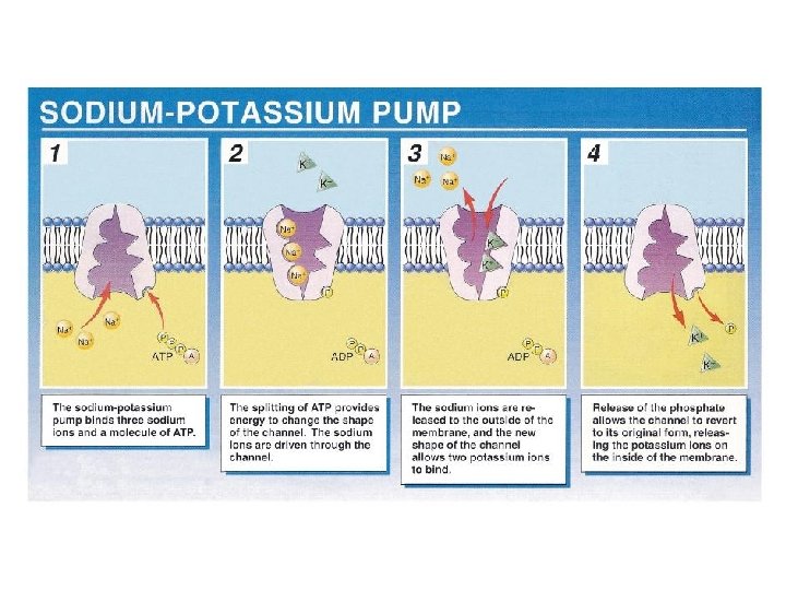
Membrane Transport • Active transport: – Uses ATP – Goes against the concentration gradient • Animal cells have higher K+ concentration inside than outside, and have greater Na+ concentration outside than inside • Balanced maintained by sodium-potassium pump

Membrane Transport • Endocytosis – Fluidity dependent – Allows macromolecules to enter cell – Part of plasma membrane pinched off to enclose macromolecule – Changes shape of membrane – Creates vesicle that enters cell

Membrane Transport • Exocytosis – Reverse of endocytosis

Specialized Transport • Pinocytosis- intake of extracellular fluids • Phagocytosis- intake of large particulate matter – Ex. Macrophage engulfing antigen

Cell Division • The Cell Cycle: – Describes the behavior of cells as they grow and divide. – Interphase: G 1, S, G 2 – G 1 -smallest cell will ever be, major event is growth – S- replication of cellular DNA – G 2 -organelles increase in number, DNA begins to condense from chromatin chromosomes, microtubules begin to form.

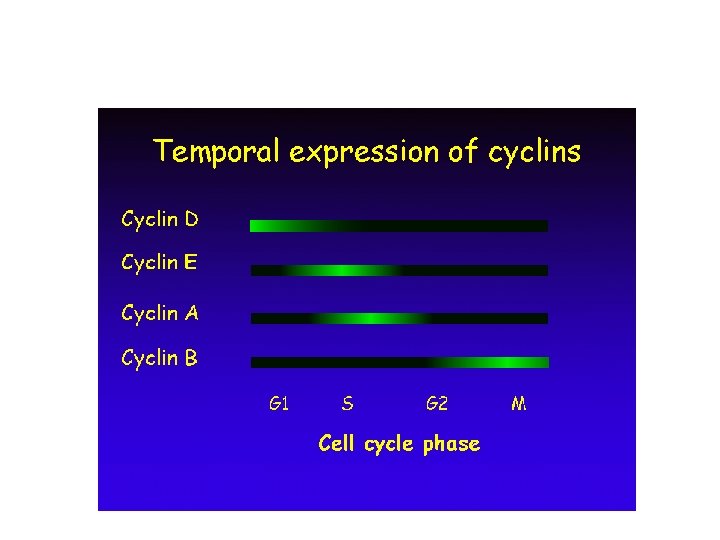
Cell Division • Cyclins: – Proteins that control cell’s progression through cell cycle – Bind to cyclin-dependent protein kinases (CDKs) enabling them to act like enzymes – Cause cell to go from G 1 S G 2 M – Can pause during G 1 and enter G 0 - a non-growing phase • Time remaining in this phase varies – Nerve and muscle cells do not progress past G 0

Mitosis • M phase: – Replicated chromosomes move to opposite poles of cell – Cytoplasm divides – Result: identical daughter cell • Phases of Mitosis: – – Prophase Anaphase Metaphase Telophase

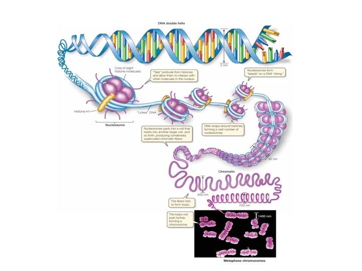
Chromosome • Before replication in S phase, cells have 1 molecule of DNA • After- 2 molecules of DNA held on center by centromere • Condensation of DNA: – Super-coiling – DNA wraps around histone proteins- “nucleosome” – Nucleosomes solenoid chromosome

Prophase • The chromatin fibers become more tightly coiled • The nuclear envelope distinegrates, nucleoli disappear • Mitotic spindle forms • Centromere of chromosomes attach to spindle fibers by kinetochores • Centrosomes move to opposite poles of cell lengthening the microtubules

Metaphase • Chromosomes move to the middle of the cell. • Centromere of chromosomes lie on the plate. • Movement of chromosomes controlled by spindle • Centrosomes at opposite poles

Anaphase • Shortest phase of mitosis • Chromosomes move to opposite parts of the cell. • Movement due to shortening of the microtubules of spindle • Centromeres attached to microtubules, so they move first • Result: each pole has complete, identical set of chromosomes.

Telophase • Chromosomes are at each pole • Nuclear membrane begins to reform • Chromosomes chromatin • Spindle disappears • Cell elongates, ready for cytokinesis

Cytokinesis • Cleavage of animal cell • Cell plate formation in plant cell https: //www. youtube. com/watch? v=ofjyw 7 A RP 1 c https: //www. youtube. com/watch? v=ZEwddr 9 ho-4

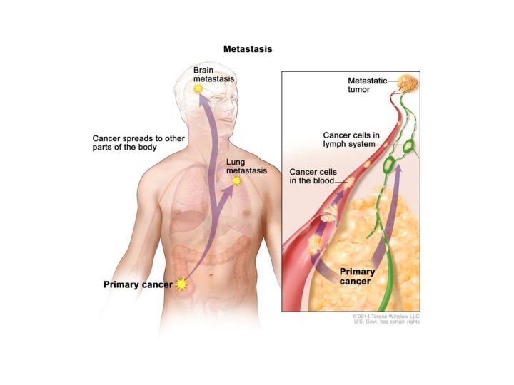
Cancer • Uncontrolled cell growth • Primary tumor: – Occurs at original site of cancer • Secondary tumor: – Metastatic- has spread from original site to another location – Can be found in many locations

Henrietta Lacks and He. La Cells • http: //www. cbsnews. com/news/the-immortalhenrietta-lacks/ • Henrietta Lacks was an African American woman who died in 1951 of cervical cancer. – Her cancerous tumor cells were of interest to doctors, because they were the first cancer cells that would grow under laboratory conditions. The cells have been studied and cultured world- wide for decades without her knowledge, permission, or family compensation. – She is the most important contributor to cancer research to date.

Cancer, cont. • Reasons formation of primary tumors: – Gene mutation – Abnormally high gene expression – Oncogenes: • Section of genes that contributes to conversion of normal cell cancer cell • Oncogenes: – Can mutate in response to environmental trigger • Ex: Cigarette smoke
- Slides: 28