
IAEA Regional Training Course on Radiation Protection of

- Slides: 35
IAEA Regional Training Course on Radiation Protection of patients for Radiographers, Accra, Ghana, 11 -15 July 2011 Optimization of Protection in Dental Radiology IAEA International Atomic Energy Agency
Introduction • Dental radiology makes use of specific types of equipment, needed for different purposes. • Frequent exposures (though each with low dose) involve a risk for the practitioner and for the patient IAEA 22: Optimization of Protection in Dental Radiology 2
Topics üDental X-ray equipment üRadiation protection in dental radiology üQuality control for dental equipment IAEA 22: Optimization of Protection in Dental Radiology 3
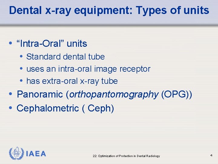
Dental x-ray equipment: Types of units • “Intra-Oral” units • Standard dental tube • uses an intra-oral image receptor • has extra-oral x-ray tube • Panoramic (orthopantomography (OPG)) • Cephalometric ( Ceph) IAEA 22: Optimization of Protection in Dental Radiology 4
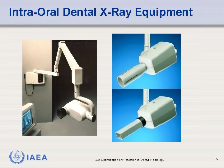
Intra-Oral Dental X-Ray Equipment IAEA 22: Optimization of Protection in Dental Radiology 5
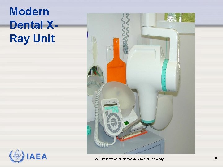
Modern Dental XRay Unit IAEA 22: Optimization of Protection in Dental Radiology 6
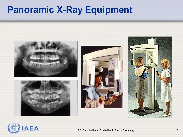
Panoramic X-Ray Equipment IAEA 22: Optimization of Protection in Dental Radiology 7
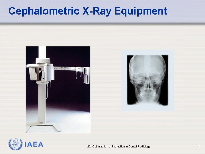
Cephalometric X-Ray Equipment IAEA 22: Optimization of Protection in Dental Radiology 8
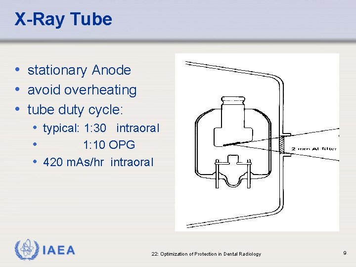
X-Ray Tube • stationary Anode • avoid overheating • tube duty cycle: • typical: 1: 30 intraoral • 1: 10 OPG • 420 m. As/hr intraoral IAEA 22: Optimization of Protection in Dental Radiology 9
Generators & Pre-Heat • Medium frequency - stable waveform • Single phase (SP) - pulsed • Pre-Heat: separate circuit for heating filament • Single Phase units without a pre-heat circuit • initial pulses of variable k. V IAEA 22: Optimization of Protection in Dental Radiology 10
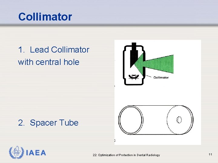
Collimator 1. Lead Collimator with central hole 2. Spacer Tube IAEA 22: Optimization of Protection in Dental Radiology 11
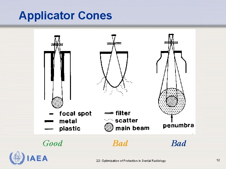
Applicator Cones Good IAEA Bad 22: Optimization of Protection in Dental Radiology Bad 12
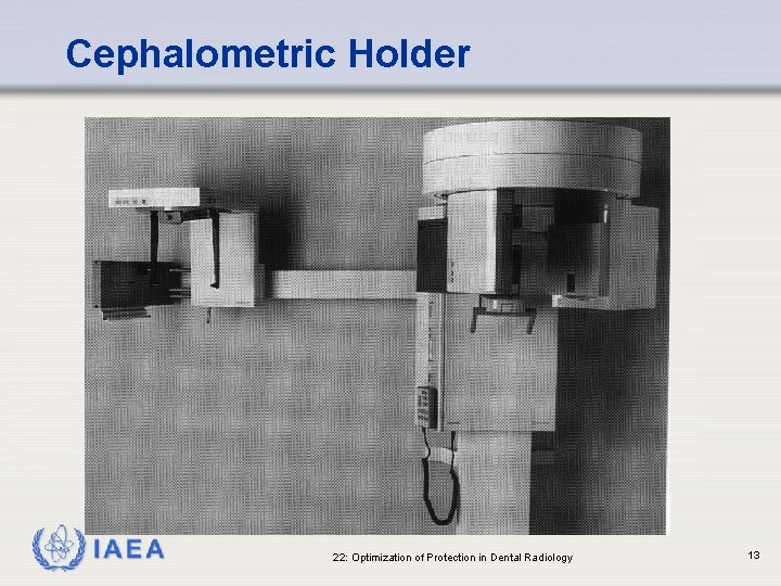
Cephalometric Holder IAEA 22: Optimization of Protection in Dental Radiology 13
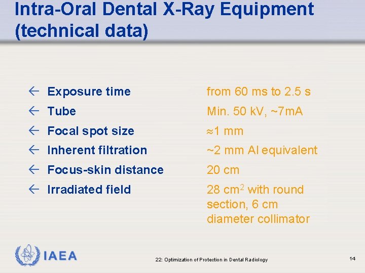
Intra-Oral Dental X-Ray Equipment (technical data) ß Exposure time from 60 ms to 2. 5 s ß Tube Min. 50 k. V, ~7 m. A ß Focal spot size 1 mm ß Inherent filtration ~2 mm Al equivalent ß Focus-skin distance 20 cm ß Irradiated field 28 cm 2 with round section, 6 cm diameter collimator IAEA 22: Optimization of Protection in Dental Radiology 14
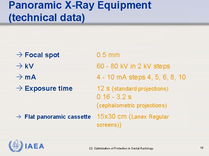
Panoramic X-Ray Equipment (technical data) à Focal spot 0. 5 mm à k. V 60 - 80 k. V in 2 k. V steps à m. A 4 - 10 m. A steps 4, 5, 6, 8, 10 à Exposure time 12 s (standard projections) 0. 16 - 3. 2 s (cephalometric projections) à Flat panoramic cassette 15 x 30 cm (Lanex Regular screens)) IAEA 22: Optimization of Protection in Dental Radiology 15
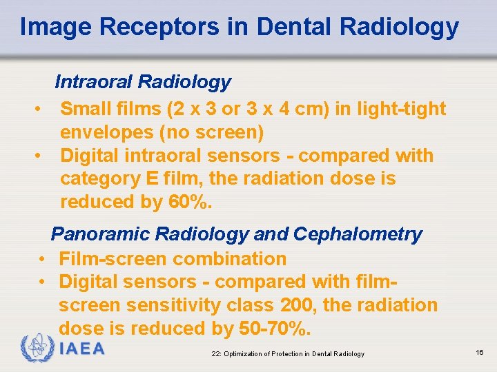
Image Receptors in Dental Radiology Intraoral Radiology • Small films (2 x 3 or 3 x 4 cm) in light-tight envelopes (no screen) • Digital intraoral sensors - compared with category E film, the radiation dose is reduced by 60%. Panoramic Radiology and Cephalometry • Film-screen combination • Digital sensors - compared with filmscreen sensitivity class 200, the radiation dose is reduced by 50 -70%. IAEA 22: Optimization of Protection in Dental Radiology 16
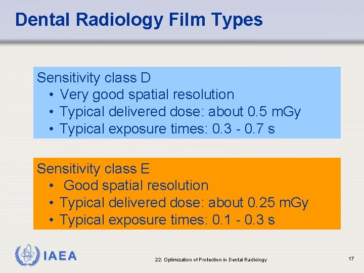
Dental Radiology Film Types Sensitivity class D • Very good spatial resolution • Typical delivered dose: about 0. 5 m. Gy • Typical exposure times: 0. 3 - 0. 7 s Sensitivity class E • Good spatial resolution • Typical delivered dose: about 0. 25 m. Gy • Typical exposure times: 0. 1 - 0. 3 s IAEA 22: Optimization of Protection in Dental Radiology 17
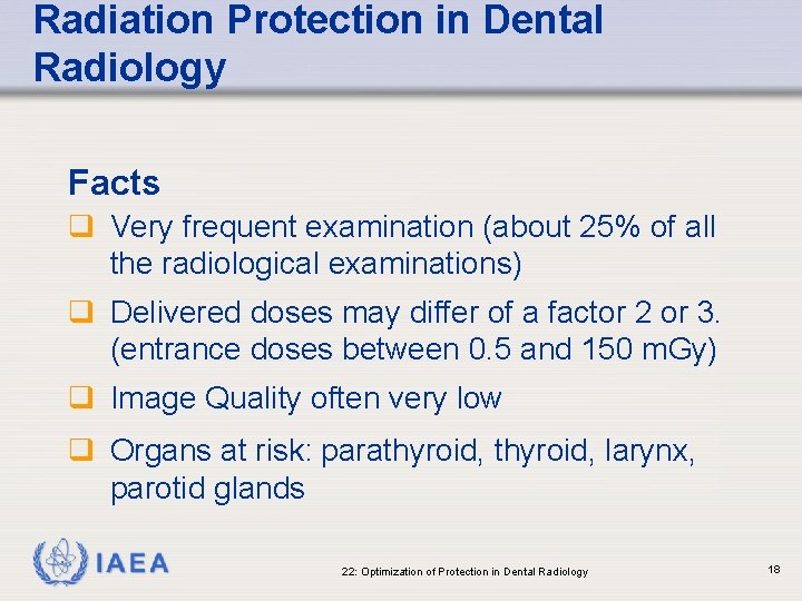
Radiation Protection in Dental Radiology Facts q Very frequent examination (about 25% of all the radiological examinations) q Delivered doses may differ of a factor 2 or 3. (entrance doses between 0. 5 and 150 m. Gy) q Image Quality often very low q Organs at risk: parathyroid, larynx, parotid glands IAEA 22: Optimization of Protection in Dental Radiology 18
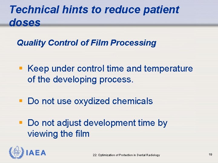
Technical hints to reduce patient doses Quality Control of Film Processing § Keep under control time and temperature of the developing process. § Do not use oxydized chemicals § Do not adjust development time by viewing the film IAEA 22: Optimization of Protection in Dental Radiology 19
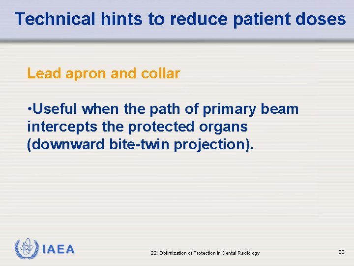
Technical hints to reduce patient doses Lead apron and collar • Useful when the path of primary beam intercepts the protected organs (downward bite-twin projection). IAEA 22: Optimization of Protection in Dental Radiology 20
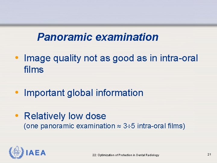
Panoramic examination • Image quality not as good as in intra-oral films • Important global information • Relatively low dose (one panoramic examination 3 5 intra-oral films) IAEA 22: Optimization of Protection in Dental Radiology 21
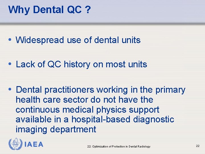
Why Dental QC ? • Widespread use of dental units • Lack of QC history on most units • Dental practitioners working in the primary health care sector do not have the continuous medical physics support available in a hospital-based diagnostic imaging department IAEA 22: Optimization of Protection in Dental Radiology 22
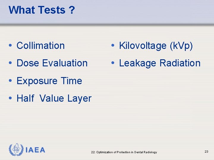
What Tests ? • Collimation • Kilovoltage (k. Vp) • Dose Evaluation • Leakage Radiation • Exposure Time • Half Value Layer IAEA 22: Optimization of Protection in Dental Radiology 23
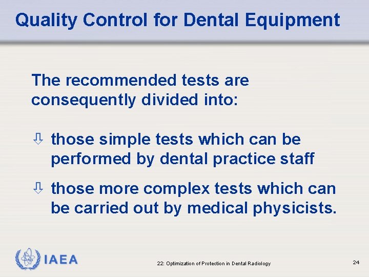
Quality Control for Dental Equipment The recommended tests are consequently divided into: ò those simple tests which can be performed by dental practice staff ò those more complex tests which can be carried out by medical physicists. IAEA 22: Optimization of Protection in Dental Radiology 24
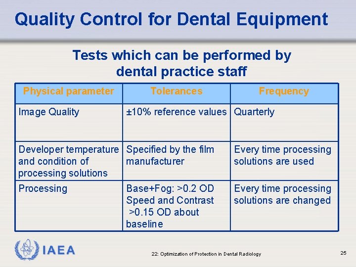
Quality Control for Dental Equipment Tests which can be performed by dental practice staff Physical parameter Image Quality Tolerances Frequency ± 10% reference values Quarterly Developer temperature Specified by the film and condition of manufacturer processing solutions Every time processing solutions are used Processing Every time processing solutions are changed IAEA Base+Fog: >0. 2 OD Speed and Contrast >0. 15 OD about baseline 22: Optimization of Protection in Dental Radiology 25
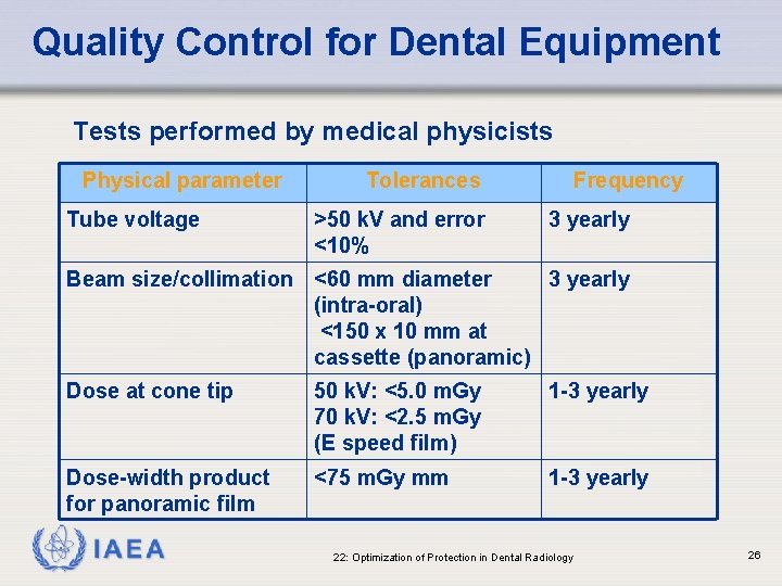
Quality Control for Dental Equipment Tests performed by medical physicists Physical parameter Tube voltage Tolerances >50 k. V and error <10% Frequency 3 yearly Beam size/collimation <60 mm diameter 3 yearly (intra-oral) <150 x 10 mm at cassette (panoramic) Dose at cone tip 50 k. V: <5. 0 m. Gy 70 k. V: <2. 5 m. Gy (E speed film) 1 -3 yearly Dose-width product for panoramic film <75 m. Gy mm 1 -3 yearly IAEA 22: Optimization of Protection in Dental Radiology 26
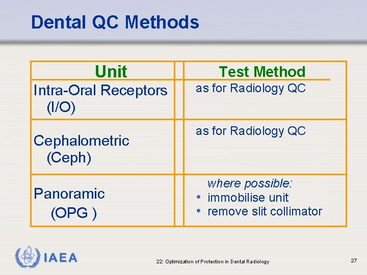
Dental QC Methods Unit Test Method Intra-Oral Receptors (I/O) Cephalometric (Ceph) Panoramic (OPG ) IAEA as for Radiology QC where possible: • immobilise unit • remove slit collimator 22: Optimization of Protection in Dental Radiology 27
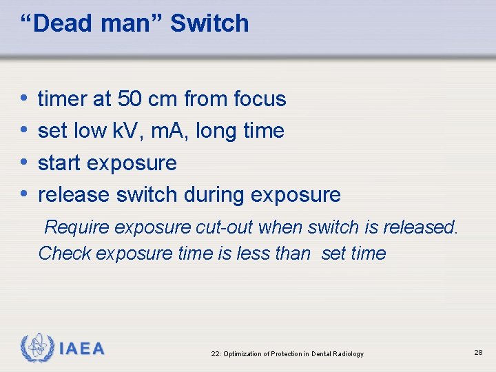
“Dead man” Switch • • timer at 50 cm from focus set low k. V, m. A, long time start exposure release switch during exposure Require exposure cut-out when switch is released. Check exposure time is less than set time IAEA 22: Optimization of Protection in Dental Radiology 28
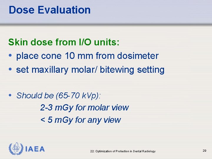
Dose Evaluation Skin dose from I/O units: • place cone 10 mm from dosimeter • set maxillary molar/ bitewing setting • Should be (65 -70 k. Vp): 2 -3 m. Gy for molar view < 5 m. Gy for any view IAEA 22: Optimization of Protection in Dental Radiology 29
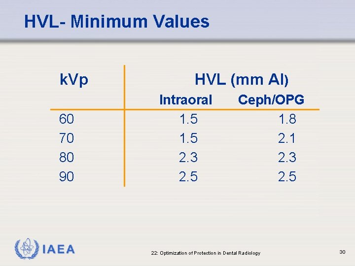
HVL- Minimum Values k. Vp 60 70 80 90 IAEA HVL (mm Al) Intraoral 1. 5 2. 3 2. 5 Ceph/OPG 1. 8 2. 1 2. 3 2. 5 22: Optimization of Protection in Dental Radiology 30
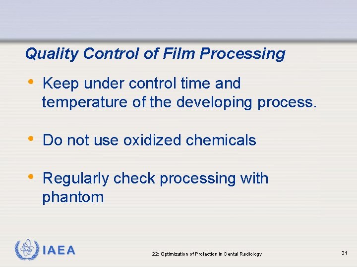
Quality Control of Film Processing • Keep under control time and temperature of the developing process. • Do not use oxidized chemicals • Regularly check processing with phantom IAEA 22: Optimization of Protection in Dental Radiology 31
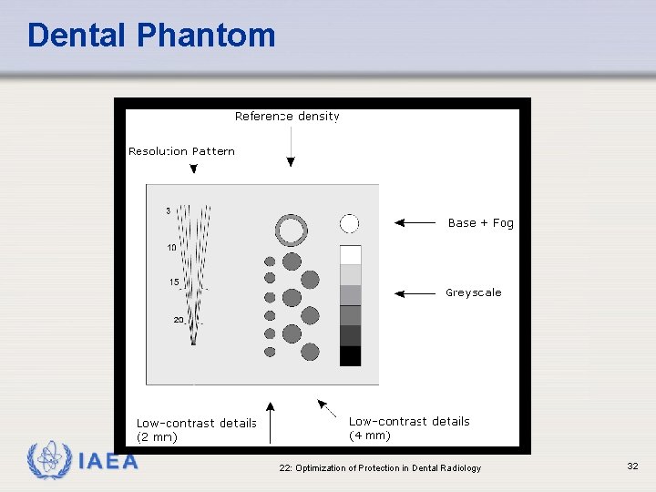
Dental Phantom IAEA 22: Optimization of Protection in Dental Radiology 32
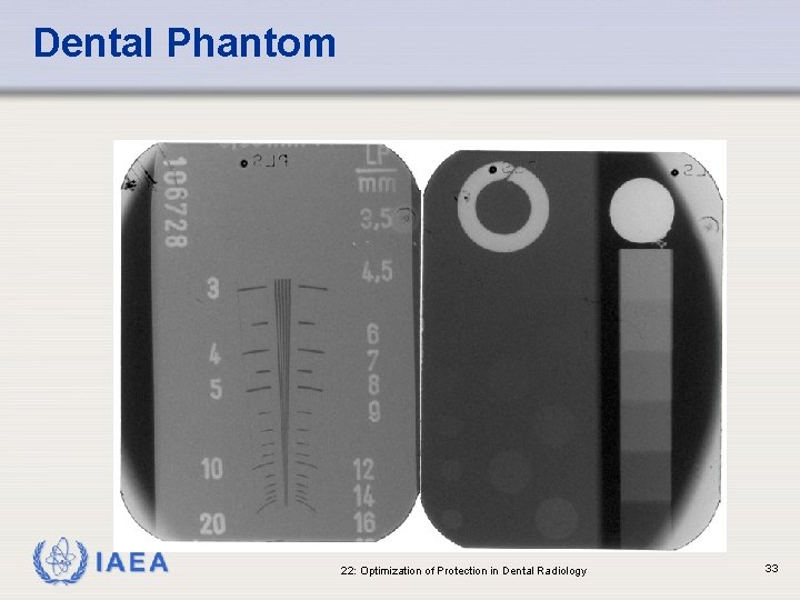
Dental Phantom IAEA 22: Optimization of Protection in Dental Radiology 33
Summary • Although doses are generally low, the high frequency of examinations requires radiation protection (for the practitioner) in dental radiology • Some tests are detailed for Quality control of dental equipment. IAEA 22: Optimization of Protection in Dental Radiology 34
THANK YOU! IAEA 22: Optimization of Protection in Dental Radiology 35