I Membrane Structure a hallmark of eukaryotic cells

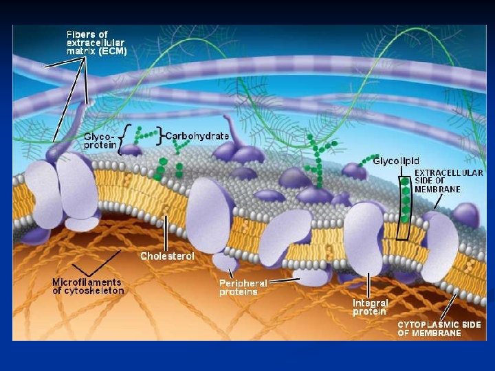
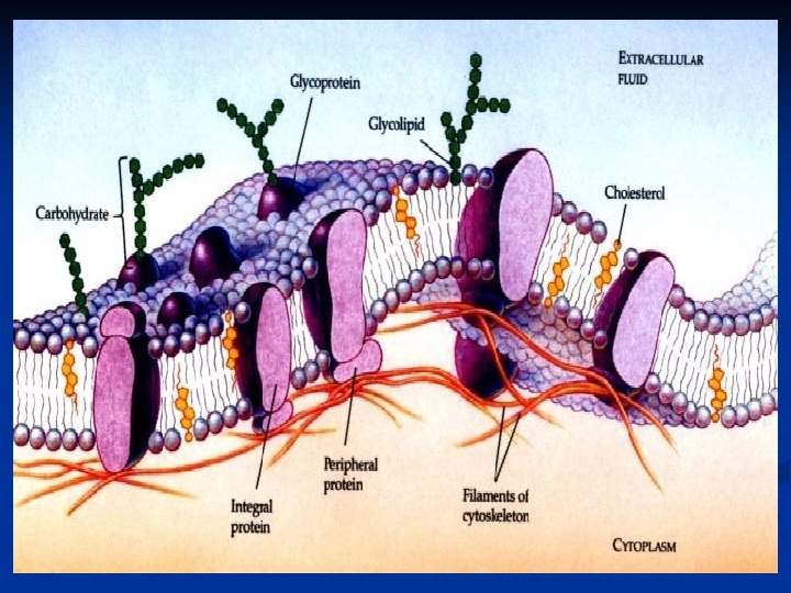
I. Membrane Structure a hallmark of eukaryotic cells is the abundance of membranes. Their ability to act as a barrier is the fact that it is selectively-permeable - some material passes through more readily than others Fluid-Mosaic Model -- Membranes are composed of phospholipids w/ various proteins & carbohydrates embedded w/in. These proteins have the ability to move laterally from side-to-side.

A. Membrane lipids 1. the primary structure of membranes is a phospholipid bilayer hydrophilic head -- “water-loving” -- phosphate group -- polar hydrophobic tail -- “water-fearing” -- fatty acids -- nonpolar

Polar Water

The membrane has 2 layers: 1. The polar heads face the outside watery env’t 2. The nonpolar tails make-up the interior (WHY IS THIS IMPORTANT? ) v nonpolar molecules (e. g. O 2 and CO 2 )can dissolve in lipids & pass thru v Large polar molecules (proteins, ions, carbohydrates) are not compatible and don’t move across (must use proteins)

2. Cholesterol -- another lipid component that gives the membrane strength, but reduces its fluidity

NOTE: Lipid composition can change from cell to cell which affects membrane fluidity. Shorter fatty acid chains make the membrane more fluid. This can help counteract changes in temp. or concentration & regulate transport across. What type of fatty acids do you need to have a more fluid membrane, saturated or unsaturated? Unsaturated – double bonds cause folding

B. Membrane Proteins 1. Fall into two classes: a. Integral – embedded in the bilayer; many extend all the way from one surface to the other b. Peripheral – entirely outside the bilayer; found on either surface attached to integral proteins or the hydrophilic heads

2. Due to the fluidity of the membrane, proteins are able to move laterally across the membrane.

3. Every membrane has different proteins which is dependent on that membranes highly specific FXN. There are 3 main categories of membrane protein: a. Transport – regulate the mov’t of most hydrophilic substances through the membrane (ex. channel & carrier proteins)

b. Receptor – have a binding site that allow only specifically shaped molecules to attach. The binding triggers cellular response Ex. Liver cells bind insulin which triggers the breakdown of glucose

c. Recognition – used to identify specific cell types. Allows cells to recognize when under attack by pathogens

C. Membrane Carbohydrates -- attach to lipids or proteins on outer surface of cell 1. glycolipid – recognize other cells 2. glycoproteins – recognize foreign substances

II. Transport Across the Membrane The phospholipid bilayer is selectively permeable due to its hydrophilic & hydrophobic regions v THUS, nonpolar molecules (e. g. O 2 and CO 2 ) can dissolve in lipids & pass thru the hydrophobic interior v Large polar molecules (proteins, ions, carbohydrates) are not compatible and do not move through membrane (must use proteins)

Membrane transport involves the behavior of particles in a solution 1. solute – The solid material that is dissolved 2. solvent –The liquid material that dissolves the solid Biological systems exist in aqueous env’ts where water is the main solvent

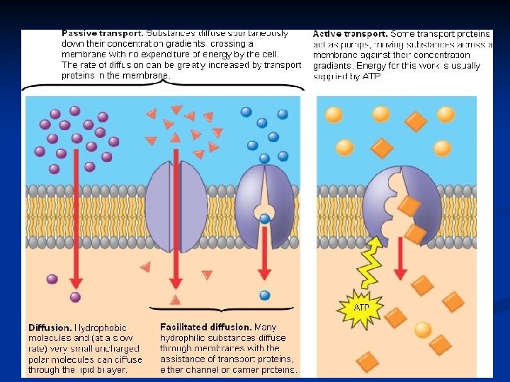
A. Passive Transport Mov’t that does NOT require NRG Concentration Gradient -- Particles move “down/with” the gradient (i. e. go from high low) -- Eventually reach equilibrium; there is still a continuous mov’t of particles, but no “net” mov’t

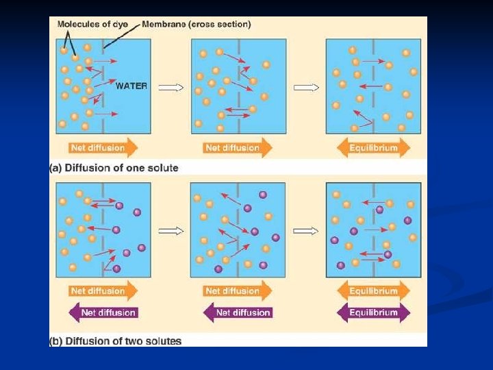
1. Simple Diffusion a. the random mov’t of small, nonpolar particles toward a state of even distribution (i. e. equilibrium b. movt’ of particles from an area of high concen. to and area of low concen. (ex. ammonia in room)

REMEMBER: polar molecules are NOT able to diffus across the membrane, why? B/C they can’t pass thru nonpolar, hydrophobic tails

c. Factors that affect Diffusion Rate 1. Particles move with the gradient, thus the steeper the grade from H L the more rapid the diffusion 2. As temp increases, rate inc. (particles moving faste 3. As pressure inc. , rate inc. (molecules keep bouncin into each other)
 or too large (e. g.")
2. Facilitated Diffusion some compounds are too hydrophilic (polar) or too large (e. g. sugars/amino acids) to pass through the membrane on their own. These compounds use intrinsic transport proteins to travel through. there are two types of transport proteins…

a. Channel proteins -- small pores through which charged ions can pass through the lipid bilayer -- there are specific channels for Na+, K+, Ca 2+, Cl-

b. Carrier proteins -- have a specific shape so can only bind specific compounds. -- when correct compounds binds, protein undergoes a shape change and carries particle across

http: //highered. mcgrawhill. com/sites/0072495855/student_view 0/cha pter 2/animation__how_facilitated_diffusion_ works. html
 through a semi-permeable membrane Also")
3. The diffusion of a solvent (i. e. water) through a semi-permeable membrane Also from an area of high low concentration

http: //highered. mcgrawhill. com/sites/0072495855/student_vi ew 0/chapter 2/animation__how_osmo sis_works. html

QUESTION 1: Can water “simply diffuse” through the cell’s plasma membrane? ANSWER: No! Water is POLAR and don’t get along with NONPOLAR fatty acid tails of phospholipid bilayer
")
FOLLOW-UP: So, how does water get through the plasma membrane? ANSWER: Protein Channels!!! (aquaporins)

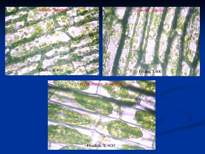
Tonicity – a term that describes the type of solution (dependent upon the concentration of dissolved solute particles). Three types of solutions: 1. Isotonic Solution (iso means equal) - concen. of solute is EQUAL on both sides
 - concen. of solute outside is LESS than")
2. Hypotonic solution (hypo means less) - concen. of solute outside is LESS than inside - CELLS SWELL -This creates turgor pressure in plant (build up of water; rigidity). -Animal cells will eventually burst b/c no cell wall to w/stand pressure
 - concen. of solute outside cell is GREATER")
3. Hypertonic solution (hyper means greater) - concen. of solute outside cell is GREATER than inside (e. g. ocean water; long showers) - CELLS SHRIVLE

ISOTONIC HYPOTONIC CELL SWELLS CYTOLYSIS HYPERTONIC CELL SHRINKS CRENATION PLASMOLYSIS

ISOTONIC HYPERTONIC

HYPOTONIC HYPERTONIC ISOTONIC
")
B. Active Transport Mov’t that REQUIRES NRG particles move AGAINST concen. gradient (low high) ex. glucose from blood into liver It is similar to facilitated diffusion in that it uses carrier proteins that act like “pumps” to transport material through membrane

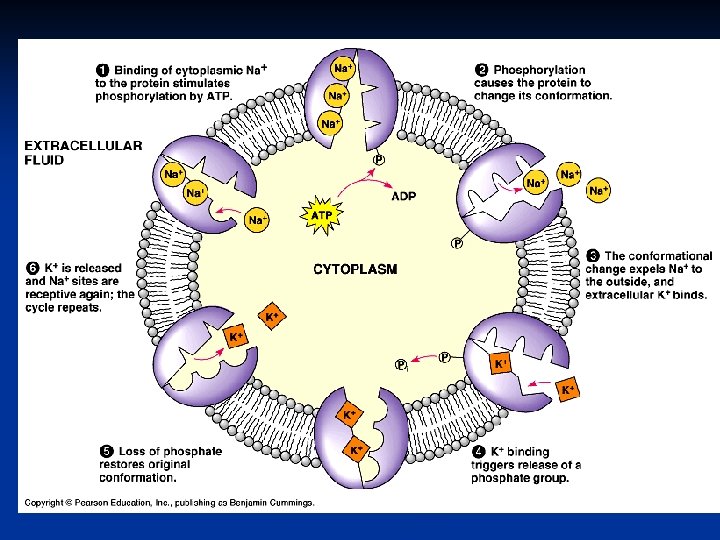
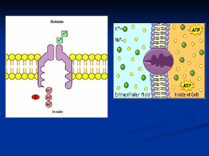
1. Na+/K+ Pump a. used in nerve cell impulses & muscle cell contractio b. needed to keep an electrochemical balance c. background: -The inside of every cell has high concen. of K+ -The outside of cells (i. e. blood) high concen. Na+ http: //highered. mcgrawhill. com/olcweb/cgi/pluginpop. cgi? it=swf: : 53 5: : 535: : /sites/dl/free/0072437316/120068/bio 0 3. swf: : Sodium. Potassium%20 Exchange%20 Pump

1. 2. 6. 3. 5. 4.
 a. Endocytosis – particles are brought into")
2. Bulk Transport (transport of large compounds) a. Endocytosis – particles are brought into the cell through a vesicle that pinches off membrane 1. phagocytosis – large particles; “eating” 2. pinocytosis – small dissolved particles; 3. “drinking”

b. Exocytosis – particles are released from the cell
 B. Facilitated Diffusion")
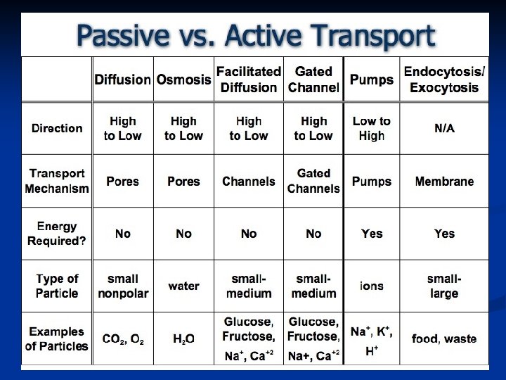
Transport Summary I. Passive Transport A. Simple Diffusion (mov’t of solute) B. Facilitated Diffusion (mov’t of solute) - uses channel/carrier proteins C. Osmosis (mov’t of solvent water) II. Active Transport A. Pumps (Na+/K+) B. Bulk Transport 1. Endocytosis a. pinocytosis b. phagocytosis 2. Exocytosis

III. Where Cells Meet Plasma membranes of adjacent cells sometimes touch, but usually there’s a space b/n the membranes. What occupies that space? A. Extracellular Matrix (ECM) 1. a meshwork of polysaccharides & proteins a. collagen – strength b. fibronectin & laminin – cell differentiation, heal wounds c. proteoglycan – regulate mov’t thru matrix 2. major FXN: to hold cells together as tissues

CYTOPLASM

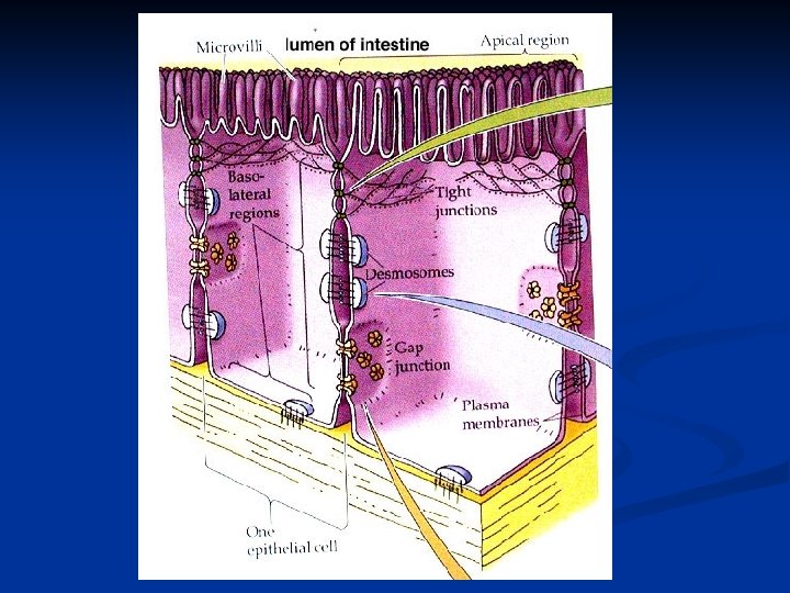
B. Junctions When cells come into direct contact, they use three binding methods: 1. Tight junctions (barriers) a. no space b/n cells b. plasma mem. of each cell will fuse together forming a barrier preventing mov’t of unwanted material Ex: intestines (waste)
 a. unlike tight junctions, these cells just tightly adher to one")
2. Desmosomes (adhere) a. unlike tight junctions, these cells just tightly adher to one another (special cytoplasmic plaques) b. help hold cells together that are often stretched (ex. heart, stomach, bladder)
 a. allow nerve impulses to pass from one cell to")
3. Gap junctions (communication) a. allow nerve impulses to pass from one cell to next b. don’t have to travel thru plasma mem. Gap
- Slides: 53