
HYPOXIC ISCHEMIC ENCEPHALOPATHY IAP UG Teaching slides 2015


- Slides: 78
HYPOXIC ISCHEMIC ENCEPHALOPATHY IAP UG Teaching slides 2015 -16 1
DEFINITION OF HIE • Hypoxic-ischemic encephalopathy – Clinical and laboratory evidence of acute or subacute brain injury due to perinatal asphyxia (ie, hypoxia, acidosis) – Most often, underlying cause unknown – Exact time of brain injury often uncertain – Abnormal brain (e. g. growth failure, impaired development) might be underlying risk factor IAP UG Teaching slides 2015 -16 2
DEFINITION: PERINATAL ASPHYXIA • Insult to the fetus or the newborn due to – lack of oxygen (hypoxia) and / or – a lack of perfusion (ischemia) to various organs • Asphyxia occurs when – organ of gas exchange fails • Those that develop hypoxic ischemic encephalopathy – Mental retardation, cerebral palsy, coordination disorders etc IAP UG Teaching slides 2015 -16 3
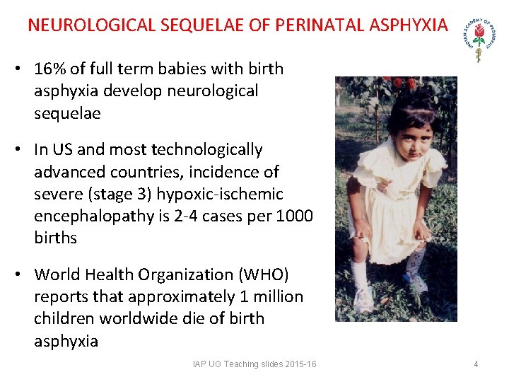
NEUROLOGICAL SEQUELAE OF PERINATAL ASPHYXIA • 16% of full term babies with birth asphyxia develop neurological sequelae • In US and most technologically advanced countries, incidence of severe (stage 3) hypoxic-ischemic encephalopathy is 2 -4 cases per 1000 births • World Health Organization (WHO) reports that approximately 1 million children worldwide die of birth asphyxia IAP UG Teaching slides 2015 -16 4
MORTALITY & MORBIDITY • In severe HIE, mortality rate - 50 -75%. • Most deaths (55%) occur in first week of life due to multiple organ failure or redirection of care. • Among infants who survive severe HIE, sequelae include mental retardation, epilepsy, and cerebral palsy of varying degrees. • CP - hemiplegia, paraplegia, or quadriplegia IAP UG Teaching slides 2015 -16 5
MORTALITY & MORBIDITY • Among the infants who survive moderately severe HIE, 30 -50% may have serious long-term complications, and 10 -20% have minor neurological morbidities. • Infants with mild HIE tend to be free from serious CNS complications. • Even in absence of obvious neurologic deficits in newborn period, long-term functional impairments may be present. IAP UG Teaching slides 2015 -16 6
ANATOMICAL ASPECTS IAP UG Teaching slides 2015 -16 7
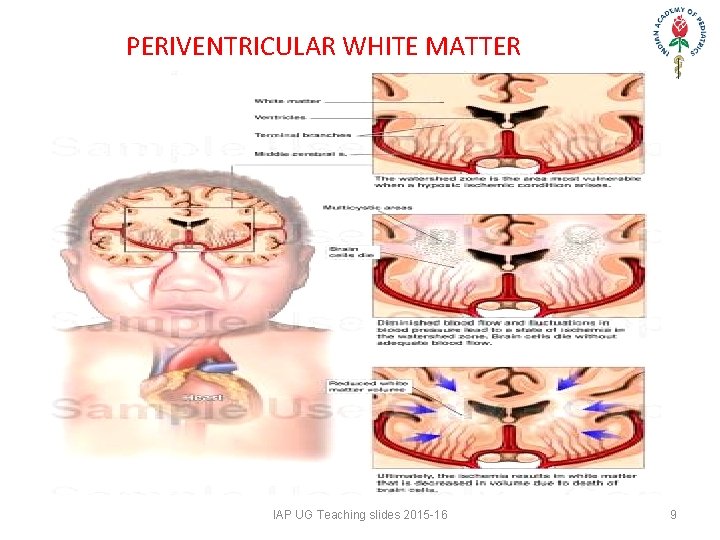
PECULIARITIES OF THE NEONATAL BRAIN More susceptible to injury because: • Watershed areas of blood supply • Specific areas where maximal development is occurring are more susceptible • Asphyxia at birth > hypoxemia, hypercarbia > blood redistribution to brain, heart and adrenals from GIT, liver, lungs and kidneys > if persists > hypotension > cerebral ischemia. Watershed areas • FT babies - parasagittal cerebral cortex • PT babies - periventricular white matter. IAP UG Teaching slides 2015 -16 8
PERIVENTRICULAR WHITE MATTER IAP UG Teaching slides 2015 -16 9
THE GERMINAL MATRIX • Subependymal • Overlies head of caudate nucleus • Huebner artery, MCA, ICA • Contains neurons • Supported by glial cells, fragile vasculature in a stroma • Most prominent between 24 to 34 wk POG • Disappears around 34 -36 wk POG • Most cases hemorrhage in groove between thalamus and head of caudate nucleus IAP UG Teaching slides 2015 -16 10
WHY IS IT VULNERABLE? • Vascular – Immature capillaries • Abrupt termination of tunica media proximal to germinal matrix • Poor perivascular support – Watershed area • Intravascular – Sudden surges of BP, Poor cerebral autoregulation – Relatively large volume of blood flow – Acute angle of internal cerebral vein • Extravascular – Physiologic coagulopathy in NB – Fibrinolytic enzymes in germinal matrix IAP UG Teaching slides 2015 -16 11
EVENTS DURING PERINATAL ASPHYXIA IAP UG Teaching slides 2015 -16 12
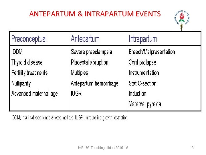
ANTEPARTUM & INTRAPARTUM EVENTS IAP UG Teaching slides 2015 -16 13
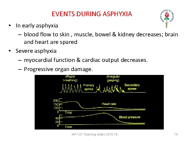
EVENTS DURING ASPHYXIA • In early asphyxia – blood flow to skin , muscle, bowel & kidney decreases; brain and heart are spared • Severe asphyxia – myocardial function & cardiac output decreases. – Progressive organ damage. IAP UG Teaching slides 2015 -16 14
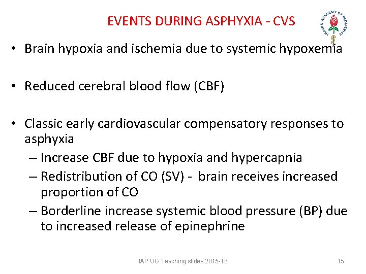
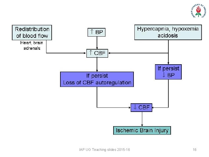
EVENTS DURING ASPHYXIA - CVS • Brain hypoxia and ischemia due to systemic hypoxemia • Reduced cerebral blood flow (CBF) • Classic early cardiovascular compensatory responses to asphyxia – Increase CBF due to hypoxia and hypercapnia – Redistribution of CO (SV) - brain receives increased proportion of CO – Borderline increase systemic blood pressure (BP) due to increased release of epinephrine IAP UG Teaching slides 2015 -16 15
IAP UG Teaching slides 2015 -16 16
EVENTS DURING ASPHYXIA - CVS • Limited data on the preterm infant suggest that CBF is stable over a range of BPs. • BP range at which CBF autoregulation is maintained is quite narrow (perhaps between 10 -20 mm Hg, compared with the 40 mm Hg range in adults) • The precise upper and lower limits of the BP values above and below which the CBF auto regulation is lost remains unknown for human newborn. • In the fetus and newborn suffering from acute asphyxia, CBF can become pressure-passive IAP UG Teaching slides 2015 -16 17
EVENTS DURING ASPHYXIA - CVS • As BP falls, CBF falls below critical levels, and brain suffers from diminished blood supply and lack of sufficient oxygen to meet its needs. • This leads to intracellular energy failure. • During early phases of brain injury, brain temperature drops, and local release of neurotransmitters • These changes reduce cerebral oxygen demand, transiently minimizing the impact of asphyxia. IAP UG Teaching slides 2015 -16 18
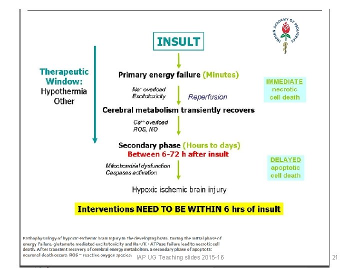
EVENTS DURING ASPHYXIA - CELLULAR • At the cellular level, neuronal injury in hypoxic-ischemic encephalopathy is an evolving process. • The magnitude of final neuronal damage depends on – Severity of initial insult – Damage due to energy failure, reperfusion injury, and apoptosis • Extent, nature, severity, and duration of primary injury important in affecting magnitude of residual neurological damage. • Following initial phase of energy failure, cerebral metabolism may recover, only to deteriorate in secondary phase, or reperfusion IAP UG Teaching slides 2015 -16 19
EVENTS DURING ASPHYXIA - CELLULAR • This new phase of neuronal damage • Starts at about 6 -24 hours after initial injury • Characterized by cerebral edema and apoptosis • “Delayed phase of neuronal injury. " • Duration of delayed phase is not precisely known • In human fetus and newborn appears to increase over first 24 -48 hours and then start to resolve thereafter IAP UG Teaching slides 2015 -16 20
IAP UG Teaching slides 2015 -16 21
PATHOLOGY OF HIE IAP UG Teaching slides 2015 -16 22
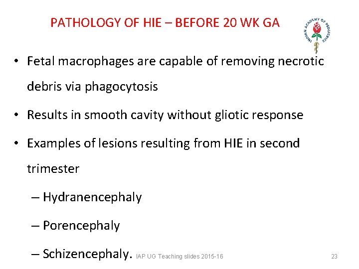
PATHOLOGY OF HIE – BEFORE 20 WK GA • Fetal macrophages are capable of removing necrotic debris via phagocytosis • Results in smooth cavity without gliotic response • Examples of lesions resulting from HIE in second trimester – Hydranencephaly – Porencephaly – Schizencephaly. IAP UG Teaching slides 2015 -16 23
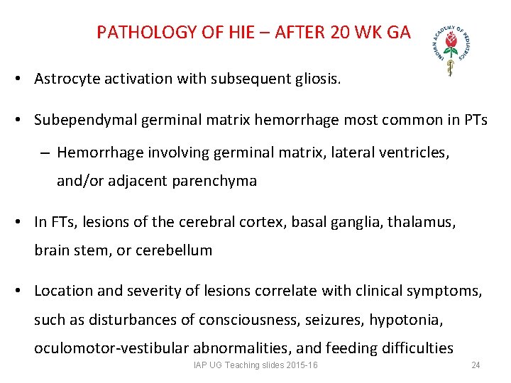
PATHOLOGY OF HIE – AFTER 20 WK GA • Astrocyte activation with subsequent gliosis. • Subependymal germinal matrix hemorrhage most common in PTs – Hemorrhage involving germinal matrix, lateral ventricles, and/or adjacent parenchyma • In FTs, lesions of the cerebral cortex, basal ganglia, thalamus, brain stem, or cerebellum • Location and severity of lesions correlate with clinical symptoms, such as disturbances of consciousness, seizures, hypotonia, oculomotor-vestibular abnormalities, and feeding difficulties IAP UG Teaching slides 2015 -16 24
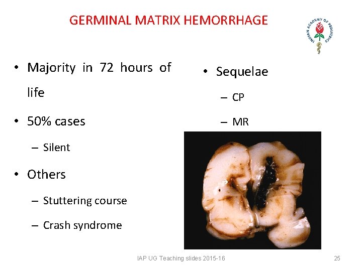
GERMINAL MATRIX HEMORRHAGE • Majority in 72 hours of life • 50% cases • Sequelae – CP – MR – Silent • Others – Stuttering course – Crash syndrome IAP UG Teaching slides 2015 -16 25
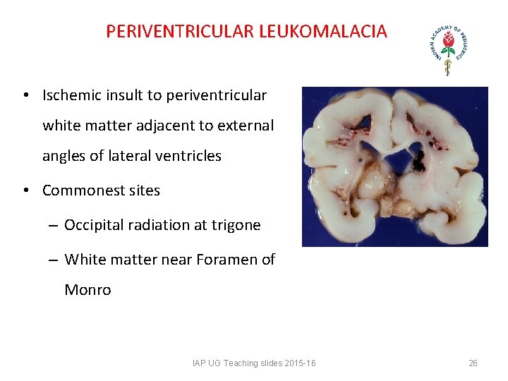
PERIVENTRICULAR LEUKOMALACIA • Ischemic insult to periventricular white matter adjacent to external angles of lateral ventricles • Commonest sites – Occipital radiation at trigone – White matter near Foramen of Monro IAP UG Teaching slides 2015 -16 26
SUBARACHNOID HEMORRHAGE • Cause – Primary - birth trauma – Secondary - extensions of subdural, intraventricular or intracerebellar bleeds • Clinically – Silent – Irritability, seizures, focal deficits, and a bulging AF and pallor • Management depends on – Extent – Clinical features • Sequelae ~ 1* / 2*, extent IAP UG Teaching slides 2015 -16 27
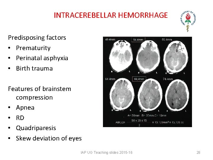
INTRACEREBELLAR HEMORRHAGE Predisposing factors • Prematurity • Perinatal asphyxia • Birth trauma Features of brainstem compression • Apnea • RD • Quadriparesis • Skew deviation of eyes IAP UG Teaching slides 2015 -16 28
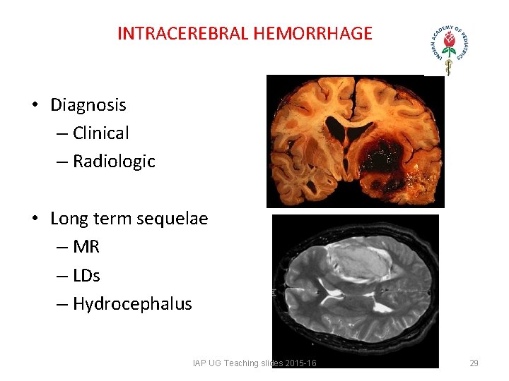
INTRACEREBRAL HEMORRHAGE • Diagnosis – Clinical – Radiologic • Long term sequelae – MR – LDs – Hydrocephalus IAP UG Teaching slides 2015 -16 29
PATTERNS OF INJURY Selective neuronal necrosis • Most common pattern of injury • Neuronal necrosis selective to areas with higher energy demands. • 5 major patterns • Diffuse neuronal necrosis - cerebral cortex (particularly the hippocampus), deep nuclear structures (thalamus, basal ganglia), brain stem, cerebellum, and anterior horn of spinal cord. • Cerebral cortex (deep nuclear) in 35 -85% of infants IAP UG Teaching slides 2015 -16 30
PATTERN OF INJURY Parasagittal cerebral injury • Typically bilateral and involves parasagittal areas of cerebral cortex • The regions of the cortex most susceptible are the end-artery zones between anterior, middle, and posterior cerebral arteries - watershed regions • Parieto-occipital cortex most susceptible • Parasagittal cerebral injury most commonly seen in FTs. • Most lesions are ischemic, approximately 25% are hemorrhagic IAP UG Teaching slides 2015 -16 31
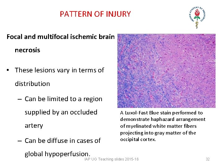
PATTERN OF INJURY Focal and multifocal ischemic brain necrosis • These lesions vary in terms of distribution – Can be limited to a region supplied by an occluded artery – Can be diffuse in cases of global hypoperfusion. A Luxol-Fast Blue stain performed to demonstrate haphazard arrangement of myelinated white matter fibers projecting into gray matter of the occipital cortex. IAP UG Teaching slides 2015 -16 32
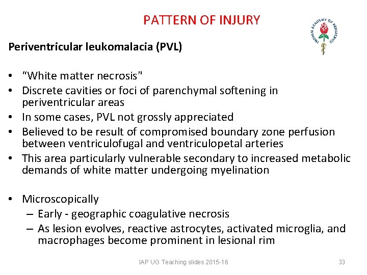
PATTERN OF INJURY Periventricular leukomalacia (PVL) • “White matter necrosis" • Discrete cavities or foci of parenchymal softening in periventricular areas • In some cases, PVL not grossly appreciated • Believed to be result of compromised boundary zone perfusion between ventriculofugal and ventriculopetal arteries • This area particularly vulnerable secondary to increased metabolic demands of white matter undergoing myelination • Microscopically – Early - geographic coagulative necrosis – As lesion evolves, reactive astrocytes, activated microglia, and macrophages become prominent in lesional rim IAP UG Teaching slides 2015 -16 33
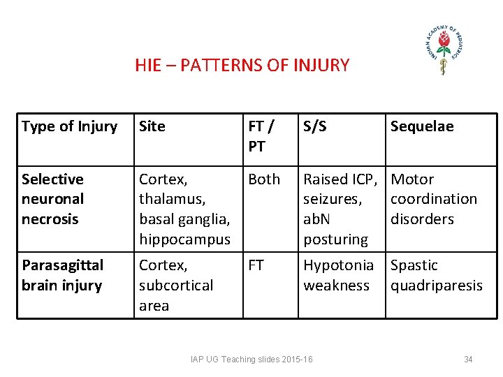
HIE – PATTERNS OF INJURY Type of Injury Site FT / PT Selective neuronal necrosis Cortex, Both thalamus, basal ganglia, hippocampus Raised ICP, Motor seizures, coordination ab. N disorders posturing Parasagittal brain injury Cortex, subcortical area Hypotonia Spastic weakness quadriparesis FT S/S IAP UG Teaching slides 2015 -16 Sequelae 34
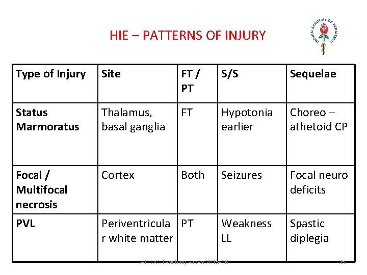
HIE – PATTERNS OF INJURY Type of Injury Site FT / PT S/S Sequelae Status Marmoratus Thalamus, basal ganglia FT Hypotonia earlier Choreo – athetoid CP Focal / Multifocal necrosis Cortex Both Seizures Focal neuro deficits PVL Periventricula PT r white matter Weakness LL Spastic diplegia IAP UG Teaching slides 2015 -16 35
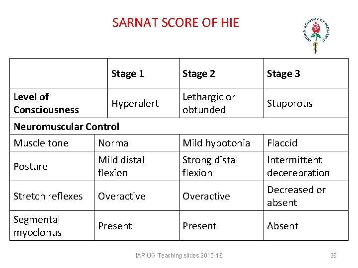
SARNAT SCORE OF HIE Level of Consciousness Stage 1 Stage 2 Stage 3 Hyperalert Lethargic or obtunded Stuporous Neuromuscular Control Muscle tone Normal Mild hypotonia Flaccid Posture Mild distal flexion Strong distal flexion Intermittent decerebration Stretch reflexes Overactive Decreased or absent Segmental myoclonus Present Absent IAP UG Teaching slides 2015 -16 36
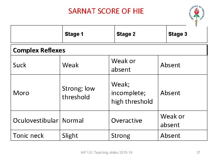
SARNAT SCORE OF HIE Stage 1 Stage 2 Stage 3 Complex Reflexes Suck Weak or absent Absent Moro Strong; low threshold Weak; incomplete; high threshold Absent Oculovestibular Normal Overactive Tonic neck Strong Slight IAP UG Teaching slides 2015 -16 Weak or absent Absent 37
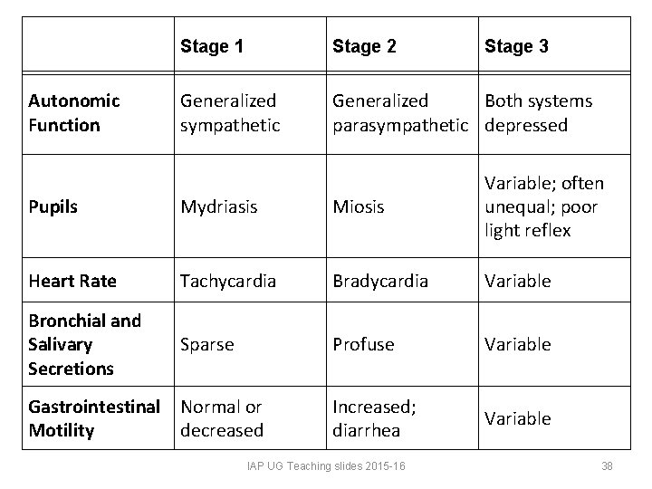
Autonomic Function Stage 1 Stage 2 Stage 3 Generalized sympathetic Generalized Both systems parasympathetic depressed Pupils Mydriasis Miosis Variable; often unequal; poor light reflex Heart Rate Tachycardia Bradycardia Variable Bronchial and Salivary Secretions Sparse Profuse Variable Increased; diarrhea Variable Gastrointestinal Normal or Motility decreased IAP UG Teaching slides 2015 -16 38
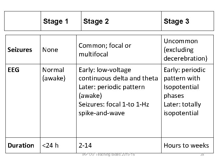
Stage 1 Stage 2 Stage 3 Common; focal or multifocal Uncommon (excluding decerebration) Seizures None EEG Normal (awake) Early: low-voltage continuous delta and theta Later: periodic pattern (awake) Seizures: focal 1 -to 1 -Hz spike-and-wave Early: periodic pattern with Isopotential phases Later: totally isopotential Duration <24 h 2 -14 Hours to weeks IAP UG Teaching slides 2015 -16 39
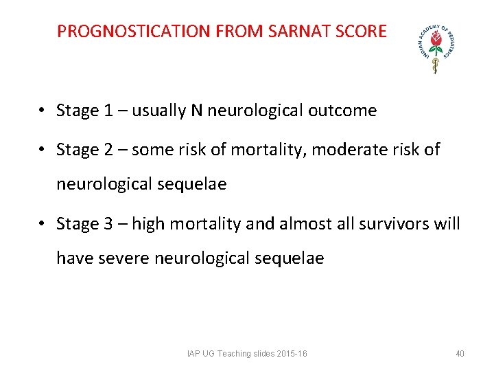
PROGNOSTICATION FROM SARNAT SCORE • Stage 1 – usually N neurological outcome • Stage 2 – some risk of mortality, moderate risk of neurological sequelae • Stage 3 – high mortality and almost all survivors will have severe neurological sequelae IAP UG Teaching slides 2015 -16 40
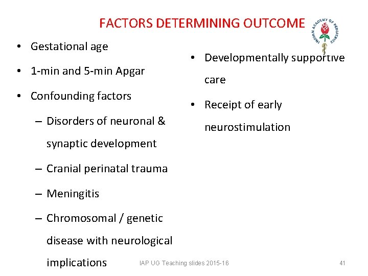
FACTORS DETERMINING OUTCOME • Gestational age • 1 -min and 5 -min Apgar • Confounding factors • Developmentally supportive care • Receipt of early – Disorders of neuronal & neurostimulation synaptic development – Cranial perinatal trauma – Meningitis – Chromosomal / genetic disease with neurological implications IAP UG Teaching slides 2015 -16 41
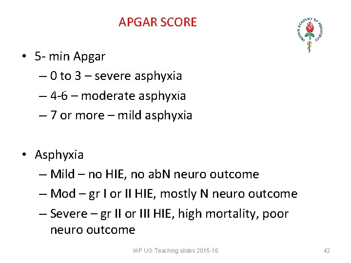
APGAR SCORE • 5 - min Apgar – 0 to 3 – severe asphyxia – 4 -6 – moderate asphyxia – 7 or more – mild asphyxia • Asphyxia – Mild – no HIE, no ab. N neuro outcome – Mod – gr I or II HIE, mostly N neuro outcome – Severe – gr II or III HIE, high mortality, poor neuro outcome IAP UG Teaching slides 2015 -16 42
HIE Investigations & Treatment IAP UG Teaching slides 2015 -16 43
CHOOSING APPROPRIATE INVESTIGATIONS IAP UG Teaching slides 2015 -16 44
LAB STUDIES • Serum electrolytes – In severe cases, daily assessment of serum electrolytes are valuable until the infant's status improves. – Markedly low serum sodium, potassium, and chloride levels in the presence of reduced urine flow and excessive weight gain may indicate acute tubular damage or inappropriate antidiuretic hormone (IADH), particularly during the initial 23 days of life. • Renal function studies: Serum creatinine levels, creatinine clearance, and BUN levels suffice in most cases. IAP UG Teaching slides 2015 -16 45
LAB STUDIES • Cardiac and liver enzymes: These values are an adjunct to assess the degree of hypoxic-ischemic injury to these other organs. • Coagulation system evaluation: This includes prothrombin time, partial thromboplastin time, and fibrinogen levels. • ABG: Blood gas monitoring is used to assess acid-base status and to avoid hyperoxia and hypoxia as well as hypercapnia and hypocapnia. IAP UG Teaching slides 2015 -16 46
LAB STUDIES • TORCH studies • Genetic studies • Chromosomal studies • Body fluids for IEMs IAP UG Teaching slides 2015 -16 47
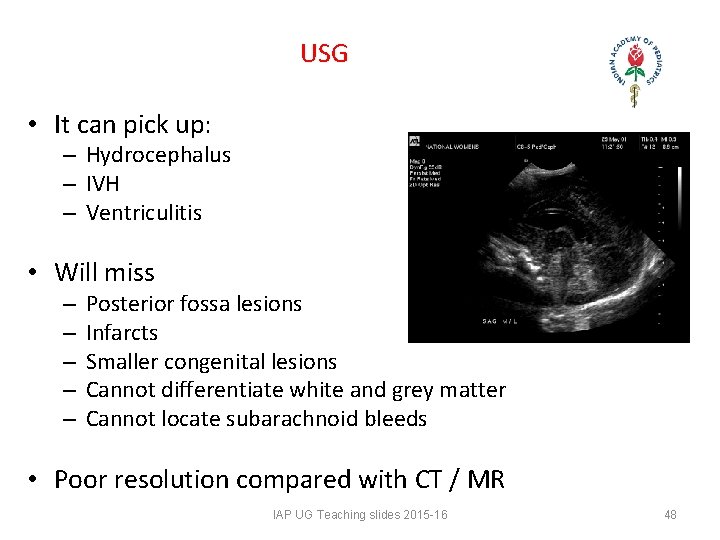
USG • It can pick up: – Hydrocephalus – IVH – Ventriculitis • Will miss – – – Posterior fossa lesions Infarcts Smaller congenital lesions Cannot differentiate white and grey matter Cannot locate subarachnoid bleeds • Poor resolution compared with CT / MR IAP UG Teaching slides 2015 -16 48
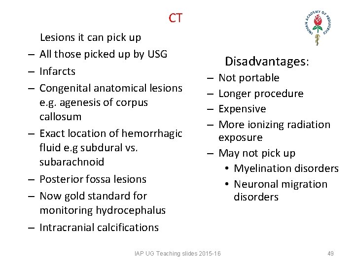
CT Lesions it can pick up – All those picked up by USG – Infarcts – Congenital anatomical lesions e. g. agenesis of corpus callosum – Exact location of hemorrhagic fluid e. g subdural vs. subarachnoid – Posterior fossa lesions – Now gold standard for monitoring hydrocephalus – Intracranial calcifications Disadvantages: Not portable Longer procedure Expensive More ionizing radiation exposure – May not pick up • Myelination disorders • Neuronal migration disorders – – IAP UG Teaching slides 2015 -16 49
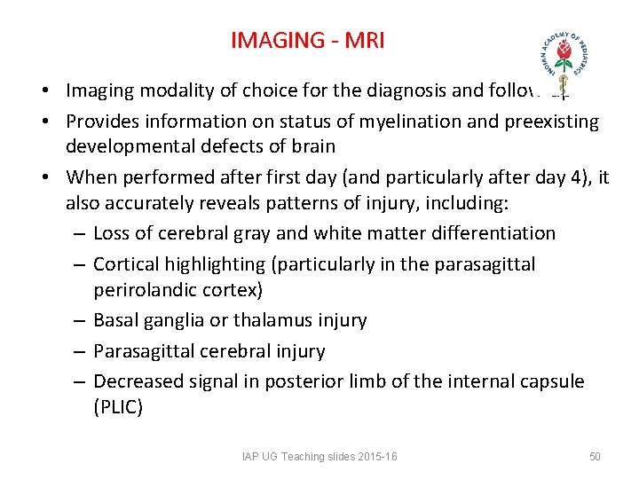
IMAGING - MRI • Imaging modality of choice for the diagnosis and follow-up • Provides information on status of myelination and preexisting developmental defects of brain • When performed after first day (and particularly after day 4), it also accurately reveals patterns of injury, including: – Loss of cerebral gray and white matter differentiation – Cortical highlighting (particularly in the parasagittal perirolandic cortex) – Basal ganglia or thalamus injury – Parasagittal cerebral injury – Decreased signal in posterior limb of the internal capsule (PLIC) IAP UG Teaching slides 2015 -16 50
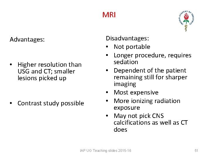
MRI Advantages: • Higher resolution than USG and CT; smaller lesions picked up • Contrast study possible Disadvantages: • Not portable • Longer procedure, requires sedation • Dependent of the patient remaining still for sharper imaging • Most expensive • More ionizing radiation exposure • May not pick CNS calcifications as well as CT does IAP UG Teaching slides 2015 -16 51
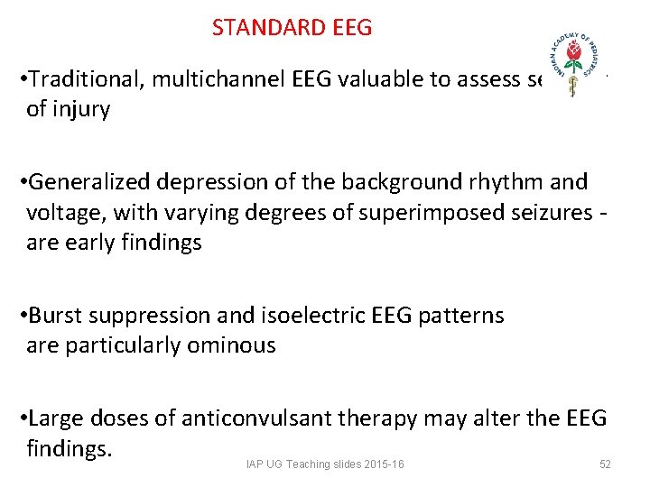
STANDARD EEG • Traditional, multichannel EEG valuable to assess severity of injury • Generalized depression of the background rhythm and voltage, with varying degrees of superimposed seizures - are early findings • Burst suppression and isoelectric EEG patterns are particularly ominous • Large doses of anticonvulsant therapy may alter the EEG findings. IAP UG Teaching slides 2015 -16 52
OTHER INVESTIGATIONS – CBF • Positron Emission Tomography • Specialized centers • Single Photon Emission • CBF Computed Tomography • Doppler Ultrasound • Cerebral infarcts, blocks and hemorrhages • MR Spectroscopy IAP UG Teaching slides 2015 -16 53
GENERAL COMMENTS • USG cannot differentiate between – Grey and white matter – Hge, infarct, hgic necrosis – CT needed • Locational correlation – Hyperdense areas inf and lat to ant horns > likely GMH – Hyperdense areas at trigone or post horns > likely PVL • Time scale correlation – Within 1 st few days > hge – After 1 wk > infarct • Ultimate radio pic – Resolution – Porencephalic cyst IAP UG Teaching slides 2015 -16 54
VISION & ROP IAP UG Teaching slides 2015 -16 55
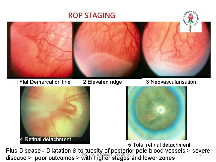
VISION - ROP • Mature retina- for 3 When to screen? months to 1 year • At 31 wks of postconceptional age or 3 -4 • Immature retina - every 2 weeks. wks after birth. (Whichever comes early). • Prethreshold ROP – every 3 rd day. How to screen? • Threshold ROP- Rx within 72 • Indirect ophthalmoscopy hrs. till retina matures. • Retinal detachment - early surgical Rx. IAP UG Teaching slides 2015 -16 56
ROP STAGING I Flat Demarcation line 4 Retinal detachment 2 Elevated ridge 3 Neovascularisation 5 Total retinal detachment Plus Disease - Dilatation & tortuosity of posterior pole blood vessels > severe IAP UG Teaching slides 2015 -16 57 disease > poor outcomes > with higher stages and lower zones
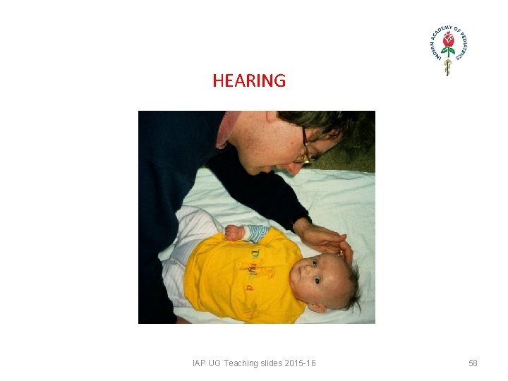
HEARING IAP UG Teaching slides 2015 -16 58
HEARING - BERA • In asphyxia neonatorum, especially if – 1 min Apgar 0 -4 – 5 min Apgar 0 -6 • Besides HIE, other indications – PT – LBW especially < 1500 g – Hyperbilirubenemia – TORCH – Meningitis IAP UG Teaching slides 2015 -16 59
HEARING - BERA • Age of screening- at the time of discharge from NICU • Or 4 -6 Weeks after discharge from NICU HEARING SCREENING SHOULD BE OVER BEFORE 6 MONTHS OF AGE IAP UG Teaching slides 2015 -16 60
MANAGEMENT IAP UG Teaching slides 2015 -16 61
SUPPORTIVE CARE • Maintain adequate ventilation. – Most infants with severe hypoxic-ischemic encephalopathy need ventilatory support during the first week. – Prevent hypoxia, hypercapnia, and hypocapnia; the latter is due to inadvertent hyperventilation, which may lead to severe hypoperfusion of the brain and cellular alkalosis. – Maintain the blood gases and acid-base status in the physiological ranges. • Maintain adequate perfusion. Maintain the mean blood pressure (BP) above 35 -40 mm Hg (for term infants). Dopamine or dobutamine can be used to maintain adequate cardiac output. IAP UG Teaching slides 2015 -16 62
SUPPORTIVE CARE • Maintain adequate metabolic status. • Fluid and glucose homeostasis should be achieved. Avoid hypoglycemia or hyperglycemia because both are known to cause brain injury. • Because of the concern for acute tubular necrosis (ATN) and inappropriate antidiuretic hormone (IADH), fluids should be started at the estimated insensible water loss (40 -60 m. L/kg/d in term infant) until the urine output is clear. • • Thereafter, fluid and electrolyte therapy need to be individualized IAP UG Teaching slides 2015 -16 63
SUPPORTIVE CARE • The role of prophylactic theophylline, given early after birth, in reducing renal dysfunction after hypoxic-ischemic encephalopathy has been evaluated in 3 small randomized controlled trials • Avoid hyperthermia IAP UG Teaching slides 2015 -16 64
TREATMENT OF SEIZURES • Seizures are generally self-limited to the first days of life but may significantly compromise other body functions, such as maintenance of ventilation, oxygenation, and blood pressure. • Additionally, seizures should be treated early and be well controlled because even asymptomatic seizures (ie, seen only on EEG) may continue to injure the brain. • Seizures should be treated with phenobarbital or lorazepam; phenytoin may be added if either of these medications fails to control the seizures IAP UG Teaching slides 2015 -16 65
TREATMENT OF SEIZURES Phenobarbital • DOC when clinical or EEG seizures are noted; is continued on basis of both EEG findings and clinical status • In most cases, can be weaned and stopped during first month of life • Rx continued for several months to 1 year in infants with persistent neurological abnormalities and clinical or EEG evidence of seizures • EEG and clinical status should guide decision IAP UG Teaching slides 2015 -16 66
CARDIOVASCULAR (INOTROPIC) AGENTS • Increase blood pressure (BP) and combat shock • Increase systemic vascular resistance, cardiac contractility, and stroke volume, thus increasing cardiac output. • Have dose and gestational age-dependent effects on vessels, particularly those of renal and GI systems • For most part, these effects are beneficial but, at higher doses, systemic side effects may be unpredictable. • No clear information is available on effects of these drugs on CBF in neonates IAP UG Teaching slides 2015 -16 67
FOLLOW UP MEDICATION • Continuation of seizure medications should depend on evolving CNS symptoms and EEG findings. • In most infants who are developing normally and have a normal EEG before hospital discharge, phenobarbital is discontinued within 3 -4 weeks of birth. • In those with significant CNS disability with or without persistent episodes of seizures, phenobarbital is continued for 3 -6 months; the decision to wean off the drug depends on later changes in EEG and clinical course. IAP UG Teaching slides 2015 -16 68
MINIMIZING BRAIN DAMAGE IAP UG Teaching slides 2015 -16 69
HYPOTHERMIA TREATMENT • Extensive experimental data suggest that mild hypothermia (3 -4°C below the baseline temperature) applied within a few hours (no later than 6 h) of hypoxia-ischemia injury is neuroprotective. • The possible mechanisms through which hypothermia is neuroprotective include – reduced metabolic rate and energy depletion; – decreased excitatory transmitter release; – reduced alterations in ion flux; – reduced apoptosis due to hypoxic-ischemic encephalopathy; – reduced vascular permeability, edema, and disruptions of blood-brain barrier functions. IAP UG Teaching slides 2015 -16 70
EARLY INTERVENTION - AIMS • To soften gap between mother’s womb and hi-tech environment of NICU • NICU babies experience – Painful procedures – Handling – Noises – Irregular, inconstant social contact – Unstable comforting mechanisms IAP UG Teaching slides 2015 -16 71
COMPONENTS OF EARLY INTERVENTION • Developmentally Supportive Care (DSC) • Follow Up Care (FC) IAP UG Teaching slides 2015 -16 72
FOLLOW UP CARE • Neurophysiological Approach • Biomechanical Approach • Functional Approach • Group Therapy • Play Therapy IAP UG Teaching slides 2015 -16 73
BIOMECHANICAL APPROACH • Orthopedic manipulation to improve – Range of movements – Muscle strength – Muscle endurance • Orthotic devices – Splints – Crutches – Walkers • Used in conjunction with NDT or proprioceptive neuromuscular facilitation IAP UG Teaching slides 2015 -16 74
OTHER APPROACHES • Functional Approach • Group Therapy • Play Therapy IAP UG Teaching slides 2015 -16 75
SUMMARY • Perinatal asphyxia and its sequelae can be prevented by – – – – – Tocolytics to prevent PT labor In utero transfer to tertiary center AN steroids reduce GMH risk by 50% Minimal handling in labor room Effective resuscitation at birth Avoid pushes of hyperosmolar IV fluids Blood and blood pdts slowly over few hrs Gentle handling – e. g. intubation etc Gentle ventilation strategies IAP UG Teaching slides 2015 -16 76
SUMMARY • Once assessed and stable, each such baby should undergo a programmed neurological intervention therapy • Ongoing re-evaluation should be done at 3, 6, 9 and 12 months with periodic assessments for sensory functions • Meanwhile growth patterns and nutrition of the baby should also be monitored IAP UG Teaching slides 2015 -16 77
THANK YOU IAP UG Teaching slides 2015 -16 78