Hypothyroidism Dr Swaapnika Introduction The thyroid gland produces

 and triiodothyronine")


 hypothyroidism. • If")


 and liothyronine (25 mic g) as a")


- Slides: 45
Hypothyroidism Dr. Swaapnika
Introduction • The thyroid gland produces two related hormones, thyroxine (T 4) and triiodothyronine (T 3) • Acting through nuclear receptors, these hormones play a critical role in cell differentiation during development and help maintain thermogenic and metabolic homeostasis in the adult.
• Autoimmune disorders of the thyroid gland • 1. stimulate the overproduction of thyroid hormones (thyrotoxicosis) • 2. cause glandular destruction and hormone deficiency (hypothyroidism)
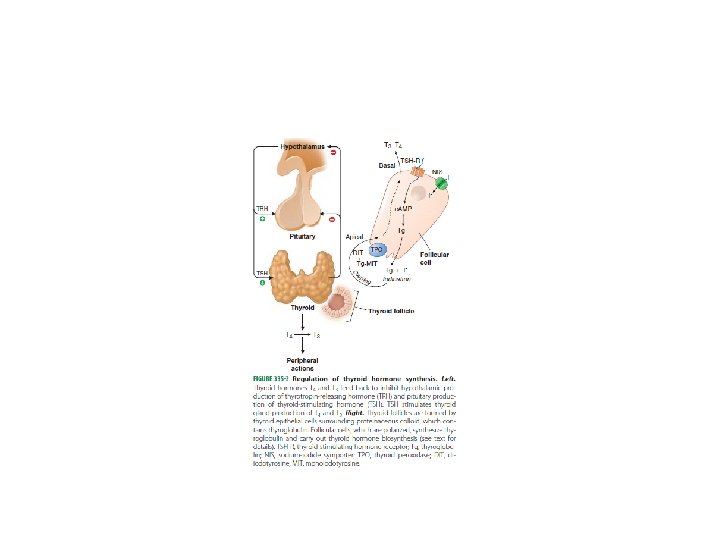
• TSH - the most useful physiologic marker of thyroid hormone action. • The thyroid axis is a classic example of an endocrine feedback loop. • Hypothalamic TRH stimulates pituitary production of TSH, which, in turn, stimulates thyroid hormone synthesis and secretion.
• Reduced levels of thyroid hormone increase basal TSH production and enhance TRHmediated stimulation of TSH. • Thyroid hormones feed back to inhibit TRH and TSH production
• High thyroid hormone levels rapidly and directly suppress TSH gene expression secretion and inhibit TRH stimulation of TSH.
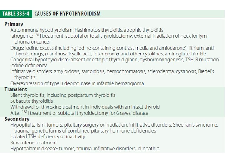
Hypothyroidism • Iodine deficiency remains the most common cause of hypothyroidism worldwide. • In areas of iodine sufficiency, autoimmune disease (Hashimoto’s thyroiditis) and iatrogenic causes (treatment of hyperthyroidism) are most common
CONGENITAL HYPOTHYROIDISM • Neonatal hypothyroidism is due to thyroid gland dysgenesis in 80– 85%, to inborn errors of thyroid hormone synthesis in 10– 15%, and is TSH-R antibody-mediated in 5% of affected
• It may be transient, especially if the mother has TSH-R blocking antibodies or has received antithyroid drugs, but permanent hypothyroidism occurs in the majority. • The majority of infants appear normal at birth, and <10% are diagnosed based on clinical features, which include
• Prolonged jaundice • Feeding problems • Hypotonia • Enlarged tongue, • Delayed bone maturation • Umbilical hernia. • Importantly, permanent neurologic damage results if treatment is delayed.
• Because of the severe neurologic consequences of untreated congenital hypothyroidism, neonatal screening programs have been established. • When the diagnosis is confirmed, T 4 is instituted at a dose of 10– 15 micro g/kg per day, and the dose is adjusted by close monitoring of TSH levels.
• Early treatment with T 4 results in normal IQ levels, but subtle neurodevelopmental abnormalities may occur in those with the most severe hypothyroidism at diagnosis or when treatment is suboptimal.
AUTOIMMUNE HYPOTHYROIDISM • Autoimmune hypothyroidism may be associated with • 1. a goiter (Hashimoto’s, or goitrous thyroiditis) • 2. at the later stages of • the disease, minimal residual thyroid tissue (atrophic thyroiditis)
• Though some patients may have minor symptoms, this state is called subclinical hypothyroidism. • Later, unbound T 4 levels fall and TSH levels rise further; symptoms become more readily apparent at this stage (usually TSH >10 m. U/L), which is referred to as clinical hypothyroidism or overt
Pathogenesis • In Hashimoto’s thyroiditis, there is a marked lymphocytic infiltration of the thyroid with germinal center formation, atrophy of the thyroid follicles accompanied by oxyphil metaplasia, absence of colloid, and mild to moderate fibrosis. • In atrophic thyroiditis, the fibrosis is much more extensive, lymphocyte infiltration is less pronounced, and thyroid follicles are almost completely absent.
• There is a relationship between autoimmune hypothyroidism and other autoimmune diseases, especially type 1 diabetes mellitus, Addison’s disease, pernicious anemia, and vitiligo. • The thyroid lymphocytic infiltrate in autoimmune hypothyroidism is composed of activated CD 4+ and CD 8+ T cells, as well as B cells.
• Antibodies to Tg and TPO are clinically useful markers of thyroid autoimmunity, but any pathogenic effect is restricted to a secondary role in amplifying an ongoing autoimmune response. • T cell–mediated injury is required to initiate autoimmune damage to the thyroid.
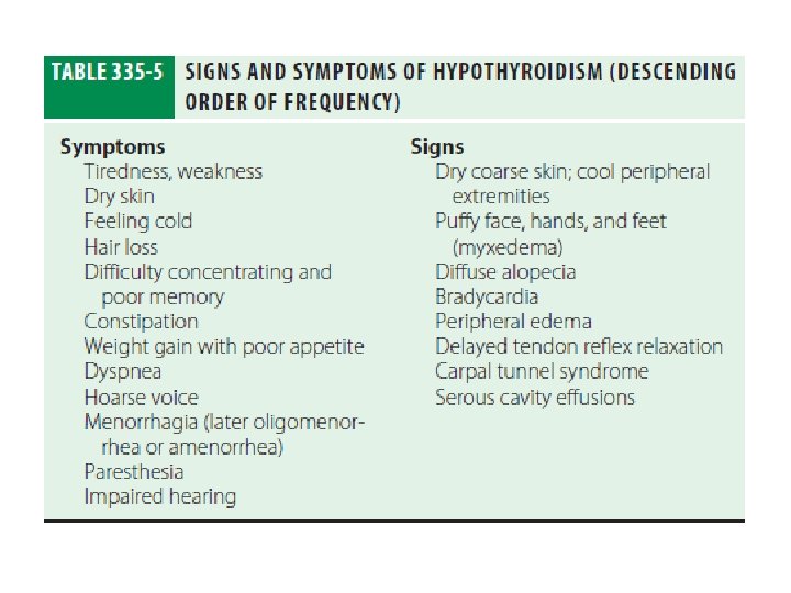
Clinical Manifestations • Patients with Hashimoto’s thyroiditis may present because of goiter rather than symptoms of hypothyroidism. • Increased dermal glycosaminoglycan content traps water, giving rise to skin thickening without pitting (myxedema). • There is pallor, often with a yellow tinge to the skin due to carotene accumulation. • The weight gain is usually modest and due mainly to fluid retention in the myxedematous tissues.
• Prolactin levels are often modestly increased and may contribute to alterations in libido and fertility and cause galactorrhea. • Myocardial contractility and pulse rate are reduced, leading to a reduced stroke volume and bradycardia.
• Increased peripheral resistance may be accompanied by hypertension, particularly diastolic. • Blood flow is diverted from the skin, producing cool extremities. • Fluid may also accumulate in other serous cavities and in the middle ear, giving rise to conductive deafness.
• Dyspnea may be caused by pleural effusion, impaired respiratory muscle function, diminished ventilatory drive, or sleep apnea. • The hoarse voice and occasionally clumsy speech of hypothyroidism reflect fluid accumulation in the
• Hashimoto’s encephalopathy has been defined as a steroid-responsive syndrome associated with TPO antibodies, myoclonus, and slow-wave activity on electroencephalography, but the relationship with thyroid autoimmunity or hypothyroidism is not established.
Laboratory Evaluation
• A normal TSH level excludes primary (but not secondary) hypothyroidism. • If the TSH is elevated, an unbound T 4 level is needed to confirm the presence of clinical hypothyroidism, but T 4 is inferior to TSH when used as a screening test, as it will not detect subclinical hypothyroidism. • Circulating unbound T 3 levels are normal in about 25% of patients,
• If there is any doubt about the cause of a goiter associated with hypothyroidism, FNA biopsy can be used to confirm the presence of autoimmune thyroiditis. • Other abnormal laboratory findings in hypothyroidism may include increased creatine phosphokinase, elevated cholesterol and triglycerides, and anemia (usually
• Ultrasound can be used to show the presence of a solitary lesion or a multinodular goiter rather than the heterogeneous thyroid enlargement typical of Hashimoto’s thyroiditis.
OTHER CAUSES OF HYPOTHYROIDISM • Iatrogenic hypothyroidism is a common cause of hypothyroidism and can often be detected by screening before symptoms develop. • In the first 3– 4 months after radioiodine treatment, transient hypothyroidism may occur due to reversible radiation damage. • Iodine deficiency is responsible for endemic goiter and cretinism but is an uncommon cause of adult hypothyroidism unless the iodine intake is very low or there are complicating factors, such as the consumption of thiocyanates in cassava or selenium deficiency.
• TSH levels are suppressed by hyperthyroidism, unbound T 4 levels are a better measure of thyroid function than TSH in the months following radioiodine treatment. • lithium, may also cause hypothyroidism. • Paradoxically, chronic iodine excess can also induce
• Secondary hypothyroidism is usually diagnosed in the context of other anterior pituitary hormone deficiencies. • TSH levels may be low, normal, or even slightly increased in secondary hypothyroidism.
• The increase is due to secretion of immunoactive but bio inactive forms of TSH. The diagnosis is confirmed by detecting a low unbound T 4 level. • The goal of treatment is to maintain T 4 levels in the upper half of the reference range, as TSH levels cannot be used to monitor therapy.
Treatment • CLINICAL HYPOTHYROIDISM • The daily replacement dose of levothyroxine is usually 1. 6 micro g/kg body weight (typically 100– 150 micro g). • Adult patients under 60 without evidence of heart disease may be started on 50– 100 micro g
• TSH responses are gradual and should be measured about 2 months after instituting treatment or after any subsequent change in levothyroxine dosage. • Patients may not experience full relief from symptoms until 3– 6 months after normal TSH levels are restored.
• Patients with a suppressed TSH of any cause, including T 4 over treatment, have an increased risk of atrial fibrillation and reduced bone density. • SUBCLINICAL HYPOTHYROIDISM : By definition, subclinical hypothyroidism refers to biochemical evidence of thyroid hormone deficiency in patients who have few or no
• Treatment is administered by starting with a low dose of levothyroxine (25– 50 micro g/d) with the goal of normalizing TSH. If thyroxine is not given, thyroid function should be evaluated annually.
• Rarely, levothyroxine replacement is associated with pseudotumor cerebri in children. Presentation appears to be idiosyncratic and occurs months after treatment has begun. • Thyroid function should be evaluated immediately after pregnancy is confirmed and at the beginning of the second and third trimesters. The dose of levothyroxine may need to
• Elderly patients may require up to 20% less thyroxine than younger patients. In the elderly, especially patients with known coronary artery disease, the starting dose of levothyroxine is 12. 5– 25 micro g/d with similar increments every 2– 3 months until TSH is normalized.
Myxedema coma • Clinical manifestations include reduced level of consciousness, sometimes associated with seizures, as well as the other features of hypothyroidism. • Hypothermia can reach 23°C (74°F). There may be a history of treated hypothyroidism with poor compliance, or the patient may be previously undiagnosed. • Precipitated by factors that impair respiration, such as drugs (especially sedatives, anesthetics, antidepressants), pneumonia, congestive heart failure, myocardial infarction, gastrointestinal bleeding, or cerebrovascular accidents. Sepsis should also be suspected.
• Hypoventilation, leading to hypoxia and hypercapnia, plays a major role in pathogenesis; hypoglycemia and dilutional hyponatremia also contribute to the development of myxedema coma. • Levothyroxine can initially be administered as a single intravenous bolus or oral bolus of 500 micro g, which
• Combine levothyroxine (200 �� g) and liothyronine (25 mic g) as a single, initial intravenous bolus followed by daily treatment with levothyroxine (50– 100 mic g/d) and liothyronine (10 mic g every 8 h). • This treatment has been advocated because T 4 to T 3 conversion is impaired in myxedema coma.
• External warming is indicated only if the temperature is <30°C, as it can result in cardiovascular collapse. • Parenteral hydrocortisone (50 mg every 6 h) should be administered, as there is impaired adrenal reserve in profound hypothyroidism. • Ventilatory support is usually needed during the first
• Hypertonic saline or intravenous glucose may be needed if there is severe hyponatremia or hypoglycemia. • Hypotonic intravenous fluids should be avoided because they may exacerbate water retention secondary to reduced renal perfusion and inappropriate vasopressin secretion. • Early broad spectrum antibiotics.
Thank you