
Hypoplastic Left Heart Syndrome and the single ventricle





- Slides: 70
Hypoplastic Left Heart Syndrome (and the single ventricle repair) Henaro Sabino, MD Sibley Heart Center Cardiology at Children’s Healthcare of Atlanta; Emory University
CHEST PAIN, SYNCOPE • “I’M COMIN’ HOME!”
GOALS • Appreciation of history of Hypoplastic Left Heart Syndrome. • Basic anatomy & physiology. • (DE-mystify Hypoplastic Left Heart Syndrome. ) • Understand the LOGIC behind the management of HLHS (& single ventricle lesions in general). • KEEP YOU ALL AWAKE FOR THE NEXT 1. 25 HOURS.
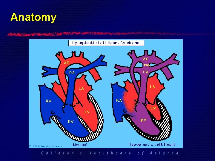
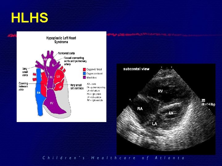
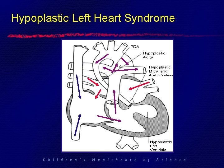
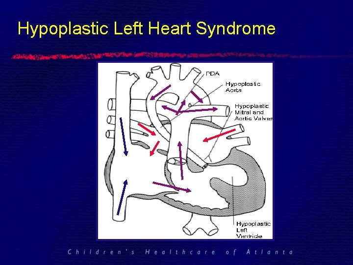
Hypoplastic Left Heart Syndrome • Spectrum of underdevelopment of the left ventricular cavity. § Have underdeveloped aortic & mitral valves (stenosis or atresia). § Left ventricle is unable to support systemic circulation (and, therefore, right ventricle is used as the single ventricle).
History of HLHS • First described by Maurice Lev in 1952. • Term used by Noonan & Nadas in 1958. • Options offered: § Comfort care § Staged palliative repair, i. e. “Norwood procedure” ØFirst successful 3 -stage completion in 1983 (after multiple surgeries from 1979). § Cardiac transplant ØFirst successful cardiac transplant: Bailey, Nov. 1985
1980 s
Xenotransplantation, “Baby Fae” • Dr. Leonard Bailey, Loma Linda University Medical Center, November 1984. • http: //www. babyfae. com
Anatomy
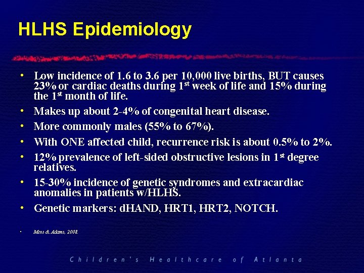
HLHS Epidemiology • Low incidence of 1. 6 to 3. 6 per 10, 000 live births, BUT causes 23% or cardiac deaths during 1 st week of life and 15% during the 1 st month of life. • Makes up about 2 -4% of congenital heart disease. • More commonly males (55% to 67%). • With ONE affected child, recurrence risk is about 0. 5% to 2%. • 12% prevalence of left-sided obstructive lesions in 1 st degree relatives. • 15 -30% incidence of genetic syndromes and extracardiac anomalies in patients w/HLHS. • Genetic markers: d. HAND, HRT 1, HRT 2, NOTCH. • Moss & Adams, 2008.
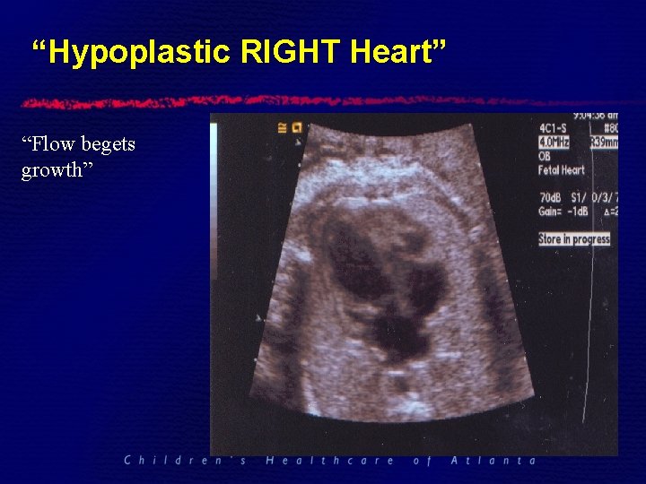
PATHOPHYSIOLOGY • Cardiac development: “Flow begets growth. ” • Altered flow through the left side of the heart: § Reduced/altered flow across the foramen ovale. § Aortic or mitral obstruction.
Typical Clinical Presentation • Known Congenital Heart Defect § Prenatal Diagnosis • Unknown Congenital Heart Defect § Normal pregnancy, labor and delivery § Clinically doing okay until the PDA closes ** § Cyanosis that does not improve with oxygen § Many have no other obvious anomalies
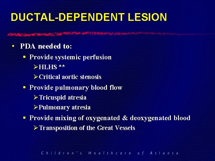
DUCTAL-DEPENDENT LESION • PDA needed to: § Provide systemic perfusion ØHLHS ** ØCritical aortic stenosis § Provide pulmonary blood flow ØTricuspid atresia ØPulmonary atresia § Provide mixing of oxygenated & deoxygenated blood ØTransposition of the Great Vessels
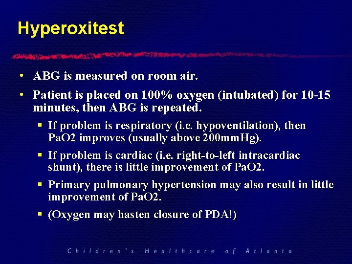
Hyperoxitest • ABG is measured on room air. • Patient is placed on 100% oxygen (intubated) for 10 -15 minutes, then ABG is repeated. § If problem is respiratory (i. e. hypoventilation), then Pa. O 2 improves (usually above 200 mm. Hg). § If problem is cardiac (i. e. right-to-left intracardiac shunt), there is little improvement of Pa. O 2. § Primary pulmonary hypertension may also result in little improvement of Pa. O 2. § (Oxygen may hasten closure of PDA!)
Positive Hyperoxitest • Seriously consider initiation of prostaglandin (PGE) at a low dose (0. 03 mcg/kg/min) until diagnosis is confirmed.
Initial Assessment • ALWAYS § A - Airway § B - breathing § C – circulation CXR and ECG usually not very helpful in Dx.
Physical Findings • Comfortable or in distress? § Cyanosis w/out respiratory distress is cardiac until proven otherwise • Active or lethargic? • Cyanosis? § Degree - saturation usually <85% to be seen § Anemia makes cyanosis difficult to notice • Pallor § Vasoconstriction from circulatory shock • Perfusion and Peripheral pulses • End organs (i. e. watch UOP)
Respiratory Status • Tachypnea but with minimal distress…cardiac until proven otherwise.
Respiratory Status • Respiratory distress § Inability of the respiratory system to compensate for the metabolic acidosis • Concurrent respiratory disease • Unrelenting metabolic acidosis - decreased cardiac function • Exhaustion
Assisted Ventilation • Intubate if: § Impending respiratory failure § Potentially not necessary to intubate just for PGE therapy if ground transport § Intubate for air transport in PGE dependent babies
Assisted Ventilation • Ventilation strategy § Volume ventilation if possible to maintain consistent minute ventilation in the face of changing lung compliance § Bigger tidal volumes compared to premature newborns (10 cc/kg); lower rates § No need to “over-ventilate” § “ 40/40/40 club”
Arterial Blood Gases • In congenital heart disease typically: § Compensated or partially compensated metabolic acidosis § Arterial PO 2 usually low <50 with cyanotic heart disease…but not always • If PCO 2 is rising, think respiratory failure - be ready to intubate!
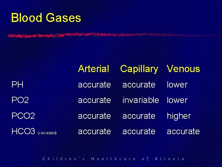
Blood Gases Arterial Capillary Venous PH accurate PO 2 accurate invariable lower PCO 2 accurate higher HCO 3 (calculated) accurate lower
Oxygen • Oxygen is a drug - use it with respect • Oxygen is a pulmonary vasodilator § May worsen pulmonary congestion • Oxygen is a stimulus for the PDA to close § May worsen ductal dependent lesions by speeding up closure of the PDA • Oxygen is not bad
Saturation Monitoring • Oxygen saturation reflects tissue oxygenation and usually does not correlate with PO 2. • With pulmonary hypertension will see differential cyanosis - shunts right to left across the PDA. • The number is not as important as the patient.
Prostaglandin Infusion • Purpose is to open the PDA if a ductal dependent lesion is suspected • Can be initiated before a definitive diagnosis is established • Need a secure IV (PIV, PIC, or UVC-central or in the liver) • Start at low dose 0. 03 mcg/kg/min
Prostaglandins continued • Side effects § Apnea - be prepared to intubate § Fever § Hypotension - have volume and inotropes available § Flushing
Access • Umbilical is preferred in a newborn § UVC – even if in suboptimal position § UAC • PIC line • PIV • AVOID groin line if possible
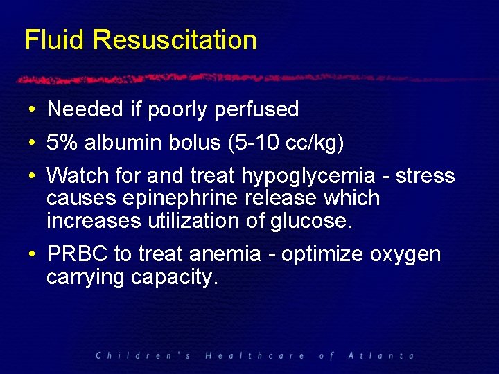
Fluid Resuscitation • Needed if poorly perfused • 5% albumin bolus (5 -10 cc/kg) • Watch for and treat hypoglycemia - stress causes epinephrine release which increases utilization of glucose. • PRBC to treat anemia - optimize oxygen carrying capacity.
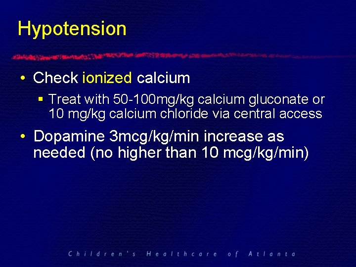
Hypotension • Check ionized calcium § Treat with 50 -100 mg/kg calcium gluconate or 10 mg/kg calcium chloride via central access • Dopamine 3 mcg/kg/min increase as needed (no higher than 10 mcg/kg/min)
Metabolic Acidosis • Treat metabolic acidosis aggressively (base deficit < -3) • 1 meq/kg Na bicarbonate • Repeat blood gas
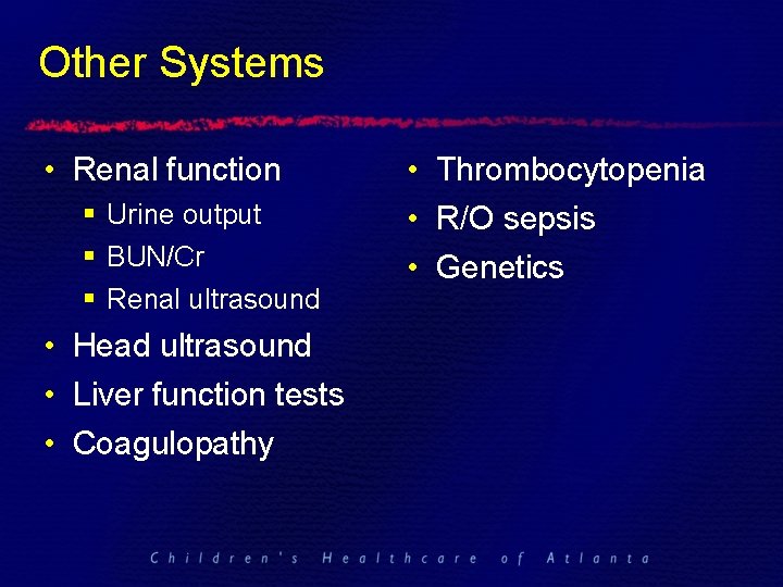
Other Systems • Renal function § Urine output § BUN/Cr § Renal ultrasound • Head ultrasound • Liver function tests • Coagulopathy • Thrombocytopenia • R/O sepsis • Genetics
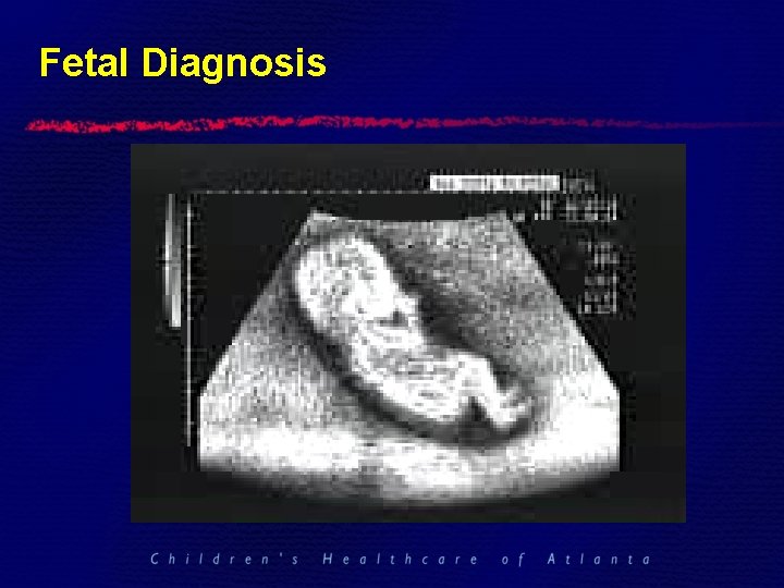
Fetal Diagnosis
Fetal Studies • Hornberger, 1995: 21 fetuses with prenatal echos that show left-sided obstruction (small mitral valve & ascending aorta) developed HLHS. • Critical aortic stenosis decreased blood flow through left heart LV dilation & dysfunction endocardial fibroelastosis (EFE) backwards flow across PFO LV stops growing & eventually shrinks
HLHS
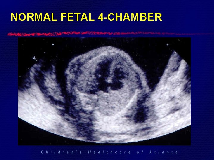
NORMAL FETAL 4 -CHAMBER
Case Presentation • • Term infant born via SVD Uncomplicated labor and delivery APGARs of 8 at 1 min. , 9 at 5 min. Tachypnea noted at 12 hrs of life.
Case Presentation • “Airway-Breathing-Circulation” § Respiratory rate (60 -90 bpm) § Work of breathing (no retractions) § Saturations (80%) § Warm extremities; good cap refill
Case Presentation No obvious dysmorphic features. More Cardiac Exam Findings: § No murmur. § Single second heart sound (S 2). § Hyperdynamic precordium.
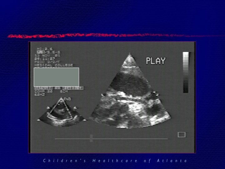
Case Presentation • Urgent Cardiology Consult 404 -256 -2593!! • Cardiac History & Physical • Echocardiogram
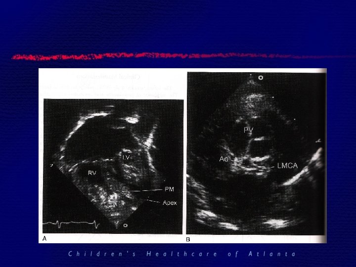
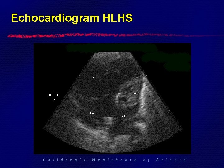
Echocardiogram HLHS
Hypoplastic Left Heart Syndrome
Case Presentation • BUT: § No beds available at Egleston immediately § Need to manage infant for 24 hours before transport • NOW what do we do?
Case Presentation • Intravenous access § UVC (double lumen) § UAC § PIV § PIC Remember: AVOID groin lines
Case Presentation • Prostaglandins § 0. 03 mcg/kg/min • Side effects § Apnea • Options ? • Intubate vs nasal cannula air
Case Presentation • Labs § § § Arterial (or venous) blood gas Electrolytes (normalize) CBC LFT Genetics Lactic acid • Head and Renal ultrasound • ECHO/EKG
Case Presentation • R/O Sepsis § If no clinical suspicion or maternal indicators no need to start antibiotics • • Follow ABG frequently (Q 4 hrs) Monitor urine output Monitor for acidosis Watch for hypotension
Blood Pressure • Blood pressure - systolic and diastolic blood pressures are equally important…not just mean!! § Coronary flow to heart dependant on diastolic BP
Case Presentation • Saturations 95% • p. O 2 50 • Decreased urine output • Metabolic acidosis • Rising lactic acid What’s going on? !?
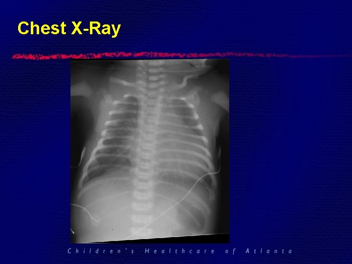
Chest X-Ray
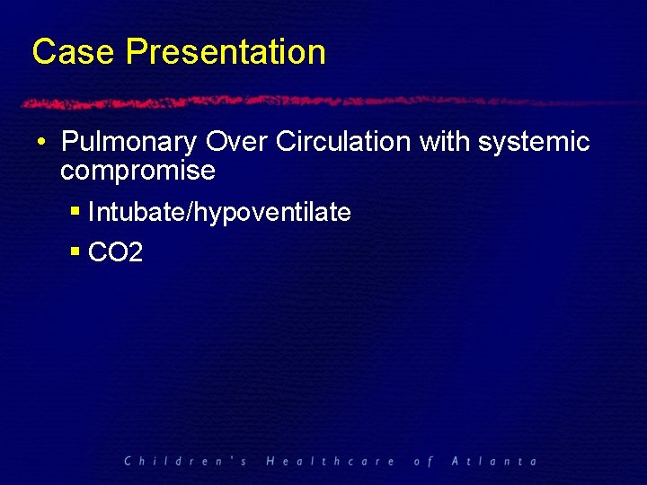
Case Presentation • Pulmonary Over Circulation with systemic compromise § Intubate/hypoventilate § CO 2
Hypoplastic Left Heart Syndrome
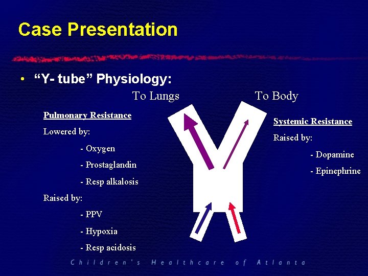
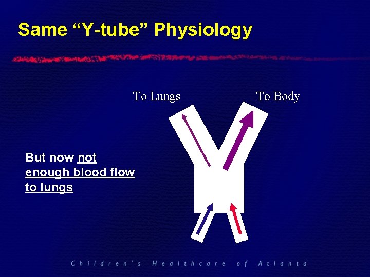
Case Presentation • “Y- tube” Physiology: To Lungs Pulmonary Resistance Lowered by: - Oxygen - Prostaglandin - Resp alkalosis Raised by: - PPV - Hypoxia - Resp acidosis To Body Systemic Resistance Raised by: - Dopamine - Epinephrine
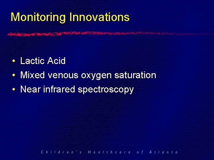
Monitoring Innovations • Lactic Acid • Mixed venous oxygen saturation • Near infrared spectroscopy
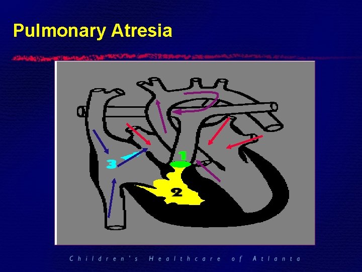
Pulmonary Atresia
“Hypoplastic RIGHT Heart” “Flow begets growth”
Same “Y-tube” Physiology To Lungs But now not enough blood flow to lungs To Body
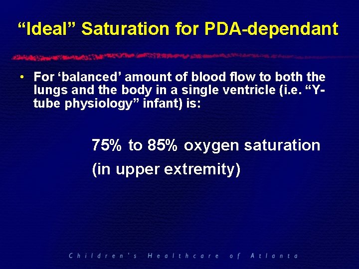
“Ideal” Saturation for PDA-dependant • For ‘balanced’ amount of blood flow to both the lungs and the body in a single ventricle (i. e. “Ytube physiology” infant) is: 75% to 85% oxygen saturation (in upper extremity)
Options for HLHS in 2010 • • Comfort Care Transplant 3 -Stage Palliative Repair Fetal Intervention
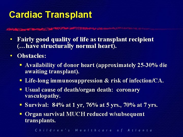
Cardiac Transplant • Fairly good quality of life as transplant recipient (…have structurally normal heart). • Obstacles: § Availability of donor heart (approximately 25 -30% die awaiting transplant). § Life-long immunosuppression & risk of infection/CA. § Usual cause of death/organ death: coronary vasculopathy. § Survival: 84% at 1 yr, 76% at 5 yrs. , 70% at 7 yrs. § Organ survival MUCH reduced w/subsequent transplants.
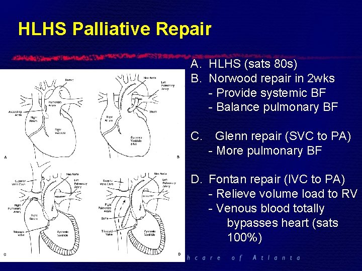
HLHS Palliative Repair A. HLHS (sats 80 s) B. Norwood repair in 2 wks - Provide systemic BF - Balance pulmonary BF C. Glenn repair (SVC to PA) - More pulmonary BF D. Fontan repair (IVC to PA) - Relieve volume load to RV - Venous blood totally bypasses heart (sats 100%)
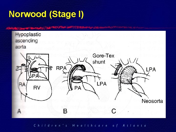
Norwood (Stage I)
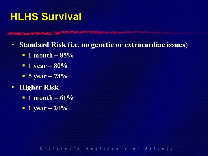
HLHS Survival • Standard Risk (i. e. no genetic or extracardiac issues) § 1 month – 85% § 1 year – 80% § 5 year – 73% • Higher Risk § 1 month – 61% § 1 year – 20%
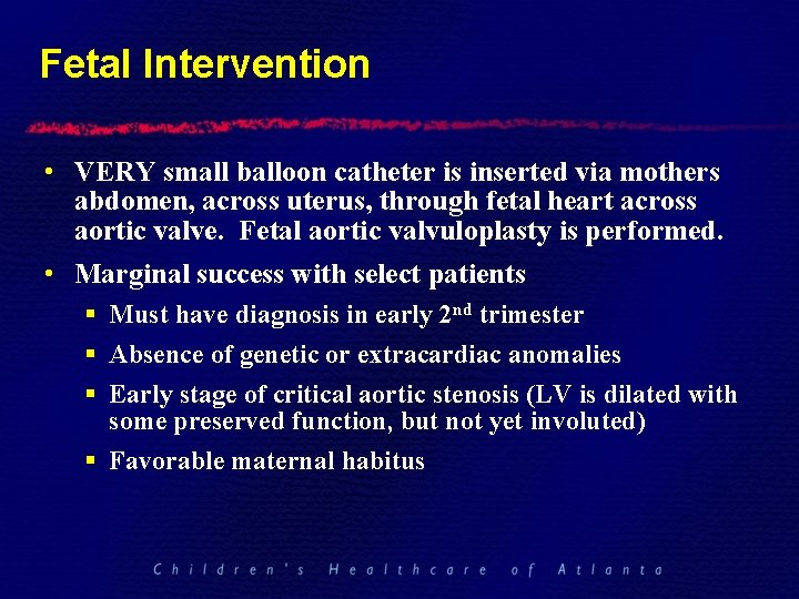
Fetal Intervention • VERY small balloon catheter is inserted via mothers abdomen, across uterus, through fetal heart across aortic valve. Fetal aortic valvuloplasty is performed. • Marginal success with select patients § Must have diagnosis in early 2 nd trimester § Absence of genetic or extracardiac anomalies § Early stage of critical aortic stenosis (LV is dilated with some preserved function, but not yet involuted) § Favorable maternal habitus
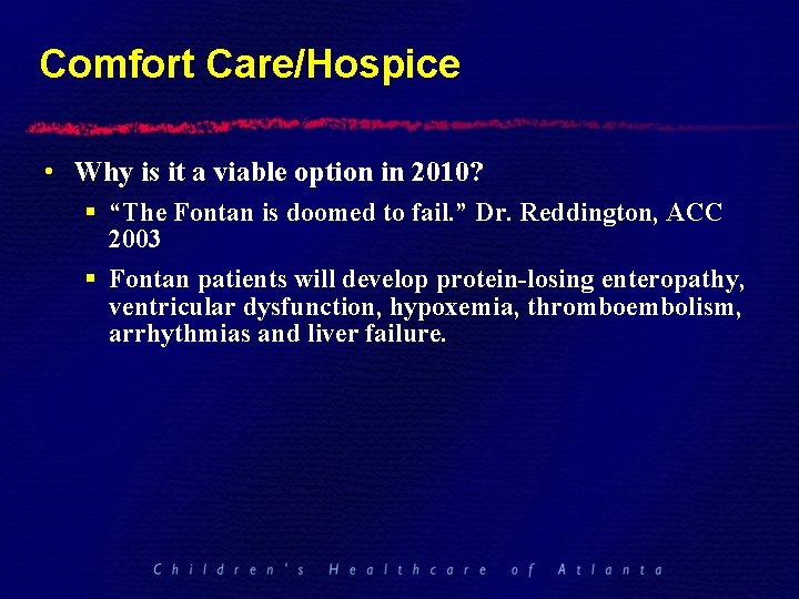
Comfort Care/Hospice • Why is it a viable option in 2010? § “The Fontan is doomed to fail. ” Dr. Reddington, ACC 2003 § Fontan patients will develop protein-losing enteropathy, ventricular dysfunction, hypoxemia, thromboembolism, arrhythmias and liver failure.
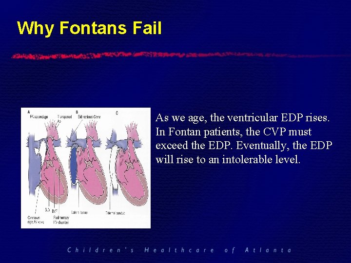
Why Fontans Fail As we age, the ventricular EDP rises. In Fontan patients, the CVP must exceed the EDP. Eventually, the EDP will rise to an intolerable level.
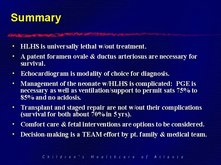
Summary • HLHS is universally lethal w/out treatment. • A patent foramen ovale & ductus arteriosus are necessary for survival. • Echocardiogram is modality of choice for diagnosis. • Management of the neonate w/HLHS is complicated: PGE is necessary as well as ventilation/support to permit sats 75% to 85% and no acidosis. • Transplant and staged repair are not w/out their complications (survival for both about 70% in 5 yrs). • Comfort care & fetal interventions are options to be considered. • Decision-making is a TEAM effort by pt. family & medical team.
Thank You!
Questions ?