
HypnagogicSleep StartEvents BY AHMAD YOUNES PROFESSOR OF THORACIC
Hypnagogic(Sleep Start)Events BY AHMAD YOUNES PROFESSOR OF THORACIC MEDICINE Mansoura Faculty of Medicine Stephan Eisenschenk, MD Department of Neurology
Different Scientific Terms • Hypnagogic defines the period when a person falls asleep. • Hypnopompic describes the period when a person wakes up. • Proprioception, is the sense of the relative position of one's own parts of the body and strength of effort being employed in movement. it is provided by proprioception in skeletal striated muscles (muscle spindles) and tendons (Golgi tendon organ) and the fibrous membrane in joint capsules. • The brain integrates information from proprioception and from the vestibular system into its overall sense of body position, movement, and acceleration. 2 Stephan Eisenschenk, MD Department of Neurology
Different Scientific Terms • kinesthesia or kinæsthesia (kinesthetic sense) strictly means movement sense, but has been used inconsistently to refer either to proprioception alone or to the brain's integration of proprioceptive and vestibular inputs. • Exteroception, by which one perceives the outside world, and interoception, by which one perceives pain, hunger, etc. , and the movement of internal organs. 3 Stephan Eisenschenk, MD Department of Neurology
Different Scientific Terms • A tremor is an involuntary, somewhat rhythmic, muscle contraction and relaxation involving twitching movements of one or more body parts. • It is the most common of all involuntary movements and can affect the hands, arms, eyes, face, head, vocal folds, trunk, and legs. • Most tremors occur in the hands. • In some people, a tremor is a symptom of another neurological disorder. • A very common tremor is the teeth chattering, usually induced by cold temperatures or by fear. 4 Stephan Eisenschenk, MD Department of Neurology
Different Scientific Terms • Rhythmic movement disorder (or RMD) is a neurological disorder characterized by involuntary (however may sometimes be voluntary), repetitive movements of large muscle groups immediately before and during sleep often involving the head and neck. • The majority of RMD episodes occur during NREM sleep, although REM movements have been reported. • RMD is often associated with other psychiatric conditions or mental disabilities. • The disorder often leads to bodily injury from unwanted movements. 5 Stephan Eisenschenk, MD Department of Neurology
Different Scientific Terms • RMD involves involuntary muscle contractions before and during sleep while Restless Legs Syndrome is the urge to move before sleep. • Myoclonus is a brief, involuntary twitching of a muscle or a group of muscles. It describes a medical sign and, generally, is not a diagnosis of a disease. • These myoclonic twitches, jerks, or seizures are usually caused by sudden muscle contractions (positive myoclonus) or brief lapses of contraction (negative myoclonus). • The most common circumstance under which they occur is while falling asleep (hypnic jerk). 6 Stephan Eisenschenk, MD Department of Neurology
Different Scientific Terms • When they appear with more persistence and become more widespread they can be a sign of various neurological disorders. • Hiccups are a kind of myoclonic jerk specifically affecting the diaphragm. When a spasm is caused by another person it is known as a provoked spasm. • Shuddering attacks in babies fall in this category. • Myoclonic jerks may occur alone or in sequence, in a pattern or without pattern. • They may occur infrequently or many times each minute. 7 Stephan Eisenschenk, MD Department of Neurology
Different Scientific Terms • Most often, myoclonus is one of several signs in a wide variety of nervous system disorders such as multiple sclerosis, Parkinson's disease, Dystonia, Alzheimer's disease, Gaucher's disease, subacute sclerosing panencephalitis, Creutzfeldt–Jakob disease (CJD), serotonin toxicity, some cases of Huntington's disease, some forms of epilepsy, and occasionally in intracranial hypotension. • In almost all instances in which myoclonus is caused by central nervous system disease it is preceded by other symptoms; for instance, in CJD it is generally a late-stage clinical feature that appears after the patient has already started to exhibit gross neurological deficits. 8 Stephan Eisenschenk, MD Department of Neurology
Different Scientific Terms • Anatomically, myoclonus may originate from lesions of the cortex, subcortex or spinal cord. • The presence of myoclonus above the foramen magnum effectively excludes spinal myoclonus; further localization relies on further investigation with electromyography (EMG) and electroencephalography (EEG). 9 Stephan Eisenschenk, MD Department of Neurology
10 Stephan Eisenschenk, MD Department of Neurology
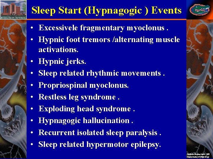
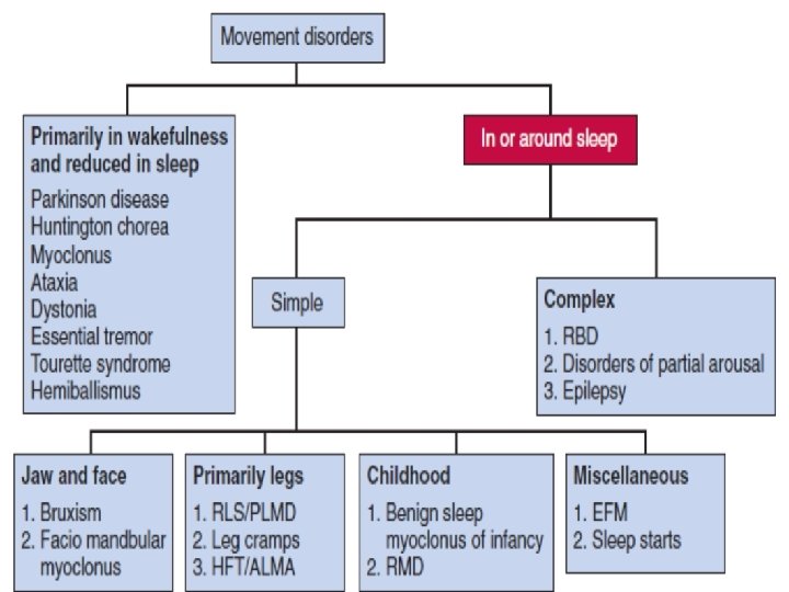
Sleep Start (Hypnagogic ) Events • Excessivele fragmentary myoclonus. • Hypnic foot tremors /alternating muscle activations. • Hypnic jerks. • Sleep related rhythmic movements. • Propriospinal myoclonus. • Restless leg syndrome. • Exploding head syndrome. • Hypnagogic hallucination. • Recurrent isolated sleep paralysis. • Sleep related hypermotor epilepsy. Stephan Eisenschenk, MD Department of Neurology
12 Stephan Eisenschenk, MD Department of Neurology
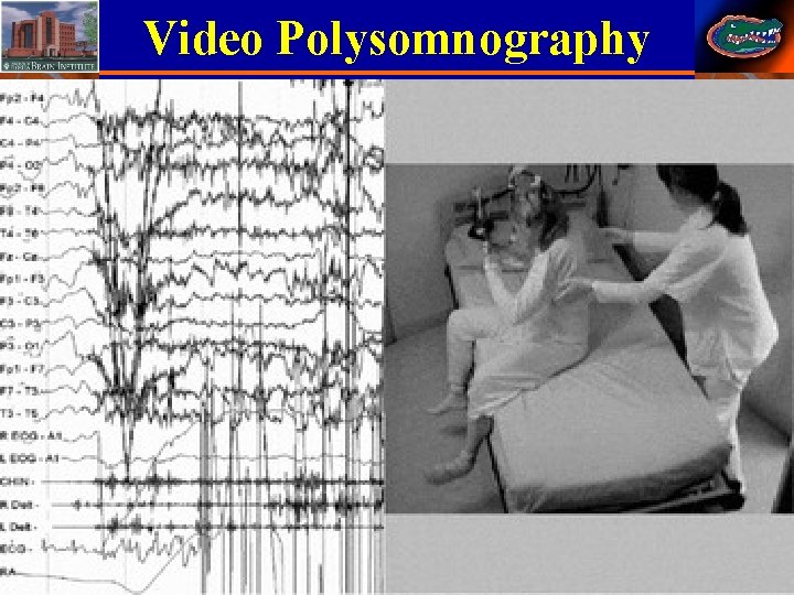
Video Polysomnography 13 Stephan Eisenschenk, MD Department of Neurology
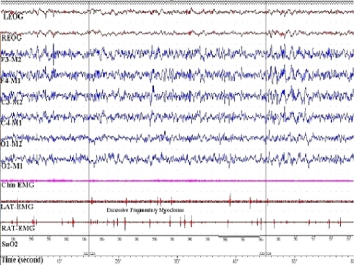
Scoring Excessive Fragmentary. Myoclonus • Maximum EMG burst duration is 0. 15 sec. • At least 20 minutes of NREM sleep must be recorded. • At least 5 EMG potentials per minutes must be recorded. • No clinical significance of Excessive Fragmentary. Myoclonus. • In many cases , no visible movements are present, if movement occure , it is like smal twitches of fingers. 14 Stephan Eisenschenk, MD Department of Neurology
15 Stephan Eisenschenk, MD Department of Neurology
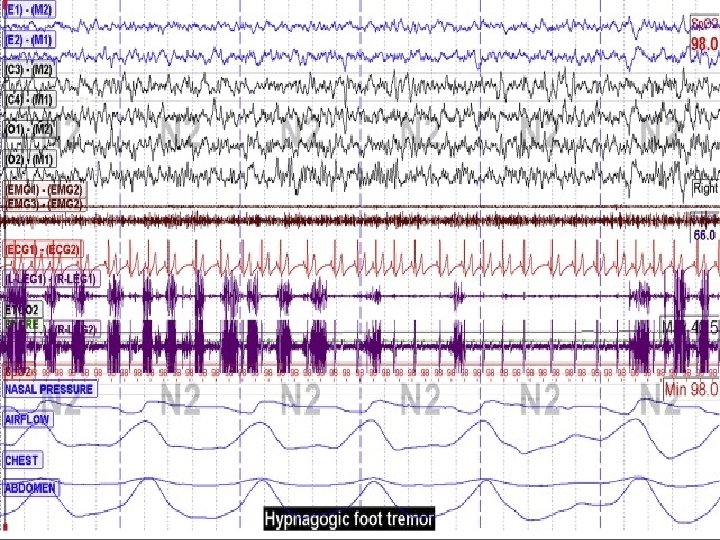
Rhythmic feet movement, formerly hypnagogic foot tremor • Rhythmic feet movement, formerly hypnagogic foot tremor (0. 5 -3 Hz), occurring during presleep wakefulness and light sleep may be considered a new kind of RMD arising in adults, in some cases associated with insomnia, sleep apnoea, periodic limb movements and RLS. 16 Stephan Eisenschenk, MD Department of Neurology
Scoring Hypnagogic Foot Tremors • Maximum number EMG burst to make a train of bursts in a HFT series is 4 HFT bursts. • Minimum frequency of EMG bursts in HFT is 0. 3 Hz. • Maximum frequency of EMG bursts in HFT is 4 Hz. • The usual duration of HFT is 0. 251 sec. • HFT is of no clinical significance. 17 Stephan Eisenschenk, MD Department of Neurology
18 Stephan Eisenschenk, MD Department of Neurology
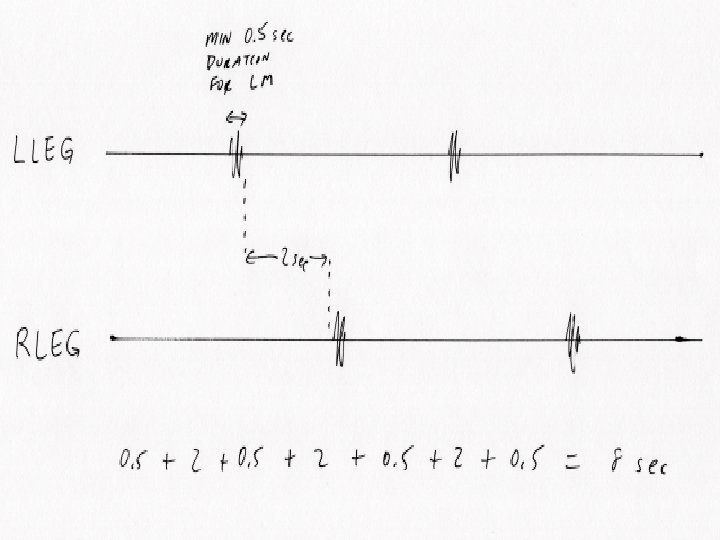
Alternating Leg Muscles Activity • Brief activation of tibialis anterior in one leg alternating with similar activation in the other leg, so-called alternating leg muscles activity (ALMA), has been described. • Such activations, similar to rhythmic feet movements while falling asleep, occur at a frequency of 1 -1. 5 Hz, each lasting up to 0. 5 s, with sequences of several to twenty seconds and recurring particularly during arousal. • ALMA has been described in patients with sleep apnoea, those taking antidepressant medication, and with periodic limb movements and RLS. 19 Stephan Eisenschenk, MD Department of Neurology
Scoring Alternating leg muscle activation • Minimum number of discrete and alternating EMG bursts of leg muscle activity events to score ALMA series is 4 ALMAs. • Minimum frequency of alternating EMG bursts in ALMA is 0. 5 Hz. • Maximum frequency of alternating EMG bursts in ALMA is 3 Hz. • ALMA alternate between legs. • The usual duration of ALMA is 0. 1 -0. 5 sec. • ALMA is of no clinical significance. 20 Stephan Eisenschenk, MD Department of Neurology
21 Stephan Eisenschenk, MD Department of Neurology
22 Stephan Eisenschenk, MD Department of Neurology
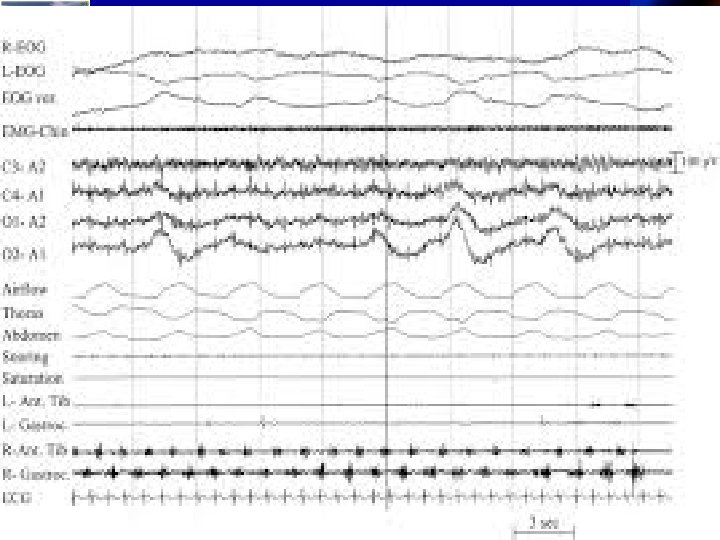
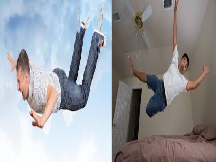
HYPNIC JERKS (HYPNAGOGIC JERKS) • Hypnic jerks represent a normal accompaniment of sleep. • Hypnic jerks consist in non periodic myoclonic movements, usually involving asynchronically different and isolated body segments, that occur mainly at sleep onset and/or with K-complexes or an EEG arousal. • They are associated with autonomic activation tachypnea and sometimes with a peculiar sensory feeling of “shock” or “falling into the void”. • These non stereotyped myoclonic jerks are often triggered by fatigue, stress, sleep deprivation, vigorous exercise and stimulants like caffeine and nicotine. 23 Stephan Eisenschenk, MD Department of Neurology
24 Stephan Eisenschenk, MD Department of Neurology
INTENSIFIED HYPNIC JERKS • The term “intensified hypnic jerks” describe accentuated hypnic jerks causing sleep onset insomnia. • Characterized by sudden transient jerks involving the limbs and the trunk, at times also the head symmetrically, but in some subjects, asymmetrically and occasionally unilaterally at the moment of falling asleep lasting for a few seconds but repeated frequently with increasing intensity. • The jerks may be clustered lasting for several seconds. • Some patients complain of similar jerks after waking up in the middle of the night and on falling asleep again. 25 Stephan Eisenschenk, MD Department of Neurology
INTENSIFIED HYPNIC JERKS • These jerks are non-stereotyped (unlike the stereotypical jerks of myoclonic seizures) but are abrupt, purposeless, and characterized by sudden flexion or extension movements. • Comorbid Disorders include obstruction sleep apnea; anxiety disorder, Multiple sclerosis, RLS, 26 Stephan Eisenschenk, MD Department of Neurology
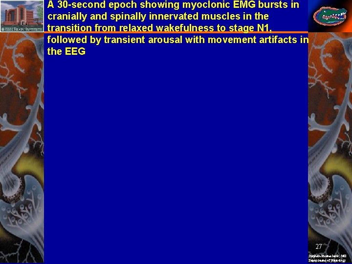
A 30 -second epoch showing myoclonic EMG bursts in cranially and spinally innervated muscles in the transition from relaxed wakefulness to stage N 1, followed by transient arousal with movement artifacts in the EEG 27 Stephan Eisenschenk, MD Department of Neurology
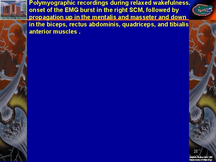
Polymyographic recordings during relaxed wakefulness. onset of the EMG burst in the right SCM, followed by propagation up in the mentalis and masseter and down in the biceps, rectus abdominis, quadriceps, and tibialis anterior muscles. 28 Stephan Eisenschenk, MD Department of Neurology
HYPNIC JERKS (HYPNAGOGIC JERKS) • HJs are a frequent sleep-related motor phenomenon in patients with Parkinson Disease and Atypical Parkinsonisms, appearing since the early stages of these diseases and unrestricted to a specific disease type. • Detailed neurophysiological analysis of the HJs in healthy subjects and patients with parkinsonism, including the lack of any ordered propagation of the spreading of muscular contraction suggested a subcortical origin of the jerks. • HJs arise from sudden descending volleys that originate in the brainstem reticular formation and are activated mostly at the transition between sleep and wake. for each HJs. 29 Stephan Eisenschenk, MD Department of Neurology
HYPNIC JERKS (HYPNAGOGIC JERKS) • The variability in motor recruitment and pattern during HJs may be related to variable spinal motor neuron excitability in the spinal cord during sleep, indicating the engagement of different unsynchronized pools of spinal motor neurons at different times for each HJs. • Usually sleep starts are common physiological phenomenon affecting up to 70% of the adult population and their course is benign, resolving without any neurological sequel. Therefore reassurance and counselling are all that is needed to reassure the patient. • Some patients may require a small dose of clonazepam (0. 5– 1 mg at bedtime) to ameliorate the symptoms on a short-term basis. 30 Stephan Eisenschenk, MD Department of Neurology
Sleep Related Rhythmic Movement Disorder • RMD consists of repetitive and stereotyped movements of the head, neck , trunk, and sometimes also legs. • The movements are predominantly sleep related, occurring near nap or bedtime, or when the individual appears drowsy or asleep. • These stereotypic movements may last a few or several minutes, repeating at a frequency of 0. 5 -2 per second. • RMD is seen in normal children, but it has also been reported in mentally retarded and autistic patients. In these patients, RMD may persist during wakefulness and into adulthood, not rarely associated with other “stereotypies” i. e. the rhythmic habit pattern. 31 Stephan Eisenschenk, MD Department of Neurology
Scoring of Rhythmic Movement Disorder • Minimum frequency of Rhythmic Movement is 0. 5 Hz. • Maximum frequency of Rhythmic Movement is 2 Hz. • Minimum number of individual movements to make a cluster of rhythmic movements is 4 movements. • Minimum amplitude of an individual rhythmic burst is 2 times the background EMG activity. • Time synchronized audio-equiped video PSG is required for diagnosis of Rhythmic Movement Disorder 32 Stephan Eisenschenk, MD Department of Neurology
Sleep Related Rhythmic Movement Disorder • Serious injuries from sleep-related RMD are rare, but include bone skull injuries. • The association of RMD with long-lasting restless legs syndrome (RLS) is well-known and RMD may also occur in RLS of recent onset. • The absence of organic causes in the majority of cases has led to behavioural and psychological theories. • Other hypothesis proposes that RMD is linked to arousal fluctuations and mediated via central motor pattern generators of the brainstem. 33 Stephan Eisenschenk, MD Department of Neurology
Sleep Related Rhythmic Movement Disorder • Provided that RMD does not substantially affect sleep quality or daytime function, the condition does not require any treatment. • For particularly violent forms of RMD, use of protective measures is appropriate. • Clonazepam (1 mg nightly), oxazepam (10 -20 mg nightly), citalopram, imipramine and behavioural therapies have been reported to produce significant improvement in some cases. 34 Stephan Eisenschenk, MD Department of Neurology
Multi-channel artifacts caused by rhythmic, violent head-slapping from N 2 sleep 35 Stephan Eisenschenk, MD Department of Neurology
Propriospinal myoclonus at the sleep wake transition • Propriospinal myoclonus (PSM) is a special type of spinal myoclonus characterized by brief, repetitive, mainly arrhythmic jerks. • The jerks usually arise in the muscles corresponding to a given thoracic myelomere and then progressively and synchronously spread in rostral and caudal directions to the adjacent myotome at low velocity along propriospinal pathways. • The jerks usually provoke a flexion movement, but less frequently an extension pattern has been reported. • Most patients with PSM have no recognizable cause. 36 Stephan Eisenschenk, MD Department of Neurology
Myoclonic jerks occur during relaxed wakefulness. The EMG bursts start in the right rectus abdominis (R Abd) and spread to the intercostal muscles (ICM). 37 Stephan Eisenschenk, MD Department of Neurology
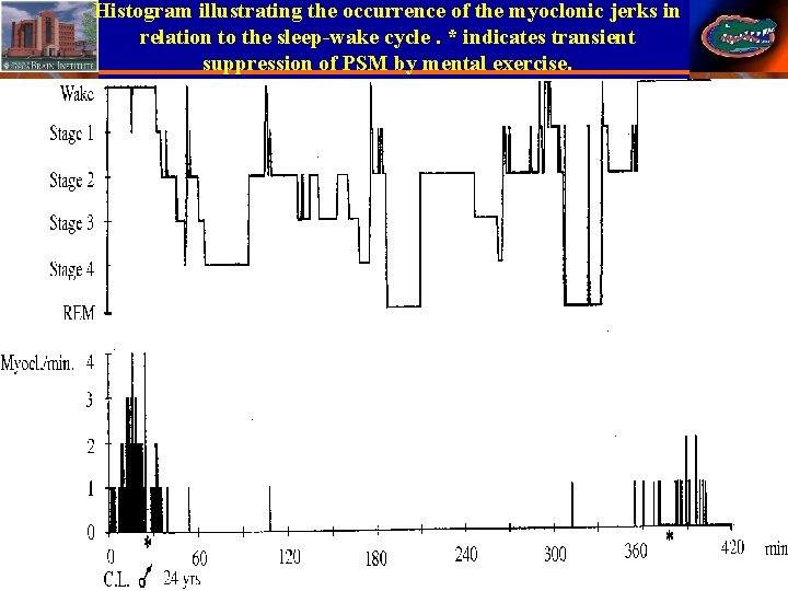
Histogram illustrating the occurrence of the myoclonic jerks in relation to the sleep-wake cycle. * indicates transient suppression of PSM by mental exercise. 38 Stephan Eisenschenk, MD Department of Neurology
Propriospinal myoclonus at the sleep wake transition • PSM at the wake-sleep transition recurs only during drowsiness preceding falling asleep and, rarely, during intrasleep relaxed wakefulness and upon awakening in the morning. • The disorder can make it very hard to fall asleep with consequent severe insomnia. • Video-polysomnography confirms that the myoclonic activity is restricted to the wakefulness period preceding sleep onset and manifests as trunk flexion or, less frequently, extension. • The EMG discharges last typically 0. 1 -0. 3 s but sometimes longer with both reciprocal and cocontracting agonist-antagonist activity. Stephan Eisenschenk, MD Department of Neurology
Propriospinal myoclonus at the sleep wake transition • The major characteristic of the EMG discharges is a simultaneous bilateral rostral and caudal slow recruitment (2 -16 m/s) from the area of spinal cord origin, sparing the cranial muscles, in agreement with a propriospinal pattern of propagation. • Jerks may recur at quasi-periodic intervals (every 5 -40 seconds). • Mental activation with the patient comfortably lying down or sitting (asking the patient to think, speak, count, and perform simple or complex motor tasks such as waving a hand or writing) desynchronizes the EEG activity and makes the jerks disappear. 40 Stephan Eisenschenk, MD Department of Neurology
Propriospinal myoclonus at the sleep wake transition • Absence of a cortical pre-movement potential, lack of involvement of cranial muscles, pattern of propagation, variable delay between different muscles, and slow propagation are all features that distinguish PSM from cortical and reticular reflex myoclonus. • “Diaphragmatic flutter”, “moving umbilicus syndrome”, “belly dancer’s dyskinesia, painful legs and moving toes and excessive fragmentary hypnic myoclonus are conditions clearly different from PSM. 41 Stephan Eisenschenk, MD Department of Neurology
Propriospinal myoclonus at the sleep wake transition • In around 80% of cases, PSM appears to be idiopathic, the remainder comprising patients with a broad range of medical conditions, including spinal lesions, namely cervical hemangioblastoma or cervical herniations, syringomyelia and dural arteriovenous fistula, paraneoplastic condition, paraproteinemic neuropathy and demyelinating lesions. • A psychogenic etiology should be also considered because the motor pattern of PSM can be mimicked voluntarily. 42 Stephan Eisenschenk, MD Department of Neurology
Propriospinal myoclonus at the sleep wake transition • There is no known effective treatment for PSM. • It responds only in part to clonazepam (up to 2 mg at night). Other treatment options includes carbamazepine (up to 400 mg/day), gabapentin (up to 800 mg/day), levetiracetam (up to 2000 mg), pramipexole (up to 0. 7 mg at night) and tramadol (up to 100 mg at night). 43 Stephan Eisenschenk, MD Department of Neurology
Diaphragmatic Flutter(Diaphragmatic Myoclonus, Respiratory Myoclonus, Belly Dancer’s Syndrome) • Diaphragmatic flutter is a rare condition characterised by rhythmic involuntary contractions of the diaphragm and other respiratory muscles (Respiratory Myoclonus ) innervated by cervical nerve roots. • Rapid rhythmical contractions of the diaphragm, averaging around 150 per minute, that resemble atrial flutter clinically and sometimes electrocardiographically. • Symptoms include thoracic or abdominal pain and dyskinetic movements in the thorax and abdominal wall, inspiratory stridor, epigastric pulsations, palpitations, dyspnoea, nausea, and vomiting. 44 Stephan Eisenschenk, MD Department of Neurology
Diaphragmatic Flutter • Abdominal movements are also frequent, which has given rise to the term belly dancer’s syndrome • Pain has sometimes led patients to undergo surgery (appendectomy, cholecystectomy) since they attributed the pain to surgical conditions. • This entity may even be confused with ischaemic heart disease due to the pain location, especially when pain affects the left hemidiaphragm: in these cases, pain radiates to the left arm and patients experience dyspnoea. • Idiopathic diaphragmatic flutter is the most common presentation. 1 4 5 MD Stephan Eisenschenk, Department of Neurology
Diaphragmatic Flutter • No precipitating factors are known and contractions fluctuate throughout the day and may persist during sleep according to some studies. • A number of causes may explain onset of diaphragmatic flutter as in conjunction with encephalitis and phrenic nerve irritation, pleurisy, mediastinal disorders (adenopathies), intraabdominal disorders (peritonitis), heart diseases (rheumatic fever), cardiac and thoracic surgeries (myocardial revascularisation), idiopathic cervical spine trauma. 46 Stephan Eisenschenk, MD Department of Neurology
Diaphragmatic Flutter • Diagnosis can be based on fluoroscopy since this technique shows real-time diaphragm movement and allows doctors to evaluate its amplitude and rate. • Electrophysiological by needle electromyography is particularly useful: needle electrodes should be inserted into the diaphragm. Surface electrophysiological studies are less accurate since thoracic wall movements may interfere. • Electrophysiological studies have shown that diaphragmatic flutter can maintain adequate ventilation despite suppressing normal breathing. 47 Stephan Eisenschenk, MD Department of Neurology
Diaphragmatic Flutter • Treatment is based on pharmacological treatment with phenytoin and carbamazepine, and invasive procedures as phrenic nerve block at the C 4 level with bupivacaine or methylprednisolone infiltrations or surgical procedure to crush the phrenic nerve. 48 Stephan Eisenschenk, MD Department of Neurology
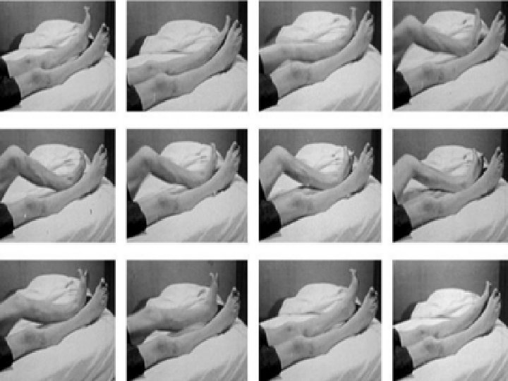
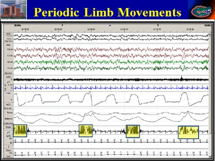
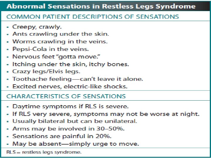
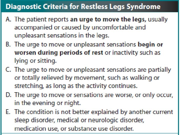
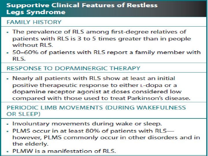
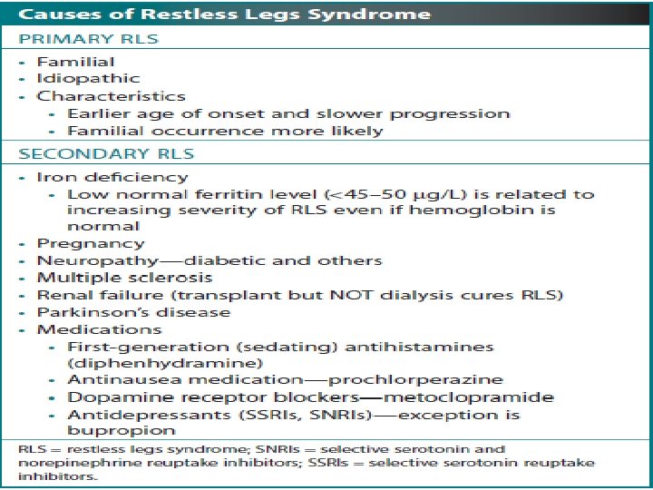
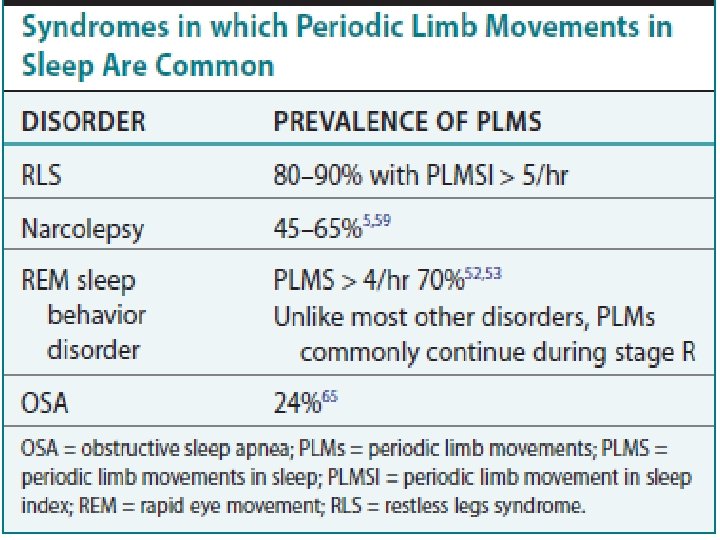
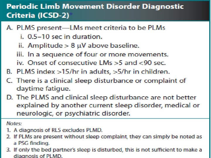
Periodic limb movements / Restless Leg Syndrome • Periodic limb movements also may appear during relaxed wakefulness(PLMW), but in such cases they continue during light sleep stages (PLMS). • PLMS consist of dorsiflexion of the big toe and foot and flexion of the knee and hip, even if they may coexist with Propriospinal myoclonus (PSM) in some patients with RLS. In such cases, PLMS and PSM could coexisting either by chance but due to different pathophysiological mechanisms as suggested by the temporal relationship of these two motor phenomena: PSM appears only during relaxed wakefulness and disappears with sleep onset, when PLMS in turn arise. Stephan Eisenschenk, MD Department of Neurology
50 Stephan Eisenschenk, MD Department of Neurology
51 Stephan Eisenschenk, MD Department of Neurology
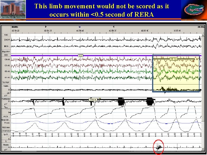
This limb movement would not be scored as it occurs within <0. 5 second of RERA 52 Stephan Eisenschenk, MD Department of Neurology
53 Stephan Eisenschenk, MD Department of Neurology
54 Stephan Eisenschenk, MD Department of Neurology
55 Stephan Eisenschenk, MD Department of Neurology
56 Stephan Eisenschenk, MD Department of Neurology
57 Stephan Eisenschenk, MD Department of Neurology
58 Stephan Eisenschenk, MD Department of Neurology
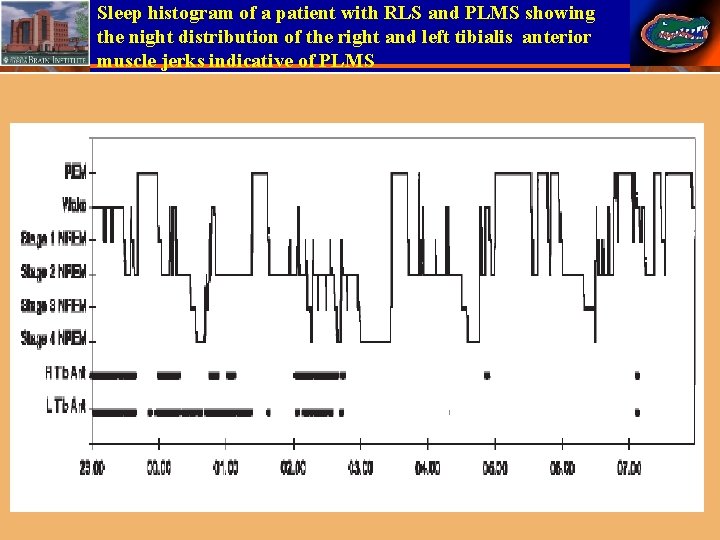
Sleep histogram of a patient with RLS and PLMS showing the night distribution of the right and left tibialis anterior muscle jerks indicative of PLMS 59 Stephan Eisenschenk, MD Department of Neurology
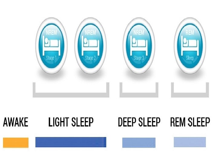
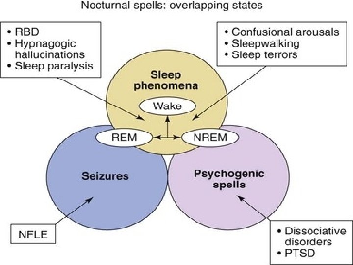
Parasomnias • Parasomnias are a category of sleep disorders that involve abnormal movements, behaviors, emotions, perceptions, and dreams that occur while falling asleep, sleeping, between sleep stages, or during arousal from sleep. • Most parasomnias are dissociated sleep states which are partial arousals during the transitions between wakefulness and NREM sleep, or wakefulness and REM sleep. 60 Stephan Eisenschenk, MD Department of Neurology
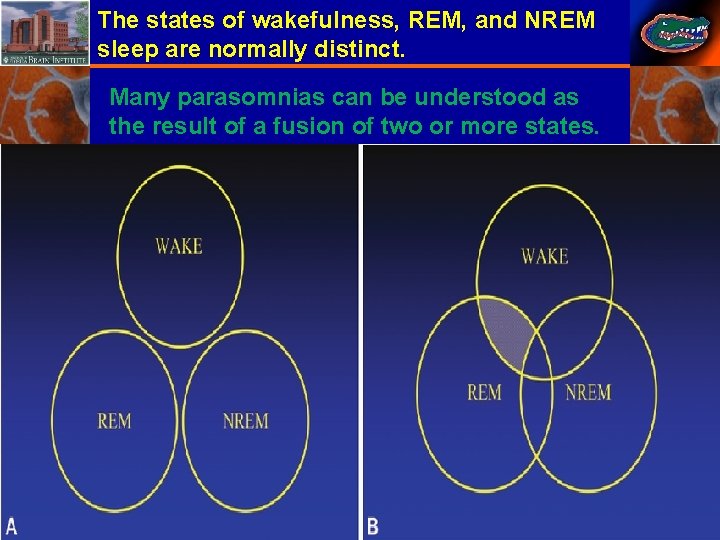
The states of wakefulness, REM, and NREM sleep are normally distinct. Many parasomnias can be understood as the result of a fusion of two or more states. 61 Stephan Eisenschenk, MD Department of Neurology
62 Stephan Eisenschenk, MD Department of Neurology
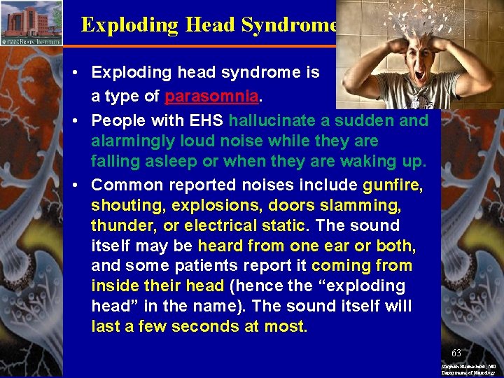
Exploding Head Syndrome • Exploding head syndrome is a type of parasomnia. • People with EHS hallucinate a sudden and alarmingly loud noise while they are falling asleep or when they are waking up. • Common reported noises include gunfire, shouting, explosions, doors slamming, thunder, or electrical static. The sound itself may be heard from one ear or both, and some patients report it coming from inside their head (hence the “exploding head” in the name). The sound itself will last a few seconds at most. 63 Stephan Eisenschenk, MD Department of Neurology
Exploding Head Syndrome • People may see flashes of bright light or feel an electrical sensation or shock traveling from their torso to their head. • Feelings of anxiety, including increased heart rate and heart palpitations, often accompany the sound. As a result, sufferers have a tougher time falling back asleep due to their heightened arousal. • In fact, even though the disorder itself is harmless and painless, the anxiety and fear of an episode occurring can create anxiety around going to sleep at all, contributing to insomnia and resulting daytime fatigue. 64 Stephan Eisenschenk, MD Department of Neurology
Exploding Head Syndrome • Fortunately, people with EHS tend to experience episodes less frequently as time goes on: 10 to 18 percent of people experience EHS at least once in their lifetime. • Individuals with isolated sleep paralysis are twice as likely to experience EHS, and people with psychiatric disorders are slightly more likely than the general population to have EHS. • Having high levels of emotional or physical stress are also associated with the disorder. 65 Stephan Eisenschenk, MD Department of Neurology
Exploding Head Syndrome • Individuals with Post trauma stress disorder may be roused by a bomb going off in a nightmare; this is distinct from EHS because the sounds associated with EHS are random and without context. • When you wake up from EHS, remembering that the sound is not real, and that it is harmless, can provide relief. • In fact, getting the reassurance from your doctor can be enough for many to go into remission. • Engage in stress-relieving activities like yoga, meditation, deep breathing exercises. Establish a warm bath or aromatherapy. 66 Stephan Eisenschenk, MD Department of Neurology
Hypnagogic Hallucination • Hypnagogic hallucinations are imagined sensations that seem very real. They occur as a person is falling asleep, and are also referred to as sleep hallucinations. • A hallucination is anything that can be sensed but is not real. • A hallucinated smell, taste, vision, or sound is only experienced in a person's mind and not by others. • They are often associated with narcolepsy. • These hallucinations are relatively common in teenagers and young adults. • A regular sleep schedule, going to bed and getting up at the same times, can help. 67 Stephan Eisenschenk, MD Department of Neurology
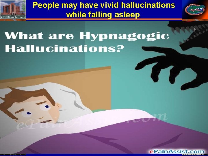
People may have vivid hallucinations while falling asleep. 68 Stephan Eisenschenk, MD Department of Neurology
Hypnagogic Hallucination • hypnagogic hallucinations may be caused by Parkinson's disease or schizophrenia. • A person will experience vivid hallucinations as they fall asleep, or just before falling asleep. • These can be images, smells, tastes, tactile sensations, or sounds. • A person may feel as if they are moving while their body is still. This sensation could be a feeling of falling or flying. • The most common hypnagogic hallucinations are visual. They may include images of people, animals, or moving objects. Images can be quite complex and detailed, and may not make any sense. 69 Stephan Eisenschenk, MD Department of Neurology
Hypnagogic Hallucination • A person knows that they are awake. • The images, sounds, or other sensations may last a number of minutes. They may prevent a person from falling asleep. • These hallucinations may happen at the same time as sleep paralysis. • The key difference between a dream and a hypnagogic hallucination is that the hallucination will feel very real. • A person may feel sure that they have seen or felt something, and this can be frightening or confusing. • They tend to occur less frequently as a person ages, and women are more likely to experience these hallucinations than men. 70 Stephan Eisenschenk, MD Department of Neurology
Hypnagogic Hallucination • If a person uses drugs or alcohol, had anxiety and insomnia , they may be more likely to experience hypnagogic hallucinations. • Hypnagogic hallucinations are not usually a risk to health EXCEPT : 1 - Symptoms of narcolepsy: Ccataplexy, being very sleepy during the day, and having a disturbed sleep at night. 2 - Symptoms of schizophrenia: hearing voices, having confused thoughts, and experiencing changes in behavior. 3 - Symptoms of Parkinson's disease: slow movement, muscle stiffness, and shaking in the hands and other parts of the body. 71 Stephan Eisenschenk, MD Department of Neurology
Hypnagogic Hallucination • If there is no underlying medical condition, getting enough sleep and avoiding drugs and alcohol can reduce their frequency. • When these hallucinations are not caused by an underlying condition, they usually do not have long-term complications. • Their most common effects are disturbed sleep, and stress or anxiety. • hypnagogic hallucinations can cause a person to wake in terror and scream or shout, which may disturb a partner or roommate. • A person experiencing a hallucination may fall out of bed or injure themselves. 72 Stephan Eisenschenk, MD Department of Neurology
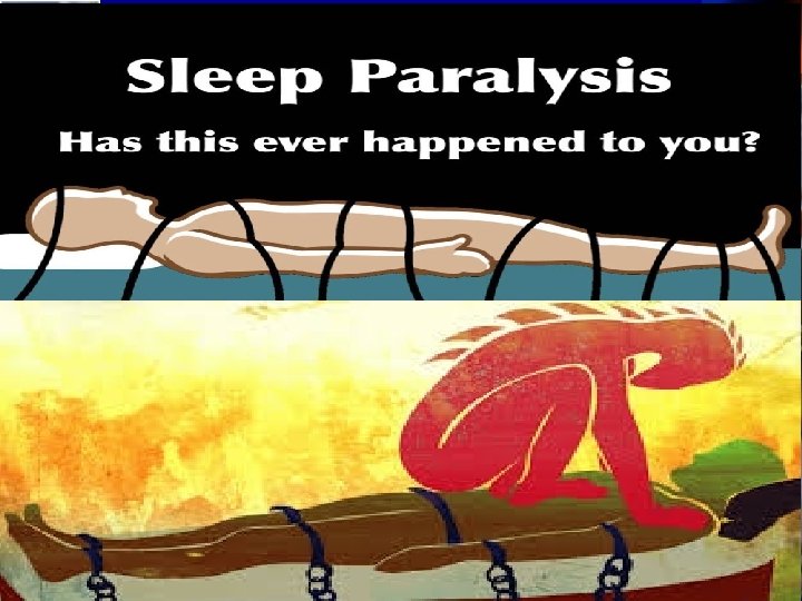
Sleep Paralysis • Sleep paralysis (SP) occurs when rapid eye movement (REM)-based atonia perseverates into wakefulness. • Most individuals with SP also experience dream activity during the conscious paralysis that is vivid, multi-sensorial, and often negatively valenced. • The combination of atonia and waking nightmares makes SP a very unpleasant experience for most people. • 7. 6% of the general population experienced at least one SP episode over the course of their life. 73 Stephan Eisenschenk, MD Department of Neurology
74 Stephan Eisenschenk, MD Department of Neurology
Sleep Paralysis • Higher lifetime prevalence rates were found in students (28. 3%) and psychiatric patients(31. 9%), withfemales experiencing SP slightly more often than males. • The presence of poor sleep and/or sleep disruption is associated with SP. Panic disorder , generalized anxiety disorder , death anxiety, and social anxiety are frequent symptomatic accompaniments to SP. • Isolated SP episodes are characterized by muscle atonia upon sleep onset or offset. • Eye movements are intact and there may be some control over respiration, but feelings of suffocation are common. 75 Stephan Eisenschenk, MD Department of Neurology
Sleep Paralysis • Episodes are usually brief (usually seconds to 20 minutes). • In order to qualify as “recurrent isolated” SP(RISP), episodes should not be better explained by other sleep disorders (eg, narcolepsy), medication effects, or other substances. • As per the current ICSD criteria, RISP consists of multiple episodes of isolated SP that are associated with clinically significant distress (eg, anxiety and/or fear related to the bedroom/sleep). • Hallucinations are often present during episodes, but are not necessary for a diagnosis. 76 Stephan Eisenschenk, MD Department of Neurology
Sleep Paralysis • These criteria for “fearful” RISP require clinically significant fear during episodes and at least two episodes in the past 6 months. • Distress during episodes is quite common, given the characteristic atonia and the possible presence of hallucinations and breathing difficulties. • Clinicians should also rule out focal epileptic seizures, atonic seizures, cataplexy, various types of familial periodic paralysis , and transient compression neuropathies. 77 Stephan Eisenschenk, MD Department of Neurology
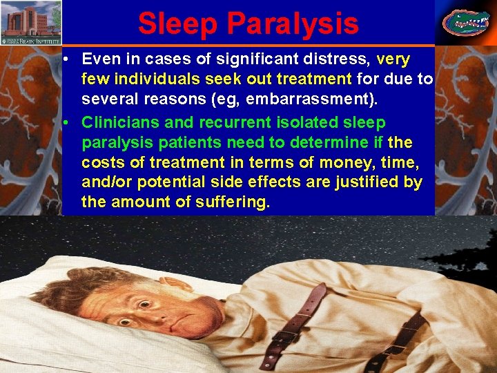
Sleep Paralysis • Even in cases of significant distress, very few individuals seek out treatment for due to several reasons (eg, embarrassment). • Clinicians and recurrent isolated sleep paralysis patients need to determine if the costs of treatment in terms of money, time, and/or potential side effects are justified by the amount of suffering. 78 Stephan Eisenschenk, MD Department of Neurology
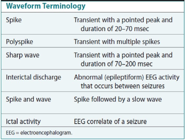
Sleep Related Hypermotor Epilepsy • Epilepsy is the chronic condition of recurrent unprovoked seizures. • The terminology and identification of epileptic seizures and interictal activity is challenging for physicians without extensive training in EEG. • A transient is any isolated wave or complex that stands out compared with background activity. • A spike is defined as a transient with a pointed peak and a duration of 20 to 70 msec. On a 30 second page, spikes look like a single vertical line. • A sharp wave is a transient with a deflection of 70 to 200 msec. 79 Stephan Eisenschenk, MD Department of Neurology
Sleep Related Hypermotor Epilepsy • A spike and wave complex is a spike followed by a slow wave (usually wide and often higher amplitude). • Polyspike complexes often consist of multiple spikes superimposed on a slow wave. • The term epileptiform activity literally means “EEG activity resembling that found in patients with epilepsy. ”This is a somewhat circular definition. • Interictal activity is defined as abnormal EEG activity that occurs between seizures. 80 Stephan Eisenschenk, MD Department of Neurology
Sleep Related Hypermotor Epilepsy • Epileptiform activity includes spikes, spike and waves, and polyspike complexes. • Abnormal sharp waves are also considered epileptiform or interictal activity. Sharp waves can be normal (e. g. , vertex sharp waves). • Artifacts can sometimes mimic spikes or spikes and waves. True spike and wave complexes have a “field”(true spike and wave activity should be seen in derivations containing several contiguous electrodes). If a spike and wave is seen only in a single derivation, the activity is usually be an artifact. 81 Stephan Eisenschenk, MD Department of Neurology
82 Stephan Eisenschenk, MD Department of Neurology

83 Stephan Eisenschenk, MD Department of Neurology
Sleep Related Hypermotor Epilepsy • Because seizures do not always appear during a given recording, the physician reading an EEG or PSG searches for spikes and/or abnormal sharp waves that may represent the interictal footprint of possible seizure activity. • It should be noted that not all patients with spikes have seizures and not all patients with seizures have detectable interictal activity. • Spikes represent abnormal brain activity that is seen as an area of negativity at the scalp. • Spikes can be localized (negativity at the scalp over one area of the brain) or appear diffusely. 84 Stephan Eisenschenk, MD Department of Neurology
Sleep Related Hypermotor Epilepsy • Focal seizures usually, though not invariably, begin at the same location as the inter-ictal spikes. • The classic spike is followed by a slow wave. Although most common post-ictally, spike and sharp waves will occur sporadically at any time and may have a slight increase pre-ictally. • Using the usual 30 -second time window, it may be difficult to appreciate spikes. A switch to a 10 -second window is indicated to scrutinize any suspicious paroxysmal activity. 85 Stephan Eisenschenk, MD Department of Neurology

86 Stephan Eisenschenk, MD Department of Neurology
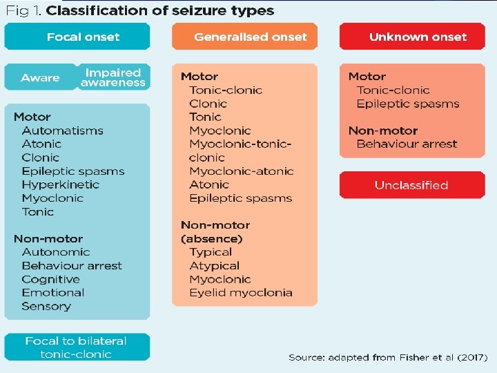
The etiologies ( structural, genetic, infectious, metabolic, immune, and unknown). The etiology and comorbidities should be considered at each stage of epilepsy classification 87 Stephan Eisenschenk, MD Department of Neurology
88 Stephan Eisenschenk, MD Department of Neurology
89 Stephan Eisenschenk, MD Department of Neurology
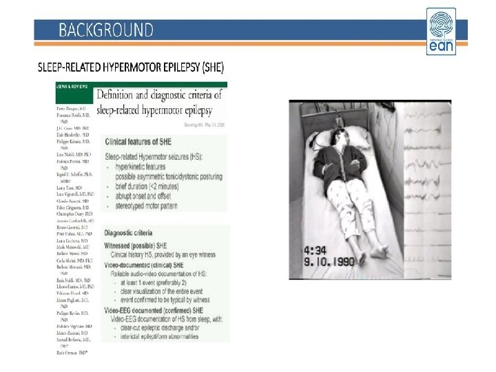
Sleep related hypermotor epilepsy • Sleep-related hypermotor epilepsy (SHE), previously known as nocturnal frontal lobe epilepsy (NFLE), is a rare form of focal epilepsy characterized by hypermotor seizures occurring predominantly during non-REM sleep. • The most common clinical expression consists of “hypermotor” events characterized by vigorous hyperkinetic features (complex body movements with kicking or cycling of limbs and rocking body movements), usually with vegetative signs, vocalization, and emotional facial expression. 90 Stephan Eisenschenk, MD Department of Neurology
Sleep related hypermotor epilepsy • Asymmetric tonic/dystonic seizures with or without head/eye deviation are also observed. Typically, patients with SHE present a series of sleep related motor events becoming increasingly long and complex, even during the same night. • Clinical features range from brusque stereotyped arousals (i. e. , paroxysmal arousals), repeated throughout the night, to complex hypermotor seizures and, more seldom, protracted ambulatory behavior known as epileptic nocturnal wandering. • Seizure onset may be at any age with a peak during childhood and adolescence. Stephan Eisenschenk, MD Department of Neurology
Sleep related hypermotor epilepsy • Seizure frequency may be very high, with occurrence either every night or almost every night, usually many times per night. • Seizures are abrupt in onset and offset, typically brief and have a highly stereotyped motor pattern within individuals. 92 Stephan Eisenschenk, MD Department of Neurology
Diagnosis of Sleep related hypermotor epilepsy 93 Stephan Eisenschenk, MD Department of Neurology
On the left NREM parasomnias, in the center REM disorders (e. g. RBD, REM behavior disorders, and nightmares), on the right Sleep Related Hypermotor Epilepsy (SHE), 94 Stephan Eisenschenk, MD Department of Neurology
Double-spike-and-slow-wave complexes are associated with myoclonic jerks of the right and left diaphragm, while no simultaneous EMG discharges are registered from the deltoid and wrist extensor muscles. 95 Stephan Eisenschenk, MD Department of Neurology
EEG-EMG recording of two hiccup seizures using an expanded time scale. The initial spikes at the left fronto -polar contact precede the onset of the right diaphragm myoclonia by 57 and 61 milliseconds, respectively. 96 Stephan Eisenschenk, MD Department of Neurology

97 Stephan Eisenschenk, MD Department of Neurology
- Slides: 97