
Hypertensive disorders in pregnancy Dr Wael Al Halaki




- Slides: 55
Hypertensive disorders in pregnancy Dr. Wael Al, Halaki Professor in Qasyoun Private university Hama University 2017
Hypertensive disorders in pregnancy
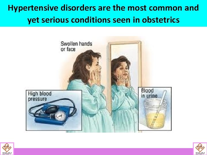
Hypertensive disorders are the most common and yet serious conditions seen in obstetrics
INTRODUCTION • Incidence is 5 -10%. • The most frequent cause of iatrogenic prematurity. • Preterm delivery • Intrauterine growth restriction (IUGR) • Perinatal death • Maternal cerebrovascular accidents ( CVA). • Placental abruption.
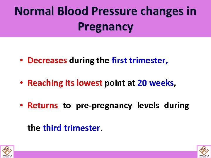
Normal Blood Pressure changes in Pregnancy • Decreases during the first trimester, • Reaching its lowest point at 20 weeks, • Returns to pre-pregnancy levels during the third trimester.
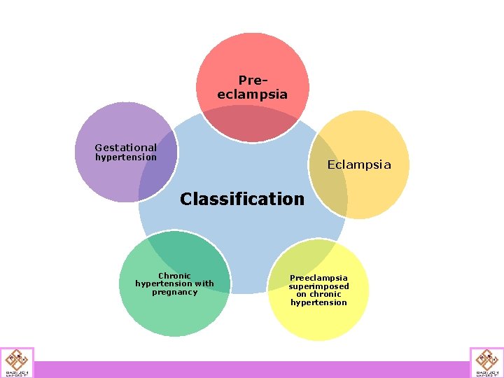
Preeclampsia Gestational hypertension Eclampsia Classification Chronic hypertension with pregnancy Preeclampsia superimposed on chronic hypertension
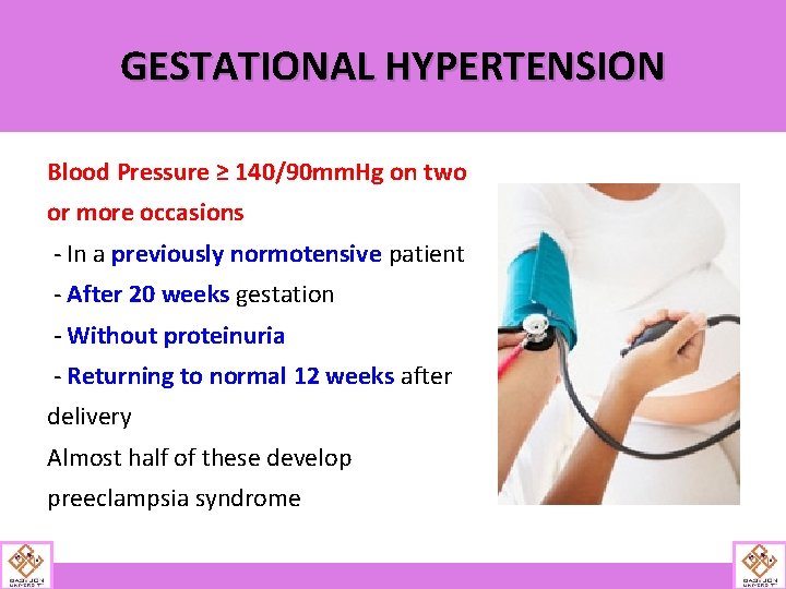
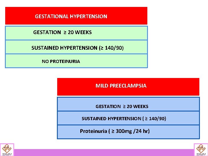
GESTATIONAL HYPERTENSION Blood Pressure ≥ 140/90 mm. Hg on two or more occasions - In a previously normotensive patient - After 20 weeks gestation - Without proteinuria - Returning to normal 12 weeks after delivery Almost half of these develop preeclampsia syndrome
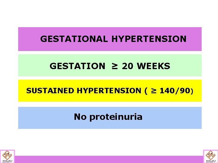
GESTATIONAL HYPERTENSION GESTATION ≥ 20 WEEKS SUSTAINED HYPERTENSION ( ≥ 140/90) No proteinuria
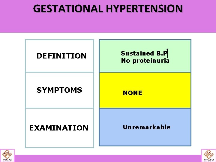
GESTATIONAL HYPERTENSION DEFINITION Sustained B. P No proteinuria SYMPTOMS NONE EXAMINATION Unremarkable
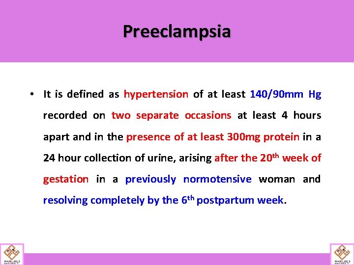
Preeclampsia • It is defined as hypertension of at least 140/90 mm Hg recorded on two separate occasions at least 4 hours apart and in the presence of at least 300 mg protein in a 24 hour collection of urine, arising after the 20 th week of gestation in a previously normotensive woman and resolving completely by the 6 th postpartum week.
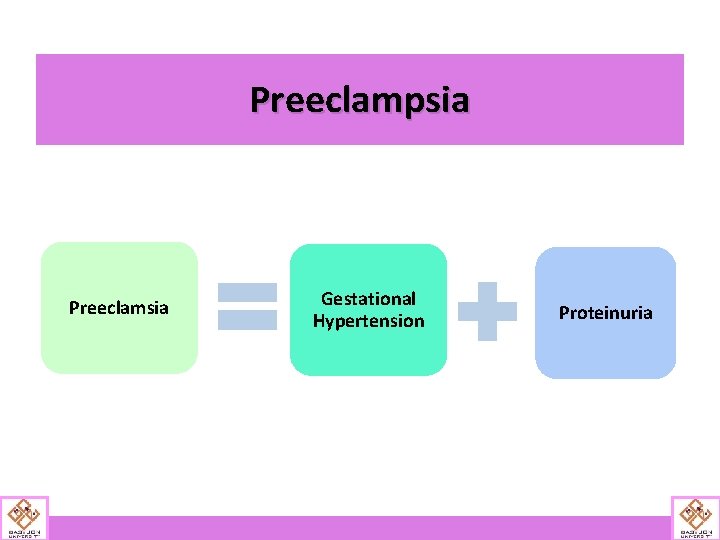
Preeclampsia Preeclamsia Gestational Hypertension Proteinuria
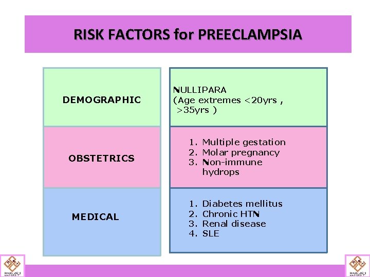
RISK FACTORS for PREECLAMPSIA DEMOGRAPHIC OBSTETRICS MEDICAL NULLIPARA (Age extremes <20 yrs , >35 yrs ) 1. Multiple gestation 2. Molar pregnancy 3. Non-immune hydrops 1. 2. 3. 4. Diabetes mellitus Chronic HTN Renal disease SLE
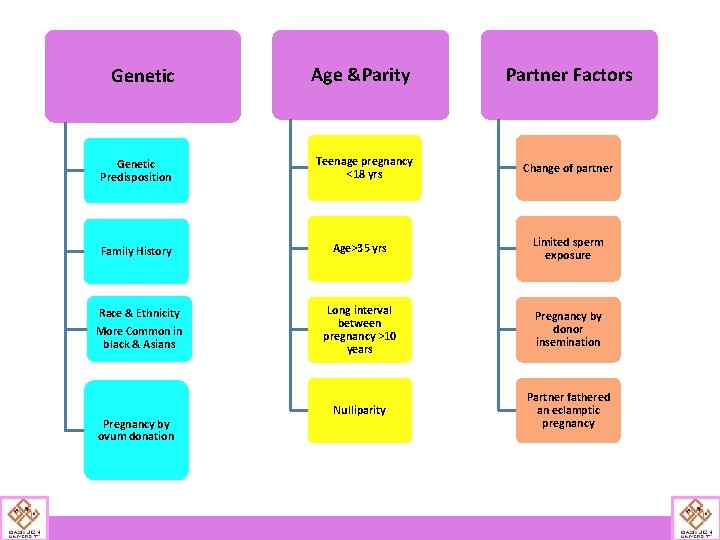
Genetic Age &Parity Genetic Predisposition Teenage pregnancy <18 yrs Partner Factors Change of partner Family History Age>35 yrs Limited sperm exposure Race & Ethnicity More Common in black & Asians Long interval between pregnancy >10 years Pregnancy by donor insemination Nulliparity Partner fathered an eclamptic pregnancy Pregnancy by ovum donation
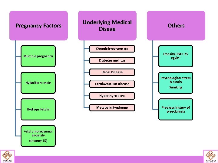
Pregnancy Factors Underlying Medical Diseae Others Chronic hypertension Multiple pregnancy Diabetes mellitus Obesity BMI> 35 kg/m 2 Renal Disease Hydatiform mole Cardiovascular disease Psychological stress & strain Smoking Hyperthyroidism Metabolic Syndrome Hydrops fetalis • Hyperhomocysteinemia , • Autoimmune disease Fetal chromosomal anomaly • Antiphospholipid antibodies, (trisomy 13) • Thrombophilia Previous history of preeclamsia
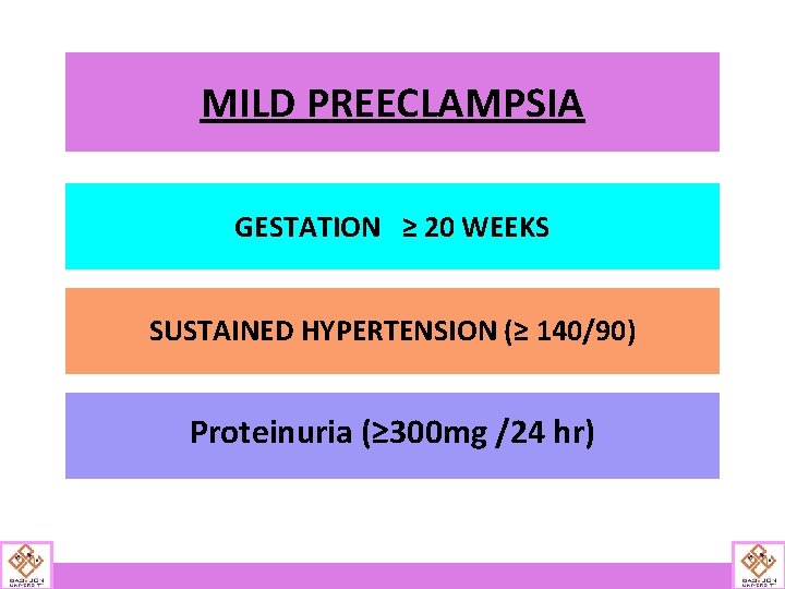
MILD PREECLAMPSIA GESTATION ≥ 20 WEEKS SUSTAINED HYPERTENSION (≥ 140/90) Proteinuria (≥ 300 mg /24 hr)
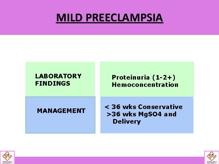
MILD PREECLAMPSIA LABORATORY FINDINGS MANAGEMENT Proteinuria (1 -2+) Hemoconcentration < 36 wks Conservative >36 wks Mg. SO 4 and Delivery
GESTATIONAL HYPERTENSION GESTATION ≥ 20 WEEKS SUSTAINED HYPERTENSION (≥ 140/90) NO PROTEINURIA MILD PREECLAMPSIA GESTATION ≥ 20 WEEKS SUSTAINED HYPERTENSION ( ≥ 140/90) Proteinuria ( ≥ 300 mg /24 hr)
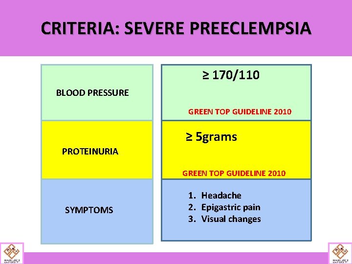
CRITERIA: SEVERE PREECLEMPSIA ≥ 170/110 BLOOD PRESSURE GREEN TOP GUIDELINE 2010 ≥ 5 grams PROTEINURIA GREEN TOP GUIDELINE 2010 SYMPTOMS 1. Headache 2. Epigastric pain 3. Visual changes
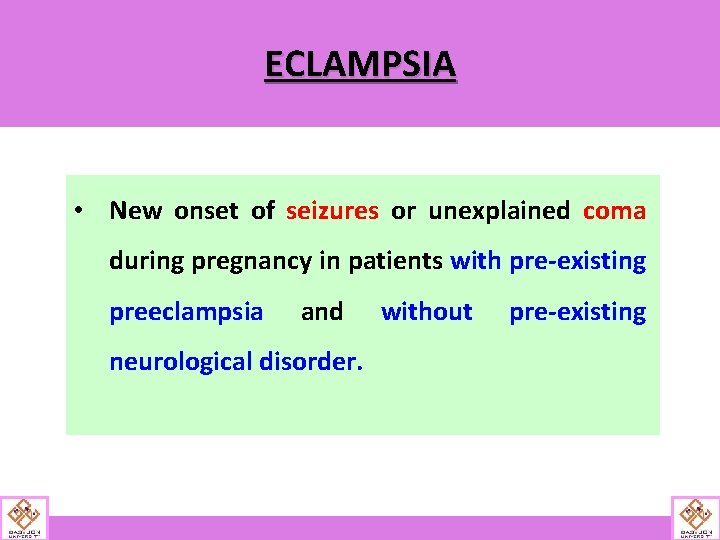
ECLAMPSIA • New onset of seizures or unexplained coma during pregnancy in patients with pre-existing preeclampsia and neurological disorder. without pre-existing
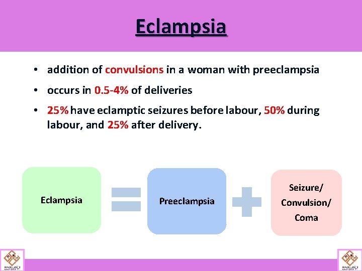
Eclampsia • addition of convulsions in a woman with preeclampsia • occurs in 0. 5 -4% of deliveries • 25% have eclamptic seizures before labour, 50% during labour, and 25% after delivery. Seizure/ Eclampsia Preeclampsia Convulsion/ Coma
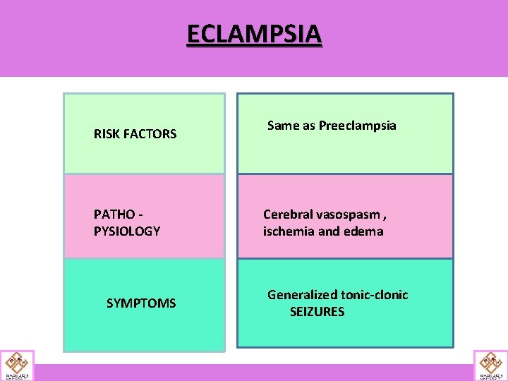
ECLAMPSIA RISK FACTORS PATHO PYSIOLOGY SYMPTOMS Same as Preeclampsia Cerebral vasospasm , ischemia and edema Generalized tonic-clonic SEIZURES
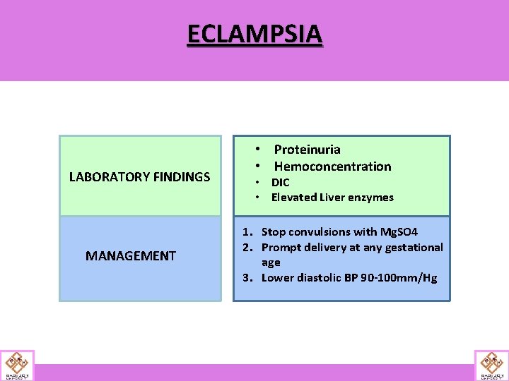
ECLAMPSIA LABORATORY FINDINGS MANAGEMENT • Proteinuria • Hemoconcentration • DIC • Elevated Liver enzymes 1. Stop convulsions with Mg. SO 4 2. Prompt delivery at any gestational age 3. Lower diastolic BP 90 -100 mm/Hg
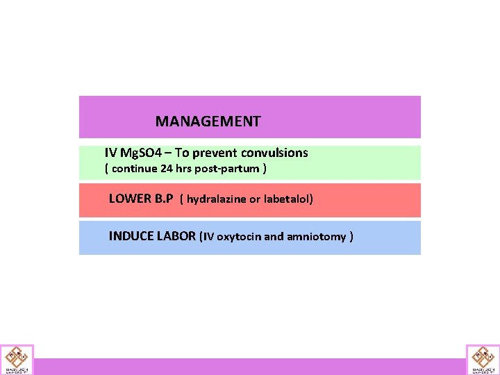
MANAGEMENT IV Mg. SO 4 – To prevent convulsions ( continue 24 hrs post-partum ) LOWER B. P ( hydralazine or labetalol) INDUCE LABOR (IV oxytocin and amniotomy )
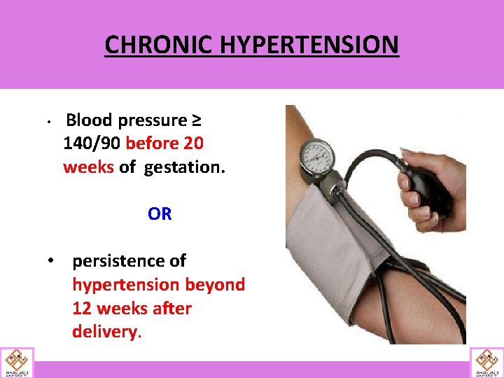
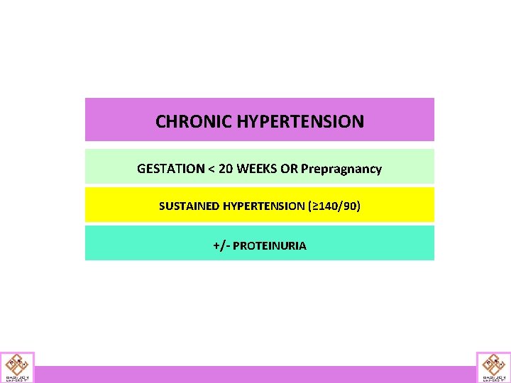
CHRONIC HYPERTENSION • Blood pressure ≥ 140/90 before 20 weeks of gestation. OR • persistence of hypertension beyond 12 weeks after delivery.
CHRONIC HYPERTENSION GESTATION < 20 WEEKS OR Prepragnancy SUSTAINED HYPERTENSION (≥ 140/90) +/- PROTEINURIA
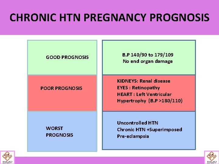
CHRONIC HTN PREGNANCY PROGNOSIS GOOD PROGNOSIS POOR PROGNOSIS WORST PROGNOSIS B. P 140/90 to 179/109 No end organ damage KIDNEYS: Renal disease EYES : Retinopathy HEART : Left Ventricular Hypertrophy (B. P >180/110) Uncontrolled HTN Chronic HTN +Superimposed Pre-eclampsia
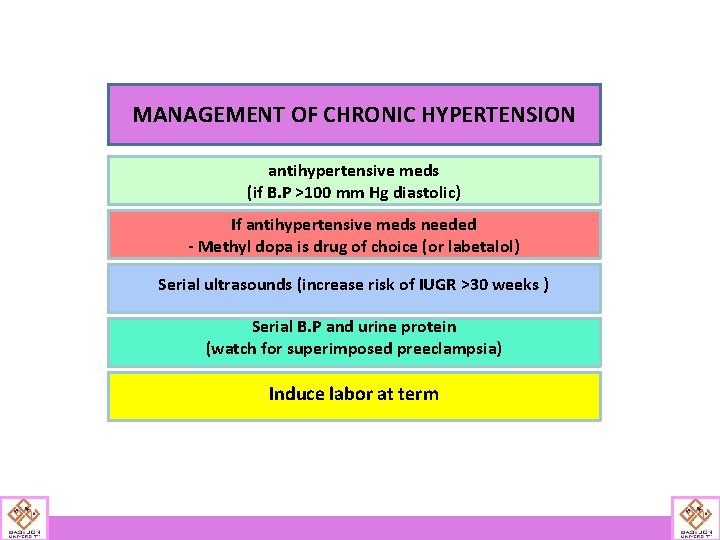
MANAGEMENT OF CHRONIC HYPERTENSION antihypertensive meds (if B. P >100 mm Hg diastolic) If antihypertensive meds needed - Methyl dopa is drug of choice (or labetalol) Serial ultrasounds (increase risk of IUGR >30 weeks ) Serial B. P and urine protein (watch for superimposed preeclampsia) Induce labor at term
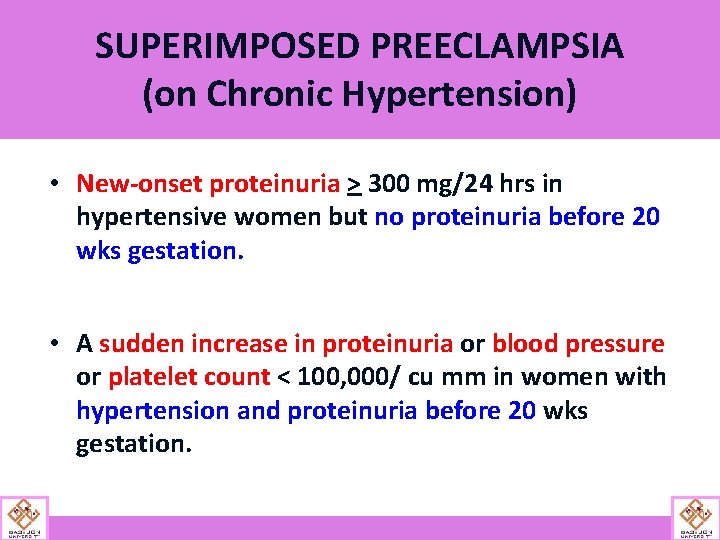
SUPERIMPOSED PREECLAMPSIA (on Chronic Hypertension) • New-onset proteinuria > 300 mg/24 hrs in hypertensive women but no proteinuria before 20 wks gestation. • A sudden increase in proteinuria or blood pressure or platelet count < 100, 000/ cu mm in women with hypertension and proteinuria before 20 wks gestation.
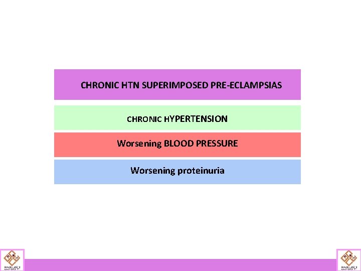
CHRONIC HTN SUPERIMPOSED PRE-ECLAMPSIAS CHRONIC HYPERTENSION Worsening BLOOD PRESSURE Worsening proteinuria
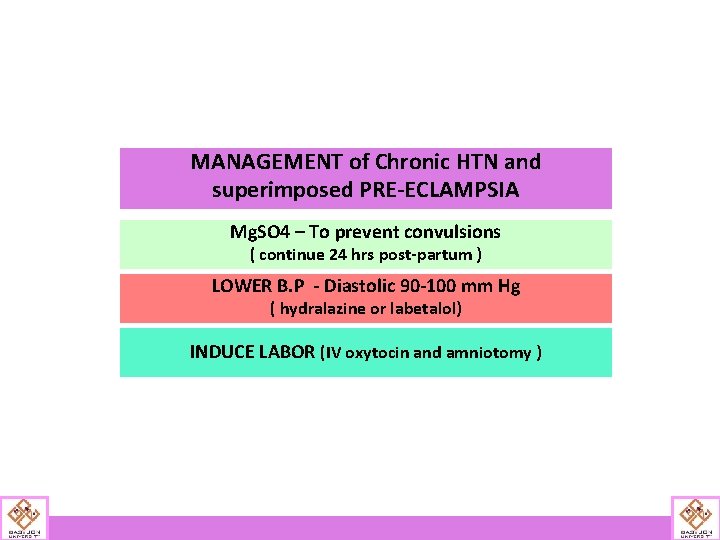
MANAGEMENT of Chronic HTN and superimposed PRE-ECLAMPSIA Mg. SO 4 – To prevent convulsions ( continue 24 hrs post-partum ) LOWER B. P - Diastolic 90 -100 mm Hg ( hydralazine or labetalol) INDUCE LABOR (IV oxytocin and amniotomy )
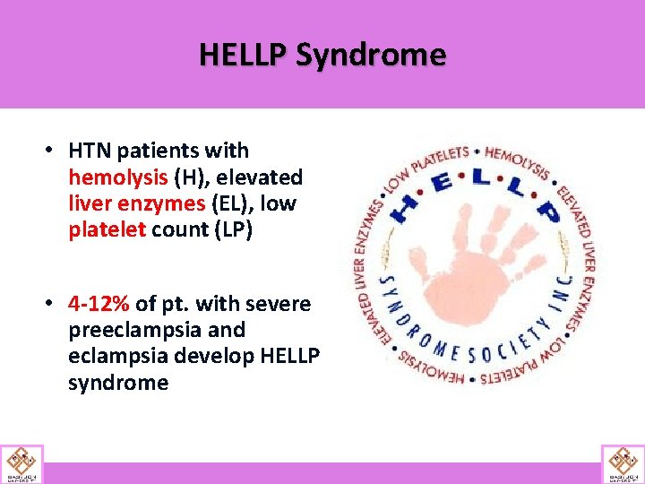
HELLP Syndrome • HTN patients with hemolysis (H), elevated liver enzymes (EL), low platelet count (LP) • 4 -12% of pt. with severe preeclampsia and eclampsia develop HELLP syndrome
HELLP Syndrome • Cardiovascular stabilization, correction of coagulation abnormalities, and delivery • PLT transfusion before or after delivery if PLT count is <20, 000/mm 3 (advised at <50, 000/mm 3 before cesarean) • <32 weeks gestation; steroid therapy may help stabilize maternal PLT count
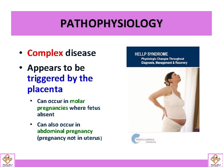
PATHOPHYSIOLOGY • Complex disease • Appears to be triggered by the placenta • Can occur in molar pregnancies where fetus absent • Can also occur in abdominal pregnancy (pregnancy not in uterus)
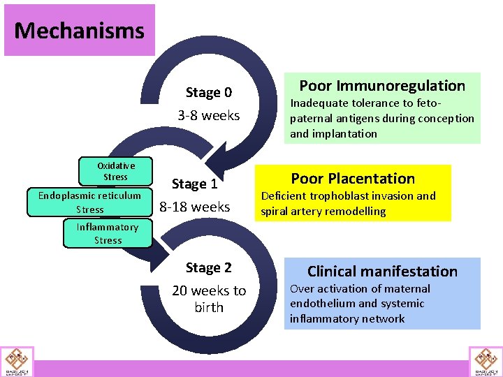
Mechanisms Stage 0 3 -8 weeks Oxidative Stress Endoplasmic reticulum Stress Stage 1 8 -18 weeks Poor Immunoregulation Inadequate tolerance to fetopaternal antigens during conception and implantation Poor Placentation Deficient trophoblast invasion and spiral artery remodelling Inflammatory Stress Stage 2 20 weeks to birth Clinical manifestation Over activation of maternal endothelium and systemic inflammatory network
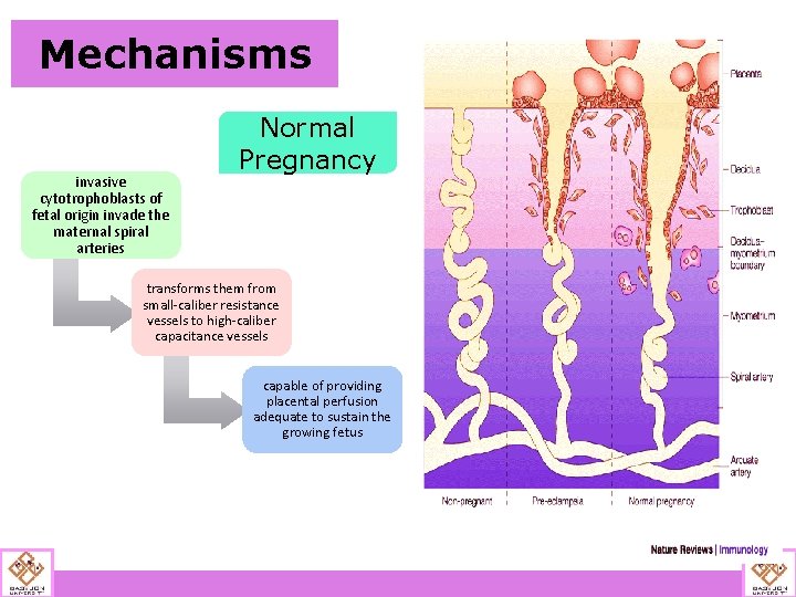
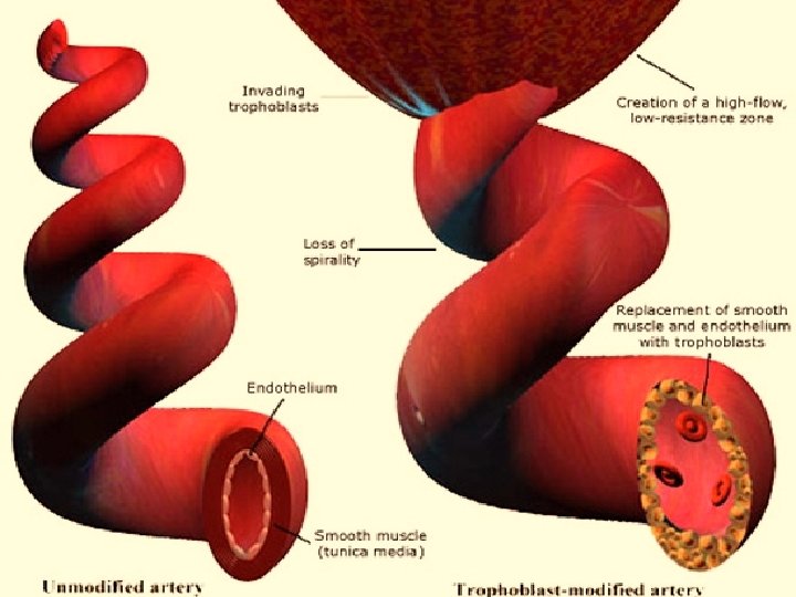
Mechanisms invasive cytotrophoblasts of fetal origin invade the maternal spiral arteries Normal Pregnancy transforms them from small-caliber resistance vessels to high-caliber capacitance vessels capable of providing placental perfusion adequate to sustain the growing fetus
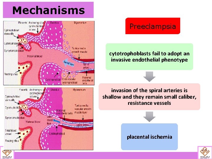
Mechanisms Preeclampsia cytotrophoblasts fail to adopt an invasive endothelial phenotype invasion of the spiral arteries is shallow and they remain small caliber, resistance vessels placental ischemia
Pre-eclampsia is two stage disease Maternal Syndrome Fetal Syndrome
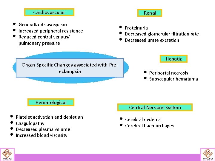
Cardiovascular • Generalized vasospasm • Increased peripheral resistance • Reduced central venous/ pulmonary pressure Organ Specific Changes associated with Preeclampsia Hematological • Platelet activation and depletion • Coagulopathy • Decreased plasma volume • Increased blood viscosity Renal • Proteinuria • Decreased glomerular filtration rate • Decreased urate excretion Hepatic • Periportal necrosis • Subscapular hematoma Central Nervous System • Cerebral oedema • Cerebral haemorrhages
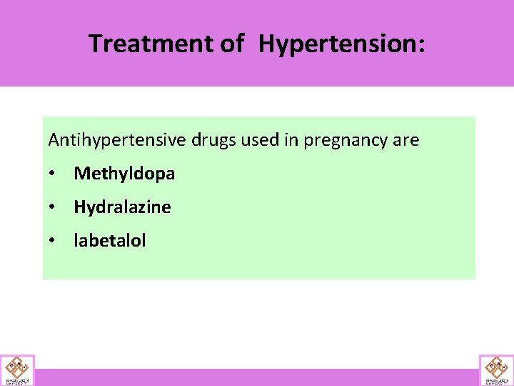
Treatment of Hypertension: Antihypertensive drugs used in pregnancy are • Methyldopa • Hydralazine • labetalol
Seizure Prophylaxis • Routinely used in severe PE. • Magnesium sulphate: most commonly used. • Initiated with onset of labor till 24 h postpartum. • For caesarean, started 2 hrs before the section till 12 hrs postpartum.
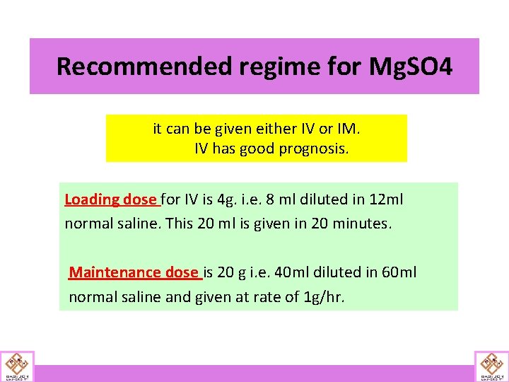
Recommended regime for Mg. SO 4 it can be given either IV or IM. IV has good prognosis. Loading dose for IV is 4 g. i. e. 8 ml diluted in 12 ml normal saline. This 20 ml is given in 20 minutes. Maintenance dose is 20 g i. e. 40 ml diluted in 60 ml normal saline and given at rate of 1 g/hr.
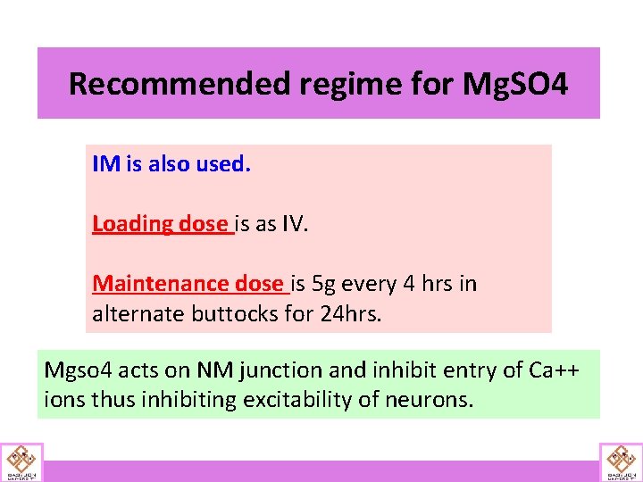
Recommended regime for Mg. SO 4 IM is also used. Loading dose is as IV. Maintenance dose is 5 g every 4 hrs in alternate buttocks for 24 hrs. Mgso 4 acts on NM junction and inhibit entry of Ca++ ions thus inhibiting excitability of neurons.
Management of Mg. SO 4 Toxicity Calcium gluconate is antagonist for Mg. SO 4. it is usually given as 10 ml of 10% Calcium gluconate in 10 minutes
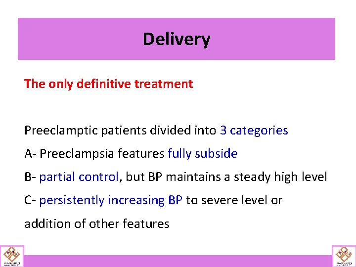
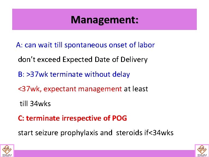
Delivery The only definitive treatment Preeclamptic patients divided into 3 categories A- Preeclampsia features fully subside B- partial control, but BP maintains a steady high level C- persistently increasing BP to severe level or addition of other features
Management: A: can wait till spontaneous onset of labor don’t exceed Expected Date of Delivery B: >37 wk terminate without delay <37 wk, expectant management at least till 34 wks C: terminate irrespective of POG start seizure prophylaxis and steroids if<34 wks
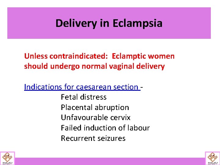
Delivery in Eclampsia Unless contraindicated: Eclamptic women should undergo normal vaginal delivery Indications for caesarean section Fetal distress Placental abruption Unfavourable cervix Failed induction of labour Recurrent seizures
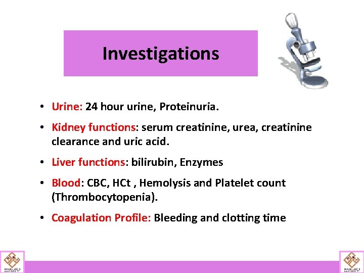
Investigations • Urine: 24 hour urine, Proteinuria. • Kidney functions: serum creatinine, urea, creatinine clearance and uric acid. • Liver functions: bilirubin, Enzymes • Blood: CBC, HCt , Hemolysis and Platelet count (Thrombocytopenia). • Coagulation Profile: Bleeding and clotting time
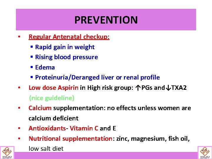
PREVENTION • • • Regular Antenatal checkup: § Rapid gain in weight § Rising blood pressure § Edema § Proteinuria/Deranged liver or renal profile Low dose Aspirin in High risk group: ↑PGs and↓TXA 2 (nice guideline) Calcium supplementation: no effects unless women are calcium deficient Antioxidants- Vitamin C and E Nutritional supplementation: zinc, magnesium, fish oil, low salt diet
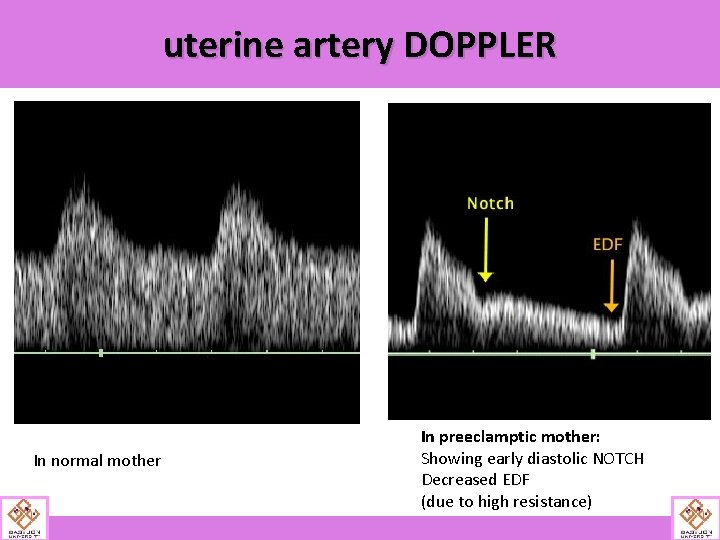
Uterine Artery Doppler Velocimetry (abnormal flow resistance/ diastolic notch in 2 nd/ 3 rd trimester) • is most promising, but currently, none of them is completely suitable for clinical use. (Conde-Agudelo, 2014; Kleinrouweler, 2012; Myatt, 2012 a). • These have value for fetal-growth restriction but not preeclampsia (ACOG, 2013 a). • As a result of these trials, some methods to prevent Preeclampsia have been theorized…
uterine artery DOPPLER In normal mother In preeclamptic mother: Showing early diastolic NOTCH Decreased EDF (due to high resistance)