Hypertension in children Introduction high BP including HTN

Hypertension in children

Introduction : high BP, including HTN, is defined from normative distribution of BP data in healthy children. This is in contrast to adult HTN, which is primarily defined by clinical outcome data (ie, risk of cardiovascular disease [CVD] and mortality) from large trials of antihypertensive therapy. However, these outcome measures cannot be applied to children because cardiovascular (CV) events other than left ventricular hypertrophy do not typically occur in childhood So Hypertension is defined as average SBP and/or diastolic BP (DBP) that is ≥ 95 th percentile for gender, age, and height on at least 3 occasions.

• In children, definitions that categorize BP values were modified by the 2017 AAP guidelines into two age groups. Of note, the newly revised definitions for adolescents are aligned with adult guidelines for the detection of chronic elevated BP. With an acute elevation of BP, it is the magnitude and the rate of increase above baseline that determines the risk of serious morbidity and, at times, mortality.

Copyrights apply

• the diagnosis of persistent childhood HTN is made when repeat blood pressure (BP) values on three separate visits are greater than the 95 th percentile for the age, gender, and height of the patient, or it is ≥ 130/80 mm. Hg. Copyrights apply

-Because height and gender are important determinants of pediatric BP, BP levels are interpreted based on gender, age, and height. Copyrights apply

Example : 5 yr old girl that measured 115 cm …. What’s the normal BP for her ? Copyrights apply

Copyrights apply

Copyrights apply

Copyrights apply

Copyrights apply

Copyrights apply

Copyrights apply

SCREENING OF BP recommendations: ●For children without risk factors or conditions associated with HTN, BP is measured beginning at three years of age during annual health supervision visits. ●For children ≥ 3 years of age with risk factors for HTN, BP measurement is recommended at every health care encounter. ●Children <3 years of age with risk factors for HTN should have BP measurements taken at each health supervision. >>Children with systolic BP (SBP) or diastolic BP (DBP) that exceeds screening thresholds for age and sex require further evaluation, starting with repeat BP measurement. Copyrights apply

Copyrights apply

Copyrights apply

Copyrights apply

MEASUREMENT OF BLOOD PRESSURE • The diagnosis of HTN is dependent on accurate blood pressure (BP) measurement. If a high BP measurement is obtained by an oscillometric device, confirmation by ausculatory measurement is required for accuracy. • The variability of BP values due to procedural differences in the BP measurement was illustrated in a comparison of normal BP readings reported by 10 different investigators in which the BP values differed by as much as 20 mm. Hg. Confounding factors included cuff size, technique used (ie, patient position and the choice of fourth or fifth Korotkoff sound to determine diastolic BP [DBP]), the number of measurements made, and/or type of instruments used. Copyrights apply

• Cuff size and placement — A variety of different cuff sizes are available, including adult, large adult, and thigh cuffs. The correct choice of cuff is important for accurate BP measurement. If too small a cuff is used, the pressure generated by inflating the cuff may not be fully transmitted to the brachial artery. In this setting, the pressure in the cuff may be considerably higher than the intra-arterial pressure, leading to overestimation of the systolic pressure. On the other hand, too wide a cuff may produce lower readings than the actual intraarterial pressure. • The cuff size should have a bladder width that is approximately 40 percent of the circumference of the upper arm, measured midway between the olecranon and the acromion. The length of the cuff bladder should encircle 80 to 100 percent of the circumference of the upper arm midway between the olecranon and the acromion. The bladder width-to-length should be at least 1: 2. Copyrights apply

Copyrights apply

Auscultation • — The preferred method of BP measurement is auscultation using a mercury sphygmomanometer as this was how normative data were obtained. The mercury sphygmomanometer is the most accurate instrument, but its availability is restricted because of the potential risk of mercury poisoning. Aneroid sphygmomanometers are an appropriate substitute for mercury-containing devices and have been shown to be accurate if regularly calibrated against mercury sphygmomanometers Copyrights apply

Technique of BP measurement — The following steps are recommended to accurately measure BP by auscultation and compare values with normative data. ●Prior to BP measurement, stimulant drugs or food should be avoided. ●The BP should be measured after three to five minutes of rest in a quiet environment. The child should be seated with his/her back and feet in a supported position. In infants, BP is measured in a supine position. ●Measure BP when the heart rate is normal and steady to minimize the likelihood of obtaining falsely elevated readings. Because anxiety acutely raises both the heart rate and BP, the most reproducible readings are obtained when the pulse rate is both steady and within the normal range. ●The BP is measured by auscultation using the correct size and placement of the BP cuff, and by placing the bell of the stethoscope over the brachial artery pulse in the cubital fossa. The BP should be taken with the patient's right arm supported at the level of the heart. The right arm is preferred in repeated measures of BP for consistency and comparison with standard tables. In addition, the possibility of coarctation of the aorta would lead to falsely low BP readings in the left arm. Allowing the arm to hang below the heart will elevate BP levels by the added hydrostatic pressure induced by gravity (as much as 10 to 12 mm. Hg in adults). The sphygmomanometer should be visible but does not have to be at the level of the heart Copyrights apply.

●The cuff should be inflated to 20 to 30 mm. Hg above the anticipated systolic BP (SBP) and then deflated slowly at a rate of 2 to 3 mm. Hg per heartbeat. The systolic BP is equal to the pressure at which the brachial pulse can first be heard by auscultation (Korotkoff phase I). As the cuff is deflated below the SBP, the pulse continues to be heard until there is abrupt muffling (Korotkoff phase IV) followed by disappearance of sound (Korotkoff phase V). ●Phase V is recommended for DBP determination in children. In some children, Korotkoff sounds can be heard to 0 mm. Hg. If this occurs, BP measurement should be repeated with less pressure on the head of the stethoscope. If phase V is still very low, phase IV (muffling) should be recorded as the DBP with the added documentation noting the use of the phase IV to determine DBP. Copyrights apply

Number of measurements • — The BP should be taken at least twice on each visit, with the measurements separated by one to two minutes to allow the release of trapped blood. If the second value is more than 5 mm. Hg different from the first, continued measurements should be made until a stable value is attained. The recorded value on the patient's chart should be the average of the last two measurements. • A new diagnosis of HTN should not be made until the SBP and/or DBP measurement is ≥ 95 th percentile or ≥ 130/80 mm. Hg on at least three separate visits. Many children have substantial reductions in BP between the first and third visits. The fall in BP with serial measurements is primarily because of two factors: an accommodation effect resulting from reduced anxiety over time and regression to the mean Copyrights apply

Oscillometric devices • — Automated oscillometric devices measure mean arterial BP based upon pressure oscillations of the brachial artery wall as the cuff is deflated. SBP and DBP measurements are calculated based on the mean BP. Manufacturers of oscillometric devices use different algorithms for these calculations. • Oscillometric devices are commonly used in practice because of their ease of use and decrease in observer bias. They are particularly helpful when auscultation is difficult, as with infants and neonates; in the intensive care setting when frequent BP measurement is needed; or to screen initial BP in a busy clinical setting. However, measurements obtained by oscillometric devices are usually higher compared with readings obtained by auscultation. As a result, we concur with the AAP recommendations that BP readings ≥ 90 th percentile obtained with an oscillometric device be confirmed by auscultation Copyrights apply

Limitations of oscillometric devices include: ●There is a wide range of BP values when different devices are compared, with 30 percent of SBP measurements varying by more than 10 mm. Hg. ●As noted above, oscillometric readings are generally higher when compared with auscultated BP measurements ●Oscillometric devices require maintenance and repeated calibration. . Copyrights apply
 changes continually in response to physiologic")
AMBULATORY BP MEASUREMENTS • — Blood pressure (BP) changes continually in response to physiologic and environmental stimuli. In adults, 24 -hour ambulatory blood pressure monitoring (ABPM) has had better reproducibility and better correlation with the risk of hypertensive cardiovascular (CV) complications and target-organ damage than office BP measurements. • Data are more limited in children, but also indicate an important role of ABPM in the evaluation of hypertension (HTN). In particular, ABPM provides multiple measurements during regular activities and is the only method that allows BP measurement during sleep, and therefore is felt to provide a more accurate description of the patient's BP than office BP measurements. It is especially useful (and the only recommended method) to identify white coat HTN in children who are anxious in the medical setting, as well as those with masked HTN with normal BP in the clinical setting and elevated BP by ABPM. For these reasons, the use of ABPM has been advocated for in the evaluation of children who have elevated BP or are at risk for elevated BP Copyrights apply

• ABPM Definition of hypertension — Of note, the threshold ambulatory blood pressure monitoring (ABPM) values used to diagnose HTN are higher than those used for casual/office thresholds, most likely because they use oscillometric technique and not auscultation and because BP is higher when individuals are active and/or ambulatory versus when they are at rest. The correlation between ABPM and office measurements is poor and normative data are limited. The normative data that are used in clinical practice are based on a single study of approximately 1000 Central European children and adolescents Copyrights apply

Copyrights apply

Copyrights apply
 or diastolic BP (DBP) >90 th to <95")
●Pre-hypertension – Office systolic BP (SBP) or diastolic BP (DBP) >90 th to <95 th percentile, mean ambulatory SBP or DBP <95 th percentile, and ambulatory SBP or DBP load of 25 to 50 percent. ●Ambulatory hypertension – Office BP >95 th percentile, mean ambulatory SBP or DBP >95 th percentile and SBP or DBP load of 25 to 50. Copyrights apply

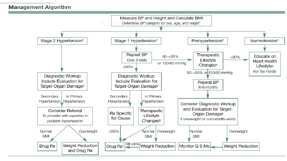
FOLLOW-UP • The decision process for follow-up, evaluation, and treatment varies with the elevation of BP. Stage 2 HTN identifies those children who need more prompt evaluation and, depending on end-organ damage and or clinical presentation, potentially immediate pharmacologic treatment. As a result, the timing of repeat blood pressure (BP) measurements is dependent upon the initial level of elevated BP, and if there are symptoms associated with HTN Copyrights apply

●Elevated BP – For children with elevated BP documented by auscultatory measurements, nonpharmacologic therapy (ie, lifestyle changes) is recommended and BP should be rechecked by auscultation in six months. • If BP remains elevated after six months, upper and lower extremity BP should be checked (right arm, left arm, and one leg) to detect coarctation of the aorta, lifestyle counseling is repeated, and auscultated BP is rechecked in six months. • If BP continues at the elevated BP level after 12 months (eg, after three auscultatory measurements), ambulatory blood pressure monitoring (ABPM) should be considered, and diagnostic evaluation should be considered if appropriate. Consider subspecialty referral (ie, nephrology or cardiology). If ABPM is not available, home BP readings with appropriate training and blood pressure cuff may be considered to help with further management decisions. • If BP normalizes at any point, return to annual BP screening at health supervisory care visits. Copyrights apply

●Stage 1 HTN Provide recommendations for nonpharmacologic measures and recheck within one to two weeks (or sooner if the patient is symptomatic). If BP remains elevated, upper and lower extremity BP should be checked (right arm, left arm, and one leg) to detect coarctation of the aorta, and BP should be rechecked in three months by auscultation. If BP remains at the stage 1 level, ABPM should be ordered (if available), and diagnostic evaluation should be considered if appropriate. Consider subspecialty referral (ie, nephrology or cardiology). Copyrights apply

Stage 2 HTN • If the patient is symptomatic, the BP is >30 mm. Hg above the 95 th percentile, or >180/120 mm. Hg (whichever is lower), the child should be referred for immediate care • If the patient is not symptomatic, upper and lower extremity BP should be checked to detect coarctation of the aorta (right arm, left arm, and one leg), lifestyle recommendations given, and the BP measurement should be repeated within one week. Alternatively, the patient could be referred to subspecialist with expertise in the evaluation and management of elevated pediatric blood pressure within one week. If at the next visit the BP is still at the stage 2 level, diagnostic evaluation, including ABPM and treatment by a specialist should be considered. Copyrights apply

Types, Causes & Clinical Picture of Hypertension
 HTN or Secondary HTN")
- It’s Either Primary (Essential ) HTN or Secondary HTN

Secondary HTN ü More common in pediatric age groups üUsualy there is an underlying condition that causes elevated BP üPt’s will have elevated BP Levels ranging from mild to severe
They are either Asymptomatic or symptoms of")
üPt’s With Secondary HTN vary in presentation: 1)They are either Asymptomatic or symptoms of the underlying causative disease process (e. g. Growth failure in CKD) 2)Presents with headache, dizziness, epistaxis, anorexia, visual changes, and seizures may occur. 3)Hypertensive encephalopathy ( suggested by the presence of vomiting, temperature elevation, ataxia, stupor, CT abnormalities, and seizures).

üThey might Present with picture of Malignant HTN ( Severely elevated BP with Cardiac failure, pulmonary edema, and renal dysfunction) ü a Hypertensive crisis that’s manifested by decreased vision (retinal hemorrhages of hypertensive retinopathy) and papilledema, encephalopathy (headache, seizures, depressed level of consciousness), heart failure, or accelerated deterioration of renal function 34
. • Higher")
When to suspect secondary HTN • A very young child (<10 years). • Higher BP readings. • No family history of HTN. • presence of symptoms related to hypertension. • Poor response to treatment (suspect non-compliance!)

Causes… 1. 2. 3. 4. 5. 6. 7. Renal (Mostly, 90% due to renovascular causes) Cardiac Vascular Endocrine Neurological Drugs Others

34 43

34 44

34 45
 HTN ü No identifiable cause can be found. üLess common than Secondary")
Primary (Essential) HTN ü No identifiable cause can be found. üLess common than Secondary HTN. • usually characterized by mild or Stage 1 hypertension. ü Hereditary factors, Diet, Stress and Obesity Plays a role in it’s development • associated with a positive family history of hypertension or cardiovascular disease (CVD).
 it becomes increasingly common üPrevalence increases")
üAs the child Grows (school aged Adolescent ) it becomes increasingly common üPrevalence increases progressively with increasing body mass index (BMI >95%) 30% have HTN. Children and adolescents with primary (essential) hypertension are commonly overweight, often have a strong family history of hypertension, and usually have BP values at or only slightly above the 95 th percentile for age
 that Further")
Overweight and high BP are components of the insulin-resistance syndrome (metabolic syndrome) that Further more exaggerated their individual CVS and diabetes risks. • In a child with primary hypertension, the presence of any comorbidity that is associated with hypertension carries the potential to increase the risk for CVD and can have an adverse effect on health outcome.

There is an association Between sleep disorders and higher blood pressure in Children. �Approximately 15 percent of children snore , and at least 1– 3 percent have sleep-disordered breathing have elevated BP. So Brief Sleep Hx should be obtained Using (BEARS) B: Bedtime problems, E: Excessive daytime sleepiness A: Awakenings during the night R: Regularity and duration of sleep S : Sleep-disordered breathing (snoring)

As Essential HTN is Asymptomatic, Doctors should always assess for Target Organ damage whenever they diagnose it.

Target-Organ Abnormalities in Childhood Hypertension Target-organ abnormalities are commonly associated with hypertension in children and adolescents. 1. Left ventricular hypertrophy (LVH) is the most prominent evidence of targetorgan damage. ■LVH has been reported in 34– 38 percent of children and adolescents with mild, untreated BP elevation. Pediatric patients with established Hypertension should have echocardiographic assessment of left ventricular mass at diagnosis and periodically thereafter. ■ The presence of LVH is an indication to Initiate Or intensify antihypertensive therapy §

2. hypertensive retinopathy. 3. Renal damage. 4. atherosclorsis At the present time, additional testing for other target-organ abnormalities (such as determination of carotid intimal-medial thickness and evaluation of urine for microalbuminuria) is not recommended for routine clinical use.

34 53
 and serum creatinine levels 54")
Azotemia : elevation of blood urea nitrogen (BUN) and serum creatinine levels 54

History and physical examination

History q. A well-taken history provides clues about the cause of hypertension and guides the selection and sequencing of the following investigations. q. Presenting symptoms and signs are not specific in neonates and are absent in most older children unless the hypertension is severe.

History q. Relevant information includes the following: • Prematurity. • Bronchopulmonary dysplasia. • History of umbilical artery catheterization. (may cause renal artery clot) • Failure to thrive. • History of head or abdominal trauma. • Family history of heritable diseases. (eg, neurofibromatosis, hypertension)

History • Medications. (eg, pressor substances, steroids, tricyclic antidepressants, cold remedies, medications for attention deficit hyperactivity disorder [ADHD]) • Episodes of pyelonephritis (perhaps suggested by unexplained fevers) that may result in renal scarring. • Dietary history, including caffeine, licorice, and salt consumption. • Sleep history, especially snoring history. • Habits, such as smoking, drinking alcohol, and ingesting illicit substances.

History q. Signs and symptoms that should alert the physician to the possibility of hypertension in neonates include the following: • Seizure. • Irritability or lethargy. • Respiratory distress. • Congestive heart failure.

History q. Signs and symptoms that should alert the physician to the possibility of hypertension in older children include all of the above, as well as the following: • Headache. • Fatigue. • Blurred vision. • Epistaxis. • Bell palsy.

Physical Examination q. A primary objective of the physical examination is to identify signs of secondary hypertension. q. The child’s height, weight, and percentiles for age (growth parameters) should be determined at the start of the physical examination. q. When hypertension is confirmed, BP should be measured in both arms and in both legs.

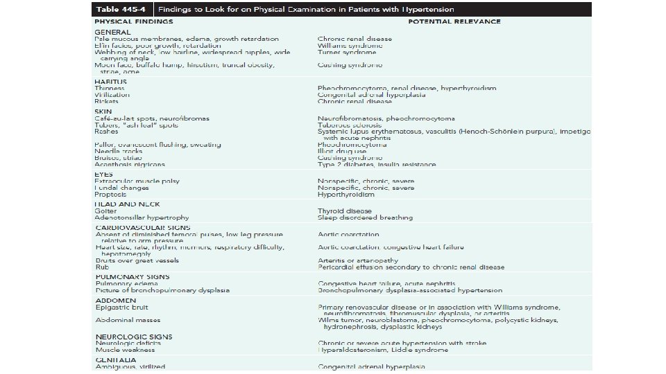
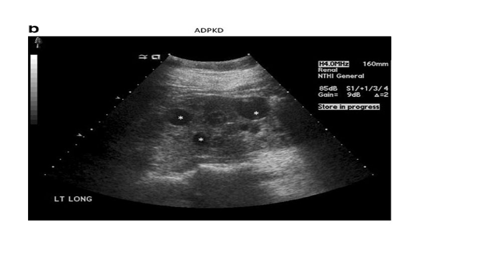
Physical Examination q. The following should be evaluated to assess for potential causes of the hypertension: • Body mass index may lead to an evaluation for metabolic syndrome. • Tachycardia may indicate hyperthyroidism, pheochromocytoma, and neuroblastoma. • Growth retardation may suggest chronic renal failure. • Café au lait spots may point to neurofibromatosis. • An abdominal mass may lead to an evaluation for Wilms tumor and polycystic kidney disease.

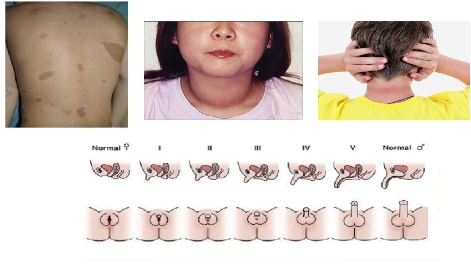
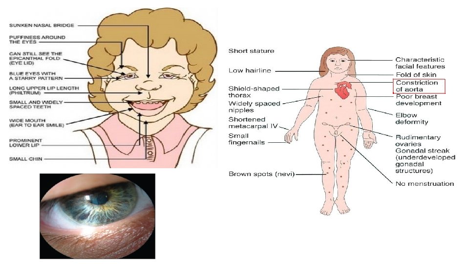
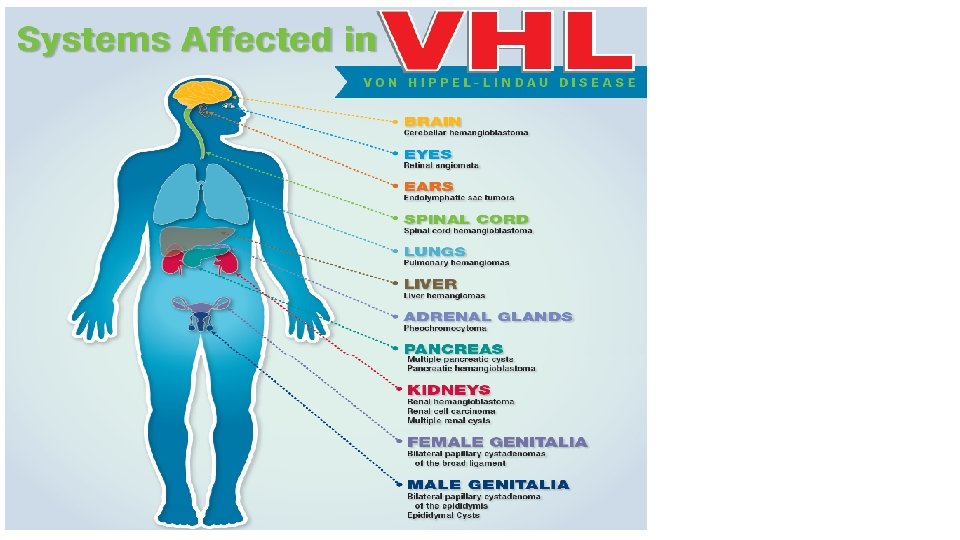
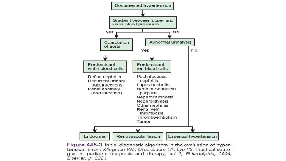
Physical Examination • Epigastric or abdominal bruit may lead to the diagnosis of coarctation of the abdominal aorta or renal artery stenosis. • BP difference between the upper and lower extremities indicates coarctation of the thoracic or abdominal aorta. • Thyromegaly may suggest hyperthyroidism. • Virilization or ambiguity may suggest adrenal hyperplasia. • Stigmata of von Hippel-Lindau, Williams, or Turner syndromes. • Acanthosis nigricans may indicate metabolic syndrome.

Investigations

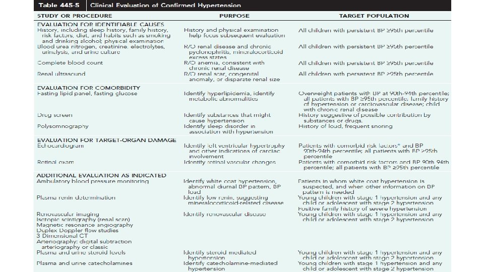
Basic Investigations • CBC : R/O anemia, consistent with chronic renal disease. • BUN, creatinine, electrolytes, urinalysis and urine culture: R/O renal disease and chronic pyelonephritis. • Renal U/S : congenital anomaly, renal size. • Renal Doppler US. • Thyroid Function Test. • Echocardiogram(LVH).

Evaluation For comorbidity • Fasting lipid panel, fasting glucose : hyperlipidemia and diabetes. • Drug screen : Identify substances that might cause hypertension. (amphetamines, Corticosteroids, decongestants) • Polysomnography : Identify sleep disorder, This test should be considered in obese children with a history of snoring, daytime sleepiness, or any sleep difficulties.

Evaluation For Target-Organ Damage • Echocardiogram : Identify LVH and other indications of cardiac involvement, Left ventricular mass measurements should be indexed to height (m 2. 7) to account for the effect of body size. The presence of LVH is an indication to treat the hypertension with pharmacologic therapy. • Left ventricular hypertrophy is detected in up to 40% of hypertensive children. • Other markers of target organ damage that have been demonstrated in hypertensive children include: increased carotid intima–media thickness, hypertensive retinopathy, and microalbuminuria.

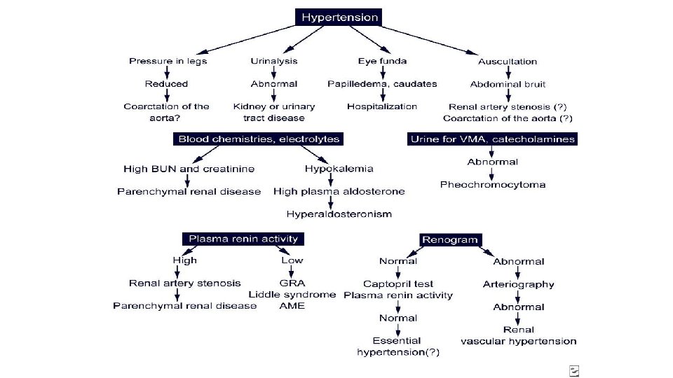
Further Evaluation As Indicated • Ambulatory BP monitoring: Identify white-coat hypertension, also useful to recognize subjects at risk, and in evaluating response to treatment. • Plasma renin determination (Plasma Renin Activity): low renin activity suggesting mineralocorticoid-related disease, while high plasma renin activity indicates renal vascular hypertension, including coarctation of the aorta. • Plasma and urine steroid levels (CAH) Identify steroid-mediated hypertension. • Plasma and urine catecholamines (neuroblastoma) : Identify catecholamine-mediated hypertension. • Urine sodium levels reflect dietary sodium intake and may be used as a marker to follow a patient after dietary changes are attempted.
. • Magnetic resonance angiography. •")
For Renal Artery Stenosis • Isotope scintigraphy (renal scan). • Magnetic resonance angiography. • Duplex Doppler flow studies. • 3 -Dimensional CT. • Arteriography: Digital subtraction angiography (DSA) or classic. • Conformation by : angiography or CT angiography. (fluoroscopic angiography may be needed, especially to detect intrarenal arterial stenosis)

CT Angiography MRA DSA

Secondary HTN-Screening Tests

Management of hypertension

Goals of management : 1 -To reduce blood pressure below the 95 th percentile for age, height and gender. 2 - To reduce blood pressure below the 90 th percentile for age, height and gender if CHD , CKD , DM , terget organs damage.

Non pharmacologic measures Intervention focus on the risk factors : 1 - weight reduction may result in a 5– 10 mm Hg reduction in systolic pressure. 2 - A reduction in sodium intake (reductions in BP, in the range of 1– 3 mm. Hg) 3 - program of aerobic exercise in patients with mild essential hypertension. 4 - Adolescents should be counseled about the adverse effects of tobacco and alcohol on blood pressure.

• When the patient is unable to cooperate with the non pharmacologic approach or the reduction in blood pressure is insufficient, antihypertensive agents should be prescribed

Pharmacologic measures Indications : . 1 - Symptomatic hypertension. 2 - Secondary hypertension. 3 - Hypertensive target-organ damage (LVH, retinopathy, proteinuria ). 4 - Diabetes ( type 1 and 2 ). 5 - Hypertension that persists despite non pharmacologic measures.

Pharmacologic measures • - pharmacotherapy should follow a step-up plan. • introducing one medication at a time at the lowest dose, then increasing the dose until therapeutic effects are seen, side effects are seen, or the maximal dose is reached. • If BP is not controlled, a drug from another class should be added. • If no control with 2 drugs, reconsider the possibility of 2 ry hypertension before adding a third drug.

Options : CCB are most frequentely chosen first line. Beta blockers , diuretics , ACEI can be also considered as first line management Alpha blockers also can be used

CCB Amlodipine. . Felodipine. They decrease systemic vascular resistence Side effects : 1 - edema 2 - flushing. . Headache 3 - reflex tachycardia 4 - hypotension

ACE inhibitors *Prefered for children with : 1 - diabetes and microalbuminuria 2 - proteinuric renal disease 3 - high-renin hypertension 2 ry to renovascular or renal parenchymal disease 4 - patients with high-renin essential hypertension. *Contraindications : 1 - bilateral renal vascular diseases 2 - single kideny 3 - renal perfusion is maintained by high level of Angiotensin 2 * You have to check serum potassium and creatinine before giving and periodically to monitor for hyperkalemia and azotemia. .

Side effects : 1 - cough … use ARBs 2 - Angioedema 3 - hyperkalemia 4 - hypotension Cough and angioedema are reportedly less common with newer members of this class than with captopril. Benazepril, enalapril, and lisinopril , captopril can prepared as suspension. FDA approval for ACE inhibitors with pediatric labeling is limited to children ≥ 6 years of age and to children with creatinine clearance ≥ 30 ml/min/1. 73 m 2.

Beta-blockers - Preferred : for children with hypertension and migraine headache - Contraindications for beta B : 1 - Asthma , reactive airway disease 2 - Heart faliure 3 - insuline dependant DM

Diuretics 1. All patients treated with diuretics should have electrolytes monitoring shortly after initiating therapy and periodically thereafter. 2. Useful as add-on therapy in patients being treated with drugs from other drug classes. * Furosemide is labeled only for treatment of edema but may be useful as add-on therapy in children with resistant hypertension, particularly in children with renal disease * Potassium-sparing diuretics (spironolactone, triamterene, amiloride) may cause severe hyperkalemia, especially if given with ACE inhibitor or ARB. * Chlorthalidone may precipitate azotemia in patients with renal diseases and should be used with caution in those with severe renal impairment.
 Used for neural crest tumors (high circulating levels of catecholamines)")
α-Adrenergic blockers (phentolamine, phenoxybenzamine) Used for neural crest tumors (high circulating levels of catecholamines) + ß-blockers to control the HR or use an agent with dual blocking action labetalol. . Can reduce systemic vascular resistence with little effect on stroke volume)
 0.")
Medications drug Dosage Range Route duration S. E Labetalol (a and ß Blockade) 0. 25_ 2. 0 mg/kg/hr 1– 3 mg/kg/24 hr IV cont PO 6 -12 hr dizziness, bronchospasm Propranolol (ßReceptor Blockade) 0. 01– 0. 1 mg/kg/dose 0. 5– 6. 0 mg/kg/24 hr, max 60 mg/24 hr IV slow push PO 6 -8 hr Bronchospasm, bradycardia, vivid dreams Hydrochlor-othiazide 1– 2 mg/kg/24 max of 100 mg/24 hr PO 12 -24 hr Hypokalemia, hyperuricemia, hypercalcemia

Drug Dosage range Route Duration S. E Furosemide 1– 2 mg/kg/dose up to 6 mg/kg/24 hr IV PO 4– 6 hr 6– 12 hr Hypokalemia, alkalosis Enalapril (ACE-I) Children: 0. 2– 1 mg/kg/24 hr Adolescents: 2. 5– 5 mg/24 hr up to 40 mg/24 hr PO 12– 24 hr Hypotension Nitroprusside Vasodilator 0. 5– 8. 0 μg/kg/min IV With infusion Thiocyanate production, rarely hypothyroidism Nifedipine CCB 0. 25– 0. 5 mg/kg/dose max of 10 mg/dose PO SL 4 -6 hr Facial flushing, tachycardia

Management of hypertensive crisis • 1 - prompt hospitalization • 2 - Use IV line for giving anti HTN 3 - select an agent with a rapid and predictable onset of action and to monitor blood pressure carefully as it is being reduced. Choices : 1 - IV Labetalol 2 - IV Nitroprusside 3 - sublingual Nifidipin 4 - Intravenous hydralazine and diazoxide are alternative but may not provide the desired gradual reduction. NOTE : Too rapid reduction in blood pressure may interfere with adequate organ perfusion, a stepwise reduction in pressure reduced by about 1/3 rd of the total planned reduction during the 1 st 6 hr and the remaining amount over the following 48 – 72 hr. should be planned.

Options for secondary HTN • 1 - Interventional cardiac catheterization procedures can be used to treat coarctation of the aorta. 2 - Balloon dilation +/- stent placement, can be used for treatment of recurrent coarctation. 3 - n children with renal artery stenosis. . percutaneous balloon angioplasty 4 - Surgery may be required for children with: • severe renal vascular hypertension • renal segmental hypoplasia • coarctation of the aorta • Wilms tumor pheochromocytoma.
- Slides: 97